. 2007 Oct 15;110(8):2855-63.
doi: 10.1182/blood-2007-04-082602. Epub 2007 Jun 14.

Hematopoietic stem-cell gene therapy of hemophilia A incorporating a porcine factor VIII transgene and nonmyeloablative conditioning regimens

Affiliations

Hematopoietic stem-cell gene therapy of hemophilia A incorporating a porcine factor VIII transgene and nonmyeloablative conditioning regimens

Lucienne M Ide et al. Blood. .

Abstract

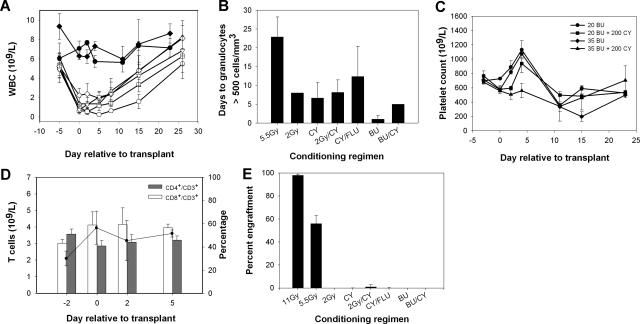
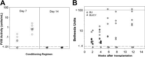
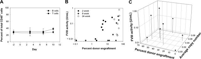
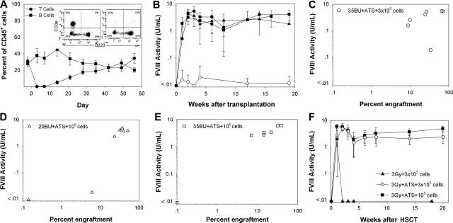
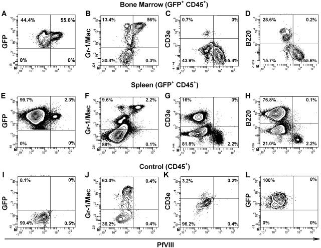
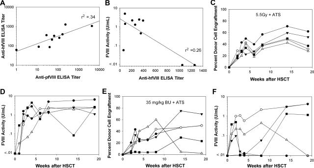
Insufficient expression of factor VIII (fVIII) is a major hurdle in the development of successful nucleic acid treatments for hemophilia. However, we recently showed that under myeloablative and reduced-intensity total body irradiation (TBI) conditioning, transplantation of hematopoietic stem cells (HSCs) transduced with recombinant retroviruses containing B domain-deleted porcine fVIII (BDDpfVIII) sequences provides curative fVIII levels in a hemophilia A mouse model. In the current study, we tested BDDpfVIII activity after nonmyeloablative conditioning with busulfan, cyclophosphamide, or fludarabine and immunosuppressive agents CTLA4-Ig + anti-CD40L or anti-(murine)thymocyte serum (ATS). ATS is similar in action to anti-(human)thymocyte globulin (ATG), which is used clinically with busulfan in bone marrow transplantations to increase donor cell engraftment. Mice conditioned with busulfan + ATS and that received a transplant of BDDpfVIII-transduced stem-cell antigen 1-positive cells exhibited moderate levels of donor cell chimerism (between 20% and 60%) and achieved sustained fVIII levels more than 1 U/mL. Similar results were observed in mice preimmunized with human fVIII and conditioned with 5 Gy TBI + ATS or busulfan + ATS. These data demonstrate that it is possible to achieve sufficient fVIII expression after transplantation of BDDpfVIII-transduced HSCs following low-toxicity pretransplantation conditioning with targeted immunosuppression, potentially even in the context of preexisting inhibitors.


Figures

Figure 1
Figure 1
Toxicity of nonmyeloablative conditioning regimens. Mice were conditioned with chemotherapeutic agents or TBI as described in “Materials and methods.” (A) White blood cell counts in mice conditioned with 2 Gy TBI (▵), 2 Gy TBI + 200 mg/kg CY (□), 200 mg/kg CY (◇), 90 mg/kg FLU + 200 mg/kg CY (▿), 20 mg/kg BU (●), and 35 mg/kg BU (♦). TBI (5.5 Gy, ○) is shown for comparison. BU + CY groups were omitted for clarity as they closely resembled the CY-only group. (B) Mean number of days to recovery of granulocytes to more than 500 per mm3. (C) Platelet counts for BU- and BU + CY-treated mice. (D) CD3+ T-cell counts (solid line) as well as the percentage of CD4+ and CD8+ subpopulations of CD3+ cells were followed by flow cytometry after conditioning with 35 mg/kg BU. (E) Percent donor chimerism measured by flow cytometry of eGFP+ donor cells in the peripheral blood following HSCT.
Figure 2
Figure 2
Transient fVIII activity and subsequent inhibitor formation following BU + CY conditioning and HSCT/ BDDpfVIII gene therapy. Mice were conditioned with 35 mg/kg BU and 200 mg/kg CY on days − 3 and − 2, then received a transplant of 3 × 10⁵ BDDpfVIII-transduced sca-1+ cells on day 0. (A) FVIII activity levels on days + 7 and + 14 as measured by chromogenic fVIII assay. Dashed line indicates lower limit of detection. Mice were assayed biweekly after day + 14 and no fVIII activity was detected in any mice. (B) Anti-BDDpfVIII inhibitory antibody titers as determined by the Bethesda assay. Solid line indicates upper limit of detection.
Figure 3
Figure 3
Toxicity, fVIII activity, and donor cell engraftment following BU conditioning with costimulation blockade. Mice were conditioned with 35 mg/kg BU on days − 3 and − 2 and then received a transplant of 3 × 10⁵ BDDpfVIII-transduced sca-1+ cells on day 0. CTLA4-Ig and anti-CD40L were administered on days 0 and + 2 relative to transplantation. (A) The percentages of B and T cells over time were determined by flow cytometry after administration of BU + costimulation blockade. (B) eGFP+ donor-cell chimerism in the peripheral blood versus fVIII activity at 2 weeks, 8 weeks, and 24 weeks after transplantation. (C) Percent eGFP+ donor-cell chimerism versus mean proviral copy number in the peripheral blood versus fVIII activity at the time of death, 6 months after transplantation.
Figure 4
Figure 4
BU + ATS conditioning facilitates sustained, therapeutic levels of fVIII. Mice were conditioned with BU on day − 3 and − 2, treated with 30 mg/kg ATS on days − 1 and 0, and then received a transplant of BDDpfVIII-transduced sca-1+ cells on day 0. (A) In vivo T-cell depletion following 2 intraperitoneal doses of ATS with representative flow cytometry analysis on days − 2 and + 3. (B) Mean fVIII plasma activity levels for mice treated with 20 mg/kg BU + ATS, 3 × 10⁵ sca-1+ cells (●), 20 mg/kg BU + ATS, 106 sca-1+ cells (▴), 35 mg/kg BU + ATS, 3 × 10⁵ sca-1+ cells (●), and 20 mg/kg BU + ATS, 106 sca-1+ cells (■). (C-E) Percentage eGFP+ donor-cell chimerism in the peripheral blood versus fVIII activity for mice conditioned with (C) 35 mg/kg BU + ATS and that received a transplant of 3 × 10⁵ sca-1+ cells, (D) 20 mg/kg BU + ATS and that received a transplant of 106 sca-1+ cells, and (E) 35 mg/kg BU + ATS and that received a transplant of 106 sca-1+ cells at 14 weeks after transplantation. (F) Mean fVIII activity levels in mice conditioned with 3 Gy TBI ± ATS on day 0.
Figure 5
Figure 5
Intracellular staining for BDDpfVIII in bone marrow and spleen after transplantation with BDDpfVIII-transduced sca-1+ cells. Four mice were killed from both the 35 mg/kg BU + ATS, 3 × 10⁵ cells and the 20 mg/kg BU + ATS, 106 cell groups. Bone marrow and spleen cells were harvested and analyzed by flow cytometry for intracellular BDDpfVIII staining, as well as surface markers for lineage differentiation. Representative data are shown for bone marrow (A-D,L) and spleen (E-H) cells from mice that underwent transplantation. Negative controls are blood from a C57BL/6 mouse (I-K) and an isotype control (L) using a biotinylated mouse anti-hfVIII antibody that is not cross-reactive to porcine or murine fVIII. Panels A-H and L are gated on CD45+ donor cells. The mouse shown was engrafted with 34% and 47% GFP+ donor cells in the bone marrow and spleen, respectively.
Figure 6
Figure 6
ATS improves circulating fVIII activity levels following BDDpfVIII-transduced HSCT of preimmunized mice. Hemophilia A mice received 6 weekly intravenous injections of recombinant hfVIII (10 U/injection). One week after the last injection, plasma was collected from each mouse (n = 5 per group) and assayed for anti-hfVIII or anti-BDDpfVIII total Ig (ELISA) and inhibitory activity (Bethesda assay). Panel A shows the anti-BDDpfVIII ELISA versus the anti-hfVIII ELISA for each mouse. Panel B shows BDDpfVIII activity following HSCT gene therapy versus pretransplantation anti-hfVIII ELISA titer for each animal. ELISA titer is defined as the reciprocal of the antibody dilution that leads to a signal 3 times background in 15 minutes. Donor cell engraftment (C) and fVIII activity (D) are shown for each animal conditioned with 5.5 Gy TBI + ATS and that received a transplant of 3 × 10⁵ BDDpfVIII-transduced sca-1+ cells. Panels E and F show the same analysis for mice conditioned with 35 mg/kg BU + ATS and that received a transplant of 3 × 10⁵ BDDpfVIII-transduced sca-1+ cells.

Similar articles

Cited by

References

    1. Lollar P. Pathogenic antibodies to coagulation factors. Part one: factor VIII and factor IX. J Thromb Haemost. 2004;2:1082–1095. - PubMed
    1. High K. Gene transfer for hemophilia: can therapeutic efficacy in large animals be safely translated to patients? J Thromb Haemost. 2005;3:1682–1691. - PubMed
    1. Doering CB, Healey JF, Parker ET, Barrow RT, Lollar P. Identification of porcine coagulation factor VIII domains responsible for high level expression via enhanced secretion. J Biol Chem. 2004;279:6546–6552. - PubMed
    1. Gangadharan B, Parker ET, Ide LM, Spencer HT, Doering CB. High-level expression of porcine factor VIII from genetically modified bone marrow-derived stem cells. Blood. 2006;107:3859–3864. - PubMed
    1. Wognum AW, Eaves AC, Thomas TE. Identification and isolation of hematopoietic stem cells. Arch Med Res. 2003;34:461–475. - PubMed

Publication types