. 2007 Aug 3;101(3):313-21.
doi: 10.1161/CIRCRESAHA.107.149047. Epub 2007 Jun 14.

Genetic manipulation of periostin expression reveals a role in cardiac hypertrophy and ventricular remodeling

Affiliations

Genetic manipulation of periostin expression reveals a role in cardiac hypertrophy and ventricular remodeling

Toru Oka et al. Circ Res. .

Abstract

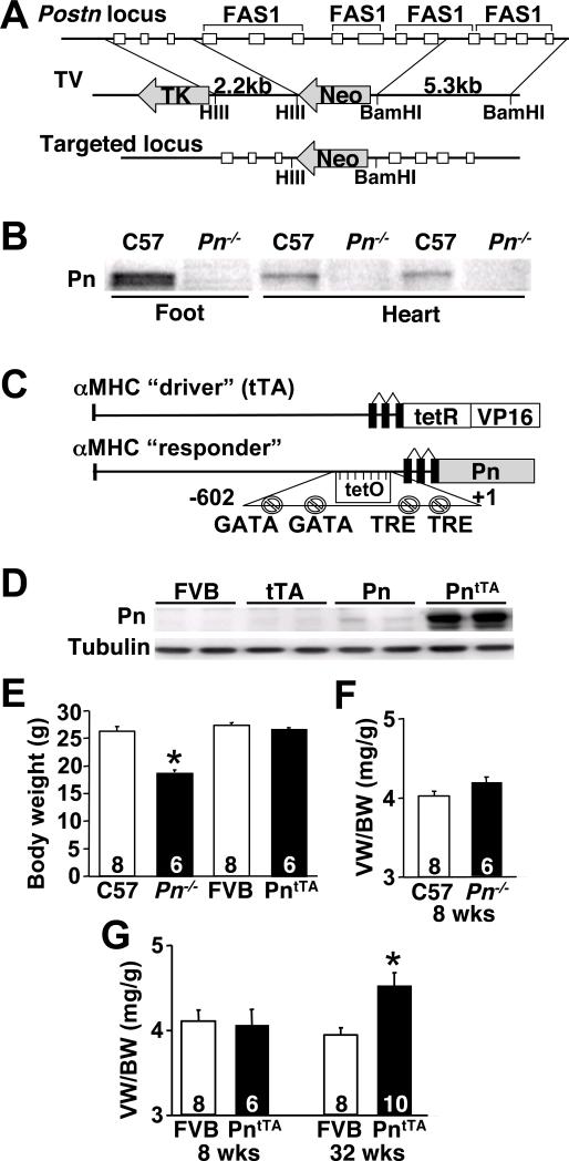
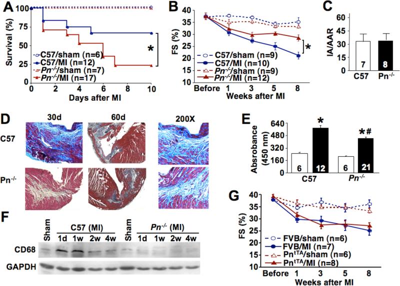
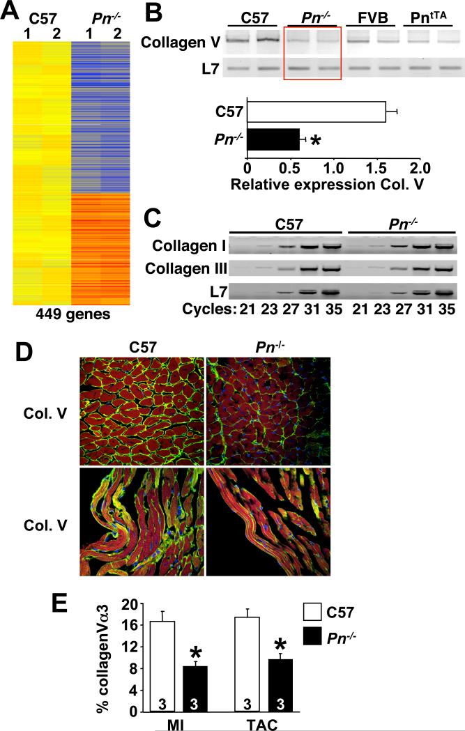
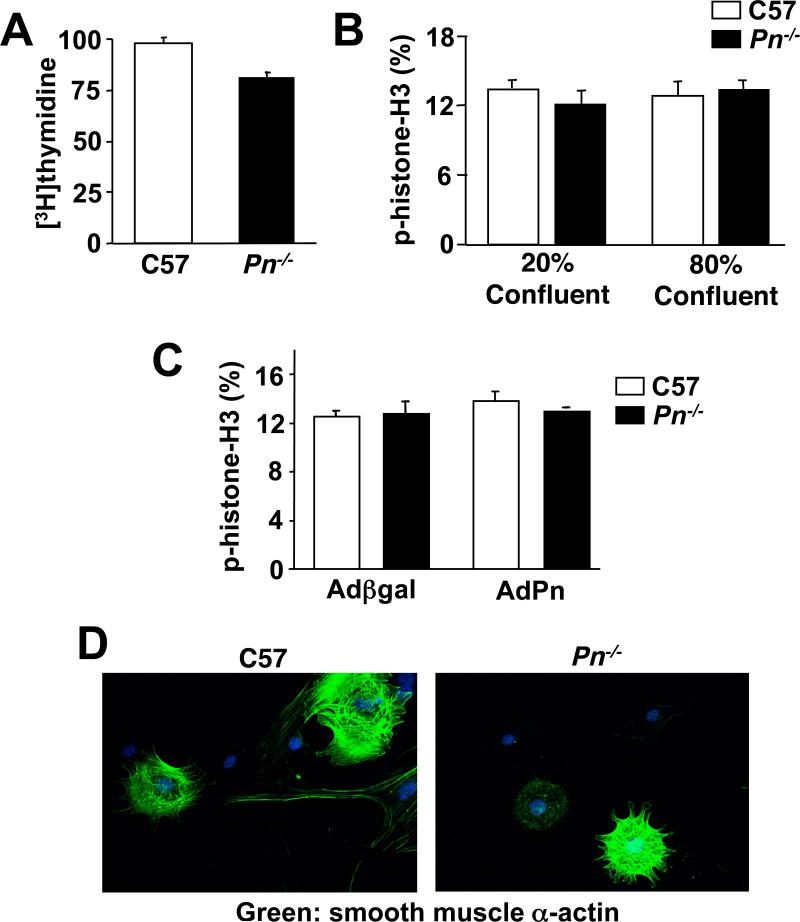
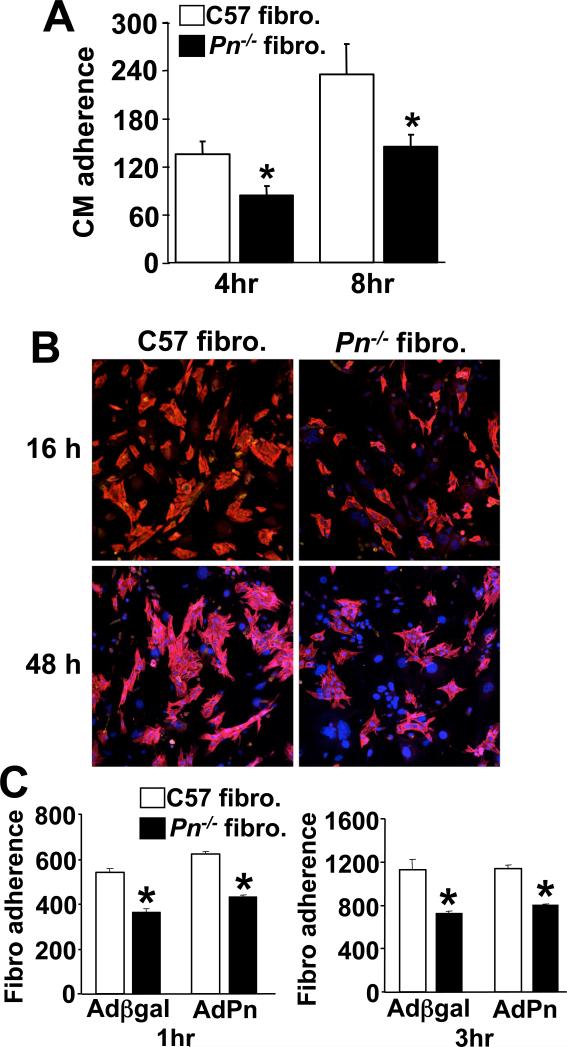
The cardiac extracellular matrix is a dynamic structural support network that is both influenced by, and a regulator of, pathological remodeling and hypertrophic growth. In response to pathologic insults, the adult heart reexpresses the secreted extracellular matrix protein periostin (Pn). Here we show that Pn is critically involved in regulating the cardiac hypertrophic response, interstitial fibrosis, and ventricular remodeling following long-term pressure overload stimulation and myocardial infarction. Mice lacking the gene encoding Pn (Postn) were more prone to ventricular rupture in the first 10 days after a myocardial infarction, but surviving mice showed less fibrosis and better ventricular performance. Pn(-/-) mice also showed less fibrosis and hypertrophy following long-term pressure overload, suggesting an intimate relationship between Pn and the regulation of cardiac remodeling. In contrast, inducible overexpression of Pn in the heart protected mice from rupture following myocardial infarction and induced spontaneous hypertrophy with aging. With respect to a mechanism underlying these alterations, Pn(-/-) hearts showed an altered molecular program in fibroblast function. Indeed, fibroblasts isolated from Pn(-/-) hearts were less effective in adherence to cardiac myocytes and were characterized by a dramatic alteration in global gene expression (7% of all genes). These are the first genetic data detailing the function of Pn in the adult heart as a regulator of cardiac remodeling and hypertrophy.


Figures

Fig 1
Fig 1
Generation of Pn−/− and PntTA inducible transgenic mice. (A) Schematic of the targeting strategy that deletes 7 exons in the Postn gene. (B) Western blot for Pn protein from 1 day-old neonatal C57 wildtype (Wt) and Pn−/− mice from the developing bones of foot and heart. (C) Schematic of the cardiac-specific Pn expressing “responder” transgene that permits inducible expression in conjunction with the tTA “driver” transgene. (D) Western blot for Pn protein from adult hearts of the indicated genotypes in the constitutively induced state. PntTA mice contain both the responder and driver transgenes required for expression. (E) Body weight of the indicated genotype of mice at 8 weeks of age. *P<0.05 vs. C57 Wt. (F,G) Assessment of ventricular weights (VW) normalized to body weights (BW) in Pn−/−, PntTA and Wt (FVB) mice at 8 or 32 weeks of age. *P<0.05 vs. Wt. The number of mice analyzed is shown in the bar.
Fig 2
Fig 2
Assessment of cardiac Pn induction by pathological stimulation. (A,B) Western blot and quantitation of Pn from cardiac protein extracts of adult mice subjected to sham or TAC for the indicated periods of time. *P<0.05 vs. sham. (C) Immunohistochemistry for Pn (green) in a Wt sham heart (left panel), after 8 weeks of TAC (middle panel), and in an adult PntTA transgenic heart (right panel). Sections were co-stained for cardiac troponin I (red). (D,E) Western blot and quantitation of Pn from cardiac protein extracts of adult mice subjected to sham or MI for the indicated periods of time in days (d) or weeks (w). *P<0.05 vs. sham. (F) Immunohistochemistry for Pn (green) 1 week after a sham or MI procedure. The infarct and border zone is shown. (G) Western blot for Pn protein from the hearts of mice subjected to swimming (left panel) or wheel running (right panel) exercise for the indicated time in days or weeks. Control (con) is from a PntTA transgenic heart. (H) Confocal immunohistochemistry for Pn (green) and vimentin (red) from adult wildtype hearts 7 days after MI injury. The arrows show areas of co-localization to fibroblasts. (I) Quantitation of fibroblast content based on vimentin staining area in hearts of wildtype (C57) and Pn−/− mice after a sham or MI procedure (7 days afterwards). *P<0.05 vs. sham; #P<0.05 vs C57 MI. Tubulin is shown as a loading control throughout the figure.
Fig 3
Fig 3
Pn−/− and PntTA mice following MI injury. (A) Survival rate of Pn−/− and C57 Wt male mice in the first 10 days after MI. *P<0.05 by Logrank test vs. C57 Wt MI mice. (B) Assessment of cardiac fractional shortening (FS, %) after sham operation or MI in the indicated groups of mice for the indicated times. *P<0.05 vs. C57 Wt MI. (C) Infarct area normalized to the area at risk (IA/AAR) in Wt and Pn−/− mice after ischemia-reperfusion injury for 24 hours. (D) Masson's trichrome staining of cardiac histological sections in the MI and border zone in Wt and Pn−/− mice for the indicted period of time. Blue staining indicates fibrosis. The 200X panels are higher magnification images to show the cellular organization and fibrosis in more detail. (E) Myeloperoxidase (MPO) assay from hearts of the indicated genotype of mice 8 weeks after a sham operation (white bar) or MI (black bar) (mice were 14−16 weeks of age). (F) Western blot for macrophage content in the heart (CD68 antibody) in Wt and Pn−/− mice after a sham operation or MI. (G) Assessment of cardiac fractional shortening (FS, %) after a sham operation or MI in Wt FVB or PntTA mice at 8 weeks of age, and followed longitudinally for the indicated time points.
Fig 4
Fig 4
Pn−/− and PntTA mice have an altered pressure overload response. (A) Ventricular weight (VW) normalized to body weight (BW) in Pn−/− and C57 Wt mice at 2 and 8 weeks after TAC or a sham operation. *P<0.05 vs. sham. #P<0.05 vs. C57 Wt at 8 weeks of TAC. (B) VW normalized to BW in PntTA and FVB Wt mice 8 weeks after TAC or a sham operation. *P<0.05 vs. sham. #P<0.05 vs. FVB Wt after TAC. (C,D) Assessment of myocyte cross-sectional area from left ventricular histological sections of the indicated genotypes (n=3 hearts each, with at least 300 cells counted in total). *P<0.05 vs. sham of the same genotype; #P<0.05 vs. Wt TAC. (E,F) Assessment of cardiac fractional shortening (FS, %) after a sham operation or TAC in Pn−/− and their strain-matched control mice, or PntTA and their respective control mice at 8 weeks of age, and followed longitudinally for the indicated time points. *P<0.05 vs. C57 Wt TAC. (G) Cardiac fibrosis assessed by hydroxyproline biochemical determination in the indicated mice 8 weeks after TAC. *P<0.05 vs. sham. #P<0.05 vs. C57 Wt TAC.
Fig 5
Fig 5
Hearts from Pn−/− mice show changes in gene expression suggestive of an altered fibroblast program. (A) Diagram of the genes that were significantly altered in expression by Affymetrix array profiling in C57 Wt and Pn−/− 8 week-old mouse hearts. Yellow is unchanged, orange and red represent increased expression, and blue represents diminished expression (n=2 hearts each). (B,C) RT-PCR for the indicted mRNA species from the hearts of the indicted genotypes. L7 was used as a control. Twenty three cycles of amplification was used in B, while panel C indicates the cycle number used. Quantitation of type V collagen α3 (Col. V) mRNA is shown in the lower panel in B. *P<0.05 vs. C57 (D,E) Immunohistochemistry and quantitiation of type V collagen α3 (Col. V) from hearts sections of Wt or Pn−/− adult mice 8 weeks after MI. Green staining in the MI border zone indicates Col. V, red indicates cardiomyocytes (troponin I), while blue indicates nuclei. Quantitation of staining in pressure overloaded hearts (TAC) is also shown in E. *P<0.05 vs. C57
Fig 6
Fig 6
Assessment of cardiac fibroblasts from Pn−/− mice. (A) Assessment of cardiac fibroblast proliferation by [3H]-thymidine incorporation in Wt or Pn−/− fibroblasts in culture. (B,C) Assessment of cardiac fibroblast proliferation by immunocytochemistry for phosphorylated-histone H3 in Wt or Pn−/− cultures at the indicated confluence. Panel C also shows the effect of Pn overexpression by adenoviral infection with AdPn, or a control, Adβgal. (D) Florescent images of smooth muscle α-actin (green) in wildtype (C57) and Pn−/− fibroblasts at low density to show myofibroblast content. Nuclei are shown in blue.
Fig 7
Fig 7
Assessment of fibroblast-cardiomyocyte adhesion in vitro. (A) The number of adherent rat neonatal cardiomyocyte (CM) normalized to an area containing 100 Wt or Pn−/− cardiac fibroblasts after 4 and 8 hrs of incubation. *P<0.05 vs. C57 Wt fibroblasts. (B) Representative immunocytochemistry for cardiomyocyte adherence on pre-plated Wt or Pn−/− cardiac fibroblasts at 16 and 48 hours. Pn−/− fibroblasts attach fewer cardiomyocytes. Red staining indicates cardiac troponin I (to show myocytes), and blue indicates nuclei. (C) Quantity of wildtype or Pn−/− fibroblasts that adhered to pre-plated neonatal cardiomyocytes cultures at 1 and 3 hrs (20 fields each). AdPn was used to generate conditioned media from Pn overexpressing myocytes and incubated on the experimental cells for 24 hrs prior to the attachment assay. Adβgal infection was used as a control. *P<0.05 vs. C57 Wt fibroblasts.

Comment in

References

    1. Levy D, Kenchaiah S, Larson MG, Benjamin EJ, Kupka MJ, Ho KK, Murabito JM, Vasan RS. Long-term trends in the incidence of and survival with heart failure. N Engl J Med. 2002;347:1397–1402. - PubMed
    1. Haldeman GA, Croft JB, Giles WH, Rashidee A. Hospitalization of patients with heart failure: National Hospital Discharge Survey, 1985 to 1995. Am Heart J. 1999;137:352–360. - PubMed
    1. Lloyd-Jones DM, Larson MG, Leip EP, Beiser A, D'Agostino RB, Kannel WB, Murabito JM, Vasan RS, Benjamin EJ, Levy D. Lifetime risk for developing congestive heart failure: the Framingham Heart Study. Circulation. 2002;106:3068–3072. - PubMed
    1. Manso AM, Elsherif L, Kang SM, Ross RS. Integrins, membrane-type matrix metalloproteinases and ADAMs: potential implications for cardiac remodeling. Cardiovasc Res. 2006;69:574–584. - PubMed
    1. Deschamps AM, Spinale FG. Matrix modulation and heart failure: new concepts question old beliefs. Curr Opin Cardiol. 2005;20:211–216. - PubMed

Publication types

MeSH terms