. 2007 Jun-Jul;28(6):1139-45.
doi: 10.3174/ajnr.A0518.

Imaging findings of bisphosphonate-associated osteonecrosis of the jaws

Affiliations

Imaging findings of bisphosphonate-associated osteonecrosis of the jaws

P M Phal et al. AJNR Am J Neuroradiol. 2007 Jun-Jul.

Abstract

Background and purpose: Bisphosphonates are drugs that decrease bone turnover by inhibiting osteoclast activity. An association between the use of bisphosphonates and osteonecrosis of the maxilla and mandible has recently been described. This study describes the imaging findings of bisphosphonate-associated osteonecrosis of the jaws.

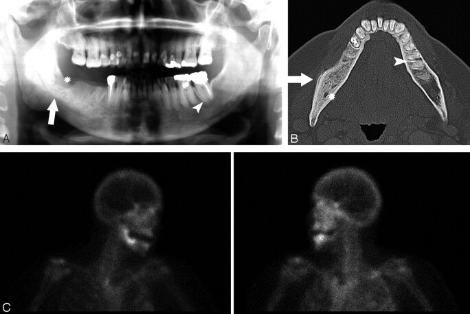
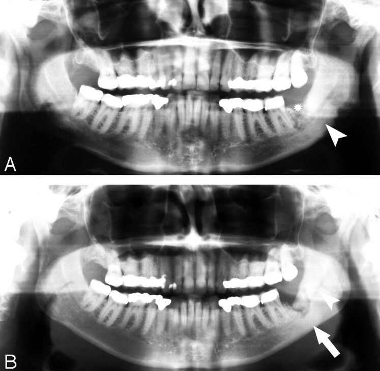
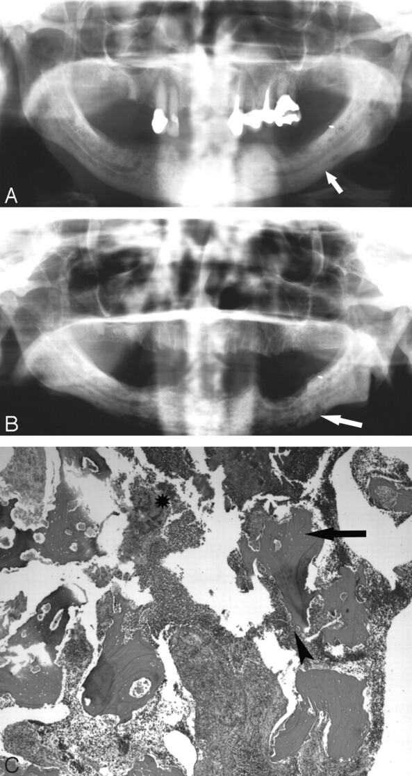
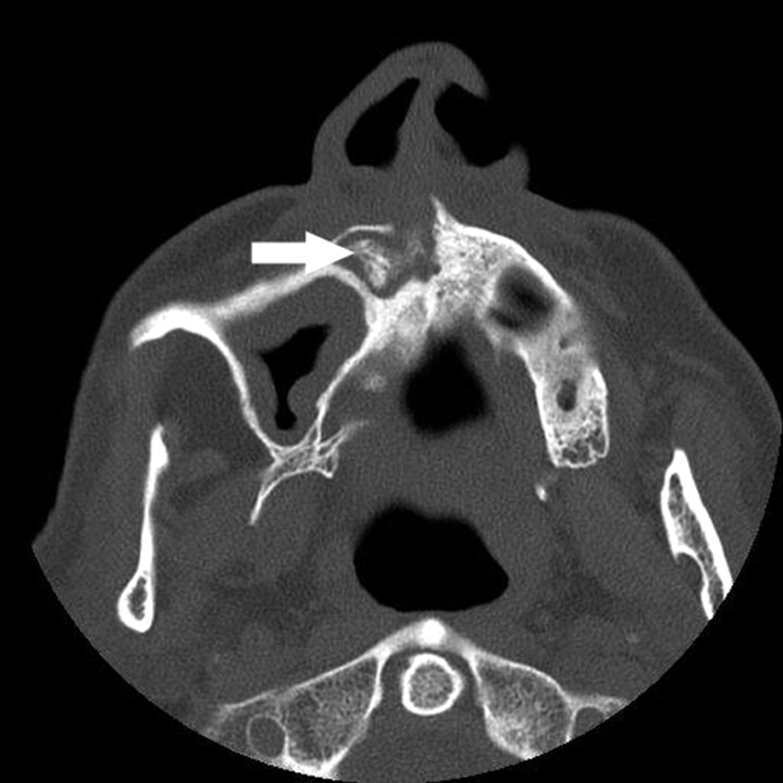
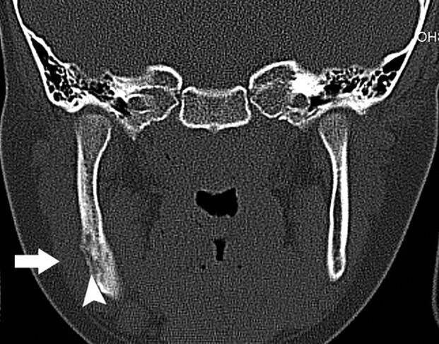
Materials and methods: This is a retrospective series of 15 clinically diagnosed patients, identified at 3 centers. Eleven patients were women, of whom 6 had breast cancer, 3 had osteoporosis, and 2 had multiple myeloma. Of the 4 male patients, 2 had prostate cancer, 1 had multiple myeloma, and 1 had osteoporosis. The age range of the patients was 52-85 years (average, 68 years). The mandible was the clinical site of involvement in 11 patients, and the maxilla was involved in 4 patients. Imaging consisted of orthopantomograms in 14 patients, CT scans in 5 patients, and radionuclide bone scan in 1 patient. Nine patients had sequential imaging. Two radiologists reviewed the images.

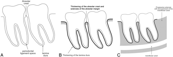
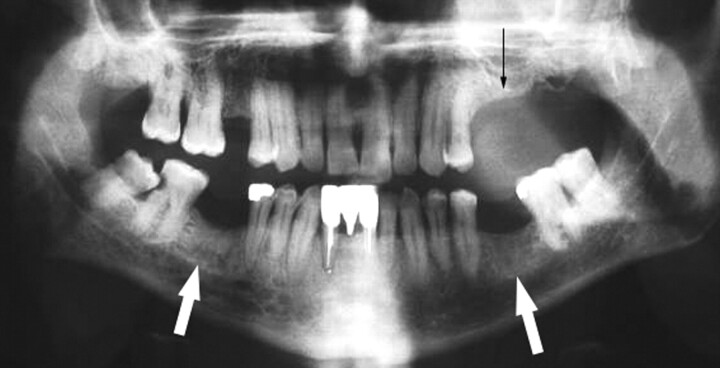
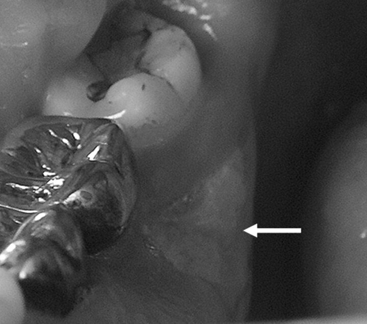
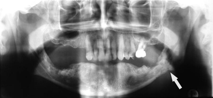
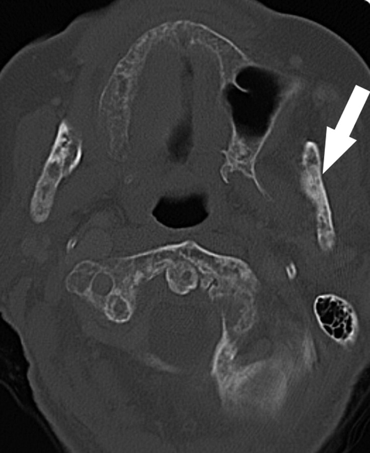
Results: All of the patients had a degree of osseous sclerosis, most commonly involving the alveolar margin, but lamina dura thickening and full-thickness sclerosis were also observed. The sclerotic change encroached on the mandibular canal in 3 patients. Less commonly encountered findings included poorly healing or nonhealing extraction sockets, periapical lucencies, widening of the periodontal ligament space, osteolysis, sequestra, oroantral fistula, soft tissue thickening, and periosteal new bone formation.

Conclusions: The most common finding in bisphosphonate-associated osteonecrosis was osseous sclerosis. This varied from subtle thickening of the lamina dura and alveolar crest to attenuated osteopetrosis-like sclerosis.


Figures

Fig 1.
Fig 1.
Radiographic findings in bisphosphonate-associated osteonecrosis of the jaws. A, Normal: the alveolar crest of the jaws is the cortical bone of the alveolar margin in between teeth and is continuous with the lamina dura around the root of the tooth. The 1- to 2-mm lucency between the root of the tooth and the lamina dura corresponds with the space for the periodontal ligament. B, In our series, osseous sclerosis was most commonly involved in the alveolar margin and lamina dura. The sclerotic changes were often diffuse rather than localized to the area of clinical involvement. C, In the patients with sequential imaging, the sclerotic changes were often progressive and may encroach on the mandibular canal. The sclerosis of the medullary cavity may be attenuated and reminiscent of osteopetrosis.
Fig 2.
Fig 2.
A 65-year-old woman (patient 1 in Table 1) with multiple myeloma treated with zoledronate presented with a nonhealing extraction socket in the left posterior maxilla (second and third molars, black arrow). The orthopantomogram demonstrates generalized sclerosis of the alveolar margin of the mandible (white arrows) and the maxilla to a lesser extent. There is also thickening of the lamina dura.
Fig 3.
Fig 3.
A 67-year-old woman (patient 2) with metastatic breast cancer treated with pamidronate and later zoledronate presented with a nonhealing extraction socket. A, The orthopantomogram demonstrates the nonhealing extraction socket in the right posterior mandible (*) with sclerosis in the adjacent body and ramus of the mandible (arrow) and generalized thickening of the lamina dura in the mandible (arrowhead) and maxilla. B, Axial CT demonstrates the osseous sclerosis, as well as narrowing the mandibular canal (*), thin periosteal new bone anteriorly (arrow) and generalized thickening of the lamina dura in the mandible (arrowhead). C, Tc99m-HDP bone scan demonstrates increased radiotracer uptake in the right hemimandible corresponding with the area of sclerosis, which had increased over the last 3 years.
Fig 4.
Fig 4.
A 60-year-old woman (patient 3) with widely metastatic breast cancer treated with pamidronate presented with a nonhealing extraction site in the left posterior mandible. A, Orthopantomogram demonstrates the nonhealing extraction site in the left posterior mandible (*) and sclerosis of the left ramus and angle of the mandible (arrowhead). B, Orthopantomogram, 23 months later with intervening curettage, demonstrates disorganized bone formation in the extraction socket of the lower left third molar, progressive sclerosis of the left ramus, and angle of the mandible (arrow) with further encroachment on the left mandibular canal (arrowhead).
Fig 5.
Fig 5.
A 77-year-old woman (patient 4) with multiple myeloma initially treated with pamidronate and subsequently zoledronate. A, The initial orthopantogram demonstrates osseous sclerosis (arrow). B, Orthopantomogram 9 months later demonstrates lytic destruction of the mandible, most prominent in the left body of the mandible (arrow). Surgical debridement was performed. C, Photomicrograph at low power (×40) of the curettage specimen stained with hematoxylin-eosin demonstrates fragments of necrotic bone with empty lacunae (arrow). There is extensive infiltration with inflammatory cells with surface resorption of bone (arrowhead) and bacteria (*).
Fig 6.
Fig 6.
A 64-year-old man (patient 5) with multiple myeloma treated with pamidronate and subsequently clodronate presented with sinusitis and chronic nasal infection. Axial CT demonstrates maxillary sclerosis, sequestrum (arrow), and mucosal thickening in the right maxillary sinus.
Fig 7.
Fig 7.
A 61-year-old man (patient 6) with metastatic prostate cancer treated with zoledronate presents with painful bone exposure of the right mandible (arrow).
Fig 8.
Fig 8.
Coronal CT in a 7-year-old girl with sclerosing osteomyelitis demonstrates osseous sclerosis, remodelling, periosteal new bone (arrowhead), and soft tissue swelling (arrow).
Fig 9.
Fig 9.
Orthopantomogram demonstrates mixed sclerotic and lytic destruction and pathologic fracture of the left body of the mandible (arrow) secondary to radiation osteonecrosis.
Fig 10.
Fig 10.
Axial CT demonstrates mixed lytic and sclerotic (arrow) metastases in the mandible and cervical spine secondary to breast carcinoma. Note the cortical thinning related to the lytic lesions.

References

    1. Sparidans RW, Twiss IM, Talbot S. Bisphosphonates in bone diseases. Pharm World Sci 1998;20:206–13 - PubMed
    1. Marx RE. Pamidronate (Aredia) and zoledronate (Zometa) induced avascular necrosis of the jaws: a growing epidemic. J Oral Maxillofac Surg 2003;61:1115–17 - PubMed
    1. Ruggiero SL, Mehrotra B, Rosenberg TJ, et al. Osteonecrosis of the jaws associated with the use of bisphosphonates: a review of 63 cases. J Oral Maxillofac Surg 2004;62:527–34 - PubMed
    1. Marx RE, Sawatari Y, Fortin M, et al. Bisphosphonate-induced exposed bone (osteonecrosis/osteopetrosis) of the jaws: risk factors, recognition, prevention, and treatment. J Oral Maxillofac Surg 2005;63:1567–75 - PubMed
    1. Migliorati CA, Schubert MM, Peterson DE, et al. Bisphosphonate-associated osteonecrosis of mandibular and maxillary bone: an emerging oral complication of supportive cancer therapy. Cancer 2005;104:83–93 - PubMed