. 2007 Jun-Jul;28(6):1179-84.
doi: 10.3174/ajnr.A0489.

Morphologic evaluation of the caudal end of the inferior petrosal sinus using 3D rotational venography

Affiliations

Morphologic evaluation of the caudal end of the inferior petrosal sinus using 3D rotational venography

Y Mitsuhashi et al. AJNR Am J Neuroradiol. 2007 Jun-Jul.

Abstract

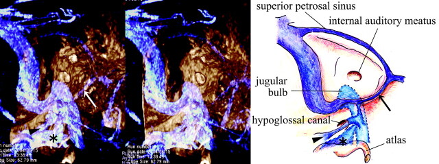
Background and purpose: The inferior petrosal sinus (IPS) is the main transvenous access route used to examine or treat lesions involving the cavernous sinus. To carry out these procedures successfully, one must have a detailed knowledge of the anatomy of the venous system around the junction of the IPS and the internal jugular vein (IJV).

Materials and methods: Eighty-three sides in 63 patients (26 men, 37 women; mean, 56.5 years of age) were examined by using 3D rotational venography (3DRV).

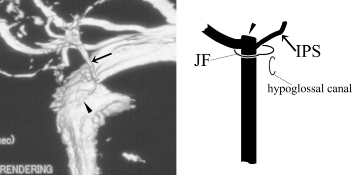
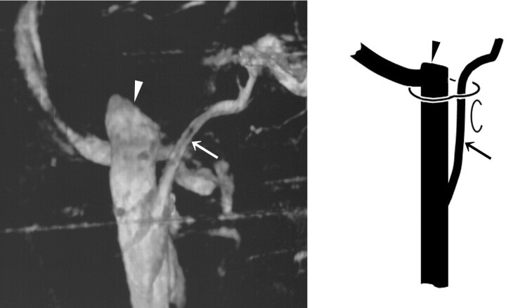
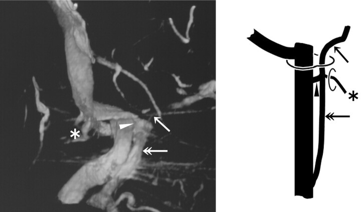
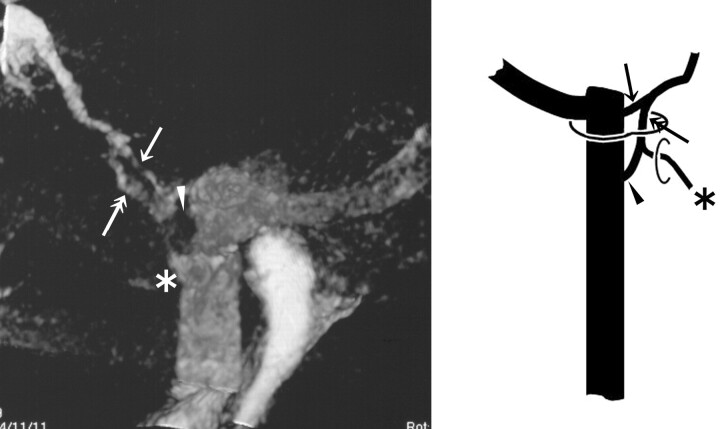
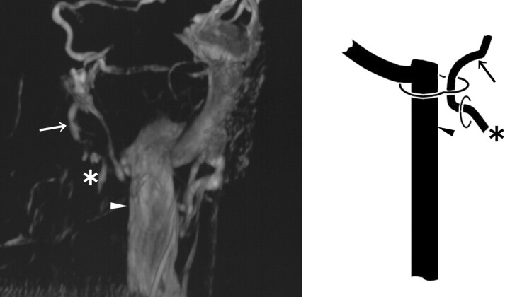
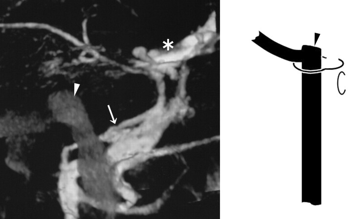
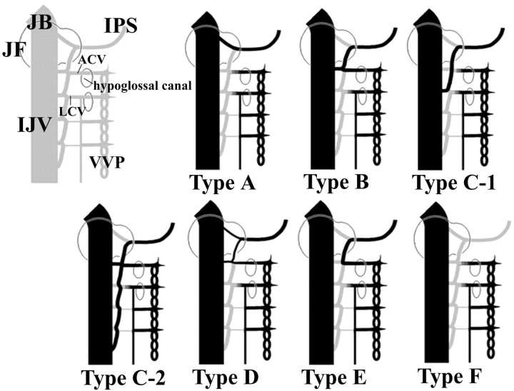
Result: The drainage patterns of the IPS could be classified into the following 6 types, with emphasis on the level of IPS-IJV junction: type A, the IPS drains into the jugular bulb in 1/83 sides (1.2%); type B, the IPS drains into the IJV at the level of the extracranial opening of the hypoglossal canal in 29/83 sides (34.9%); type C, the IPS drains into the lower extracranial IJV in 31/83 sides (37.3%); type D, the IPS forms a plexus and has multiple junctions to the IJV near the jugular foramen in 5/83 sides (6.0%); type E, the IPS drains directly into the vertebral venous plexus (VVP) with no connection to the IJV in 3/83 sides (3.6%); and type F, the IPS is absent in 14/83 sides (16.9%). Each type is also characterized by the way of anastomosis with the VVP.

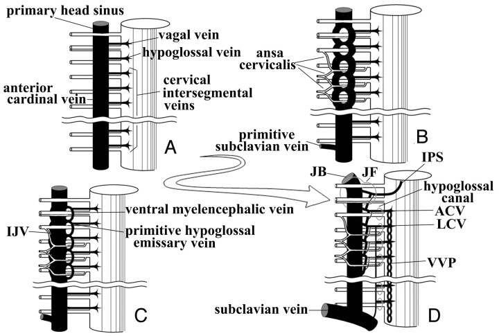
Conclusion: This classification seemed to be rational from the embryologic viewpoint, and it may be useful in establishing treatment strategies that involve endovascular manipulation via the IPS.


Figures

Fig 1.
Fig 1.
A 3DRV superimposed on a 3D rotational osteogram shows the relationship of venous structures—IPS (arrow), ACV (arrowhead), LCV (asterisk)—to bony structures (skull and upper cervical spine).
Fig 2.
Fig 2.
3DRV (anteromedial view of the right side) and schema (frontal view of the right side) of type A: The IPS (arrow) drains into the JB (arrowhead) and does not anastomose with the VVP.
Fig 3.
Fig 3.
3DRV rotation venogram (medial view of the right side) and schema (frontal view of the right side) of type B: The IPS (arrow) drains into the IJV (arrowhead) at the level of the extracranial opening of the hypoglossal canal and anastomoses with the VVP via the ACV (asterisk).
Fig 4.
Fig 4.
3DRV (frontal view of the right side) and schema (frontal view of the right side) of type C-1: The IPS (arrow) shows a long extracranial extension and joins the IJV at the level of the atlas. In this case, the IPS has no anastomosis with the VVP. The arrowhead indicates the JB.
Fig 5.
Fig 5.
3DRV (anterolateral view of the right side) and schema (frontal view of the right side) of type C-2: The IPS (arrow) shows a long extracranial extension (double arrow). Note another upper junction (arrowhead) to the IJV and the anastomosis with the VVP via the ACV (asterisk) at the level of the extracranial opening of the hypoglossal canal.
Fig 6.
Fig 6.
3DRV (medial view of the right side) and schema (frontal view of the right side) of type D: The upper route (arrow) that enters the JB has no anastomosis with the VVP, and the lower route (double arrows) anastomoses with the VVP via the ACV (asterisk) and enters the IJV (arrowhead).
Fig 7.
Fig 7.
3DRV (frontal view of the left side) and schema (frontal view of the right side) of type E: The IPS (arrow) has no connection to the IJV (arrowhead) and drains directly into the VVP via the ACV (asterisk).
Fig 8.
Fig 8.
3DRV (medial view of the left side) and schema (frontal view of the right side) of type F: The IPS is absent, and the pterygoid plexus (arrow) shows marked development. An arrowhead indicates the JB; an asterisk indicates the cavernous sinus.
Fig 9.
Fig 9.
Theoretic schematic representation of the development of the caudal portion of the IPS in the embryonic and fetal periods. A, In the early embryonic phases, the primary head sinus and the anterior cardinal vein (the future IJV) run ventromedially to the vagal, accessory, and hypoglossal nerve and to cervical nerve roots. B, The primary head sinus and the anterior cardinal vein migrate dorsolaterally to the lower cranial nerves and the ansa cervicalis, forming a venous plexus around nerves. C, The vagal vein, the hypoglossal vein, and the upper cervical intersegmental veins are stretched and anastomose with each other on the ventromedial side of the IJV. The ventral myeloencephalic vein and the primitive hypoglossal emissary vein are formed through this process. D, The ventral myeloencephalic vein is connected to the cavernous sinus with its cranial end, and the IPS is formed. The morphologic variation of the caudal portion of the IPS may be determined by the extent of development and degeneration of individual venous channels.
Fig 10.
Fig 10.
The new classification is evaluated with our theory about the development of the caudal portion of the IPS. In type A, the caudal portion of the IPS is derived from the vagal vein only and has no anastomosis with the VVP. In type B, the caudal portion of the IPS is formed by the anastomosis of the vagal vein and the hypoglossal vein, so the anastomosis with the VVP via the ACV is common. In type C-1, the caudal portion of the IPS is formed by the longitudinal anastomosis of the vagal vein, the hypoglossal vein, and the cervical intersegmental veins and drains into the extracranial lower IJV. In type C-2, the upper venous route is formed as in type B, and the another lower route develops as in type C-1. In type D, the development of the upper route drains is the same as that in type A, and the lower route develops as in type B. In type E, no venous channel connecting the IPS to the IJV develops, and the venous return drains directly into the VVP via the venous route formed by anastomosis between the vagal vein and the hypoglossal vein. In type F, the IPS is absent.
Fig 11.
Fig 11.
3DRVs (A, superomedial view of the left side; B, medial view of the left side) and schemas presenting our theory of the development of the caudal end of the IPS (ventral view of the right side) are shown. In these cases, we can find a duplicated venous channel (double arrow) with the IJV. The arrow indicates the IPS, the arrowhead indicates the JB, and the asterisk indicates the ACV. A, The duplicated channel may be formed with the longitudinal anastomosis of the hypoglossal vein and the 1st cervical intersegmental vein. B, The duplicated channel may be formed with the longitudinal anastomosis of the 1st and 2nd cervical intersegmental veins.

References

    1. Katsuta T, Rhoton AL Jr, Matsushima T. The jugular foramen: microsurgical anatomy and operative approaches. Neurosurgery 1997;41:149–202 - PubMed
    1. Ayeni SA, Ohata K, Tanaka K, et al. The microsurgical anatomy of the jugular foramen. J Neurosurg 1995;83:903–09 - PubMed
    1. Shiu PC, Hanafee WN, Wilson GH, et al. Cavernous sinus venography. Am J Roentgenol Radium Ther Nucl Med 1968;104:57–62 - PubMed
    1. Miller DL, Doppmann JL. Petrosal sinus sampling: technique and rationale (comments). Radiology 1991;178:37–47 - PubMed
    1. Miller DL, Doppman JL, Chang R. Anatomy of the junction of the inferior petrosal sinus and the internal jugular vein. AJNR Am J Neuroradiol 1993;14:1075–83 - PMC - PubMed

LinkOut - more resources