. 2007 Jun;132(7):2422-37.
doi: 10.1053/j.gastro.2007.03.101. Epub 2007 Apr 13.

Protease-activated receptor 2, dipeptidyl peptidase I, and proteases mediate Clostridium difficile toxin A enteritis

Affiliations

Protease-activated receptor 2, dipeptidyl peptidase I, and proteases mediate Clostridium difficile toxin A enteritis

Graeme S Cottrell et al. Gastroenterology. 2007 Jun.

Abstract

Background & aims: We studied the role of protease-activated receptor 2 (PAR(2)) and its activating enzymes, trypsins and tryptase, in Clostridium difficile toxin A (TxA)-induced enteritis.

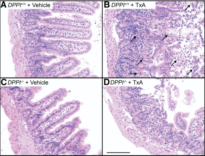
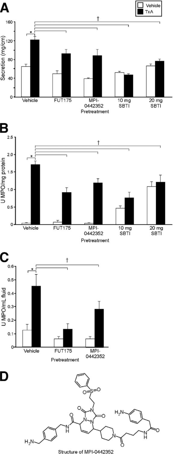
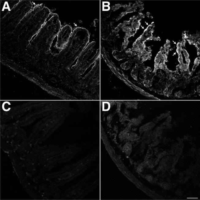
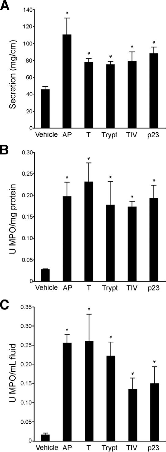
Methods: We injected TxA into ileal loops in PAR(2) or dipeptidyl peptidase I (DPPI) knockout mice or in wild-type mice pretreated with tryptase inhibitors (FUT-175 or MPI-0442352) or soybean trypsin inhibitor. We examined the effect of TxA on expression and activity of PAR(2) and trypsin IV messenger RNA in the ileum and cultured colonocytes. We injected activating peptide (AP), trypsins, tryptase, and p23 in wild-type mice, some pretreated with the neurokinin 1 receptor antagonist SR140333.

Results: TxA increased fluid secretion, myeloperoxidase activity in fluid and tissue, and histologic damage. PAR(2) deletion decreased TxA-induced ileitis, reduced luminal fluid secretion by 20%, decreased tissue and fluid myeloperoxidase by 50%, and diminished epithelial damage, edema, and neutrophil infiltration. DPPI deletion reduced secretion by 20% and fluid myeloperoxidase by 55%. In wild-type mice, FUT-175 or MPI-0442352 inhibited secretion by 24%-28% and tissue and fluid myeloperoxidase by 31%-71%. Soybean trypsin inhibitor reduced secretion to background levels and tissue myeloperoxidase by up to 50%. TxA increased expression of PAR(2) and trypsin IV in enterocytes and colonocytes and caused a 2-fold increase in Ca(2+) responses to PAR(2) AP. AP, tryptase, and trypsin isozymes (trypsin I/II, trypsin IV, p23) caused ileitis. SR140333 prevented AP-induced ileitis.

Conclusions: PAR(2) and its activators are proinflammatory in TxA-induced enteritis. TxA stimulates existing PAR(2) and up-regulates PAR(2) and activating proteases, and PAR(2) causes inflammation by neurogenic mechanisms.


Figures

Figure 1
Figure 1
Deletion of PAR2 inhibits TxA-induced fluid secretion and granulocyte infiltration. Ileal loops were injected with vehicle (vehicle control, open bars) or 0.5 μg TxA (closed bars). (A) Intestinal secretion expressed as milligrams per centimeter of loop. (B) Tissue MPO expressed as units MPO per milligram protein. (C) MPO in loop fluid expressed as units per milliliter fluid. *P < .05 between vehicle and TxA loops in Par2+/+ mice. †P < .05 between results obtained with TxA in Par2+/+ and Par2+/− or Par2−/− mice.
Figure 2
Figure 2
Deletion of PAR2 inhibits TxA-induced enteritis. Images show the ileum. (A) Par2+/+ vehicle control, (B) Par2+/+ TxA, (C) Par2+/− vehicle control, (D) Par2+/− TxA, (E) Par2−/− vehicle control, (F) Par2−/− TxA. Note the normal villous structure in control groups. In Par2+/+ mice, TxA caused pathologic changes including loss of villi, infiltration of granulocytes, and severe necrosis (arrows). Scale bar = 100 μm.
Figure 3
Figure 3
Deletion of DPPI diminishes TxA-induced fluid secretion and MPO activity in loop fluid. Ileal loops were injected with vehicle (vehicle control, open bars) or 1.0 μg TxA (closed bars). (A) Intestinal secretion expressed as milligram per centimeter of loop. (B) Tissue MPO, expressed as units MPO per milligram protein. (C) MPO in loop fluid expressed as units per milliliter of fluid. *P < .05 between buffer and TxA loops in DPPI+/+ mice. †P < .05 between results obtained with TxA in DPPI+/+ and DPPI−/− mice.
Figure 4
Figure 4
Deletion of DPPI inhibits TxA-induced enteritis. Images show the ileum. (A) DPPI+/+ vehicle control, (B) DPPI+/+ TxA, (C) DPPI−/− vehicle control, (D) DPPI−/− TxA. Note the normal villous structure in control groups. In DPPI+/+ mice, note loss of villi, infiltration of granulocytes, and severe necrosis (arrows), pathologic effects that were diminished in DPPI−/− mice. Scale bar = 100 μm.
Figure 5
Figure 5
Effect of pretreatment with tryptase and trypsin inhibitors on TxA-induced enteritis. Mice were preinjected with FUT-175, MPI-0442352, or SBTI before injection with vehicle (vehicle control, open bars) or 1.0 μg TxA (closed bars). (A) Intestinal secretion expressed as milligram per centimeter of loop. (B) Tissue MPO expressed as units per milligram protein. (C) MPO in loop fluid expressed as units per milliliter of fluid. (D) Structure of MPI-0442352. *P < .05 between vehicle control and TxA loops in vehicle-pretreated mice. †P < .05 between results obtained with TxA in mice pretreated with vehicle compared with protease inhibitors.
Figure 6
Figure 6
Effect of pretreatment with protease inhibitors on TxA-induced pathologic changes. Images show the ileum. (A) Vehicle control, (B) TxA, (C) FUT-175 plus TxA, (D) MPI-0442352 plus TxA, (E) SBTI (10 mg) plus TxA, (F) SBTI (20 mg) plus TxA. TxA caused a marked loss of villi, edema, neutrophil infiltration, and necrosis (arrows). SBTI pretreatment decreased epithelial damage and edema, but neutrophil infiltration was sometimes prominent (arrow). Scale bar = 100 μm.
Figure 7
Figure 7
Effects of TxA on PAR2 expression. Ileal loops formed in wild-type mice were injected with (A and C) vehicle or (B and D) 1.0 μg TxA. Tissue was stained for PAR2 using a mouse anti-PAR2 antibody 9717 and (A and B) a goat anti-rabbit secondary antibody conjugated to FITC or (C and D) the goat anti-rabbit secondary antibody conjugated to FITC alone. Images were acquired by confocal microscopy and are unmodified. The images shown in A and B were acquired under identical conditions to optimize the quantification of pixel intensity above baseline. The images shown in C and D were acquired under identical conditions to optimize tissue visualization. (A) Basal PAR2 expression with positive enterocytes (arrow). (B) TxA markedly up-regulated PAR2 expression in enterocytes (arrows). Omission of the mouse anti-PAR2 antibody 9717 eliminated specific staining in both (C) vehicle-treated ileum and (D) TxA-treated ileum. Scale bar = 50 μm.
Figure 8
Figure 8
Effects of TxA on NCM460 cells. (A) Real-time quantitative PCR of PAR2 levels. Results of 3 experiments are shown. After 120 minutes of exposure to TxA, PAR2 levels were increased and remained increased even after 360 minutes. (B) Real-time quantitative PCR of trypsin IV levels. Results of 3 experiments are shown. After 120 minutes of exposure to TxA, trypsin IV levels were increased and remained increased even after 360 minutes. (C) Ca2+ mobilization in response to PAR2 AP or vehicle (1/1300 dilution of 0.05 mol/L Tris in NCM culture medium) in cells pretreated with 5 nmol/L or 5 μmol/L TxA. Ca2+ responses to PAR2-AP (10−5 mol/L) are expressed as a 340/380 ratio (peak baseline; n = 38−45 cells). TxA significantly enhanced responses to PAR2-AP. *P < .05 between control or 0 minutes and TxA-exposed cells. †P = .055 between control or 0 minutes and TxA-exposed cells.
Figure 9
Figure 9
Effect of activation of PAR2 on inflammation in the ileum. Mouse ileum was injected with AP (SLIGRL), pancreatic trypsin (T), tryptase (Trypt), trypsin IV (TIV), p23, or vehicle. Inflammation was assessed as before (Figures 1, 3, and 5). (A) All agents increased intestinal secretion, expressed as milligrams per centimeter of loop, with AP having the largest effect. (B) All agents increased tissue MPO, expressed as units MPO per milligram protein, to similar levels. (C) Fluid MPO, expressed as units MPO per milliliter fluid, was most strikingly increased by AP, trypsin, and tryptase. *P < .05 between control and results obtained by activating agents.
Figure 10
Figure 10
Effect of NK1R inhibition on PAR2-induced inflammation in the ileum. Mouse ileum was injected with carrier (1% dimethyl sulfoxide) or SR140333 60 minutes before AP. (A) Intestinal secretion, expressed as milligram per centimeter of loop, (B) tissue MPO, expressed as units MPO per milligram protein, and (C) fluid MPO, expressed as units MPO per milliliter fluid, were all significantly decreased by pretreatment with SR140333. *P < .05 between AP and results obtained by pretreatment with SR140333 followed by AP.

Comment in

References

    1. Vergnolle N. Modulation of visceral pain and inflammation by protease-activated receptors. Br J Pharmacol. 2004;141:1264–1274. - PMC - PubMed
    1. Bunnett NW, Cottrell GS. Protease-activated receptors in gastrointestinal function and disease. In: Lendeckel U, Hooper NM, editors. Proteases in gastrointestinal tissues. Vol. 5. Dordrecht: Springer: 2006. pp. 1–32.
    1. Corvera CU, Dery O, McConalogue K, Bohm SK, Khitin LM, Caughey GH, Payan DG, Bunnett NW. Mast cell tryptase regulates rat colonic myocytes through proteinase-activated receptor 2. J Clin Invest. 1997;100:1383–1393. - PMC - PubMed
    1. Reed DE, Barajas-Lopez C, Cottrell G, Velazquez-Rocha S, Dery O, Grady EF, Bunnett NW, Vanner SJ. Mast cell tryptase and protein-ase-activated receptor 2 induce hyperexcitability of guinea-pig submucosal neurons. J Physiol. 2003;547:531–542. - PMC - PubMed
    1. Kong W, McConalogue K, Khitin LM, Hollenberg MD, Payan DG, Bohm SK, Bunnett NW. Luminal trypsin may regulate enterocytes through proteinase-activated receptor 2. Proc Natl Acad SciUSA. 1997;94:8884–8889. - PMC - PubMed

Publication types

MeSH terms