. 2007 Sep;1768(9):2145-56.
doi: 10.1016/j.bbamem.2007.04.027. Epub 2007 May 10.

Interactions of Plasmodium falciparum erythrocyte membrane protein 3 with the red blood cell membrane skeleton

Affiliations

Interactions of Plasmodium falciparum erythrocyte membrane protein 3 with the red blood cell membrane skeleton

Karena L Waller et al. Biochim Biophys Acta. 2007 Sep.

Abstract

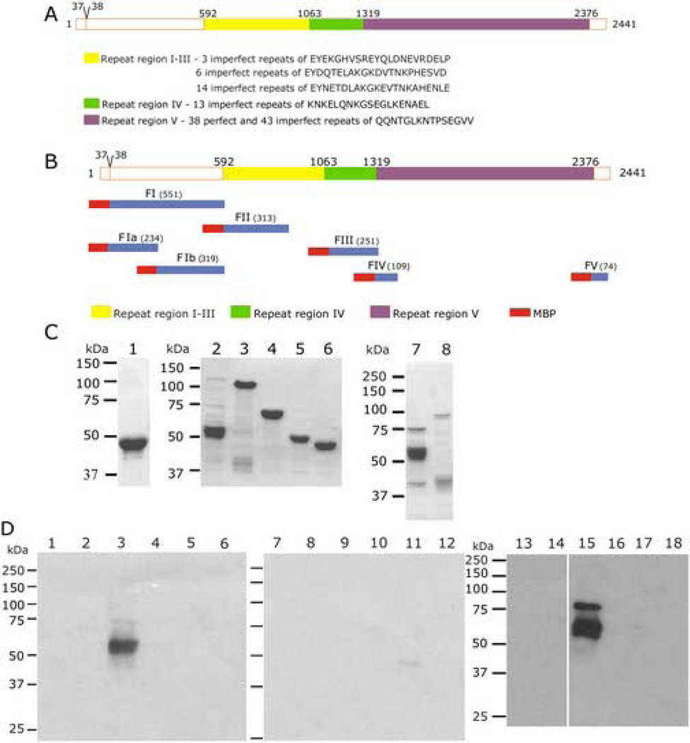
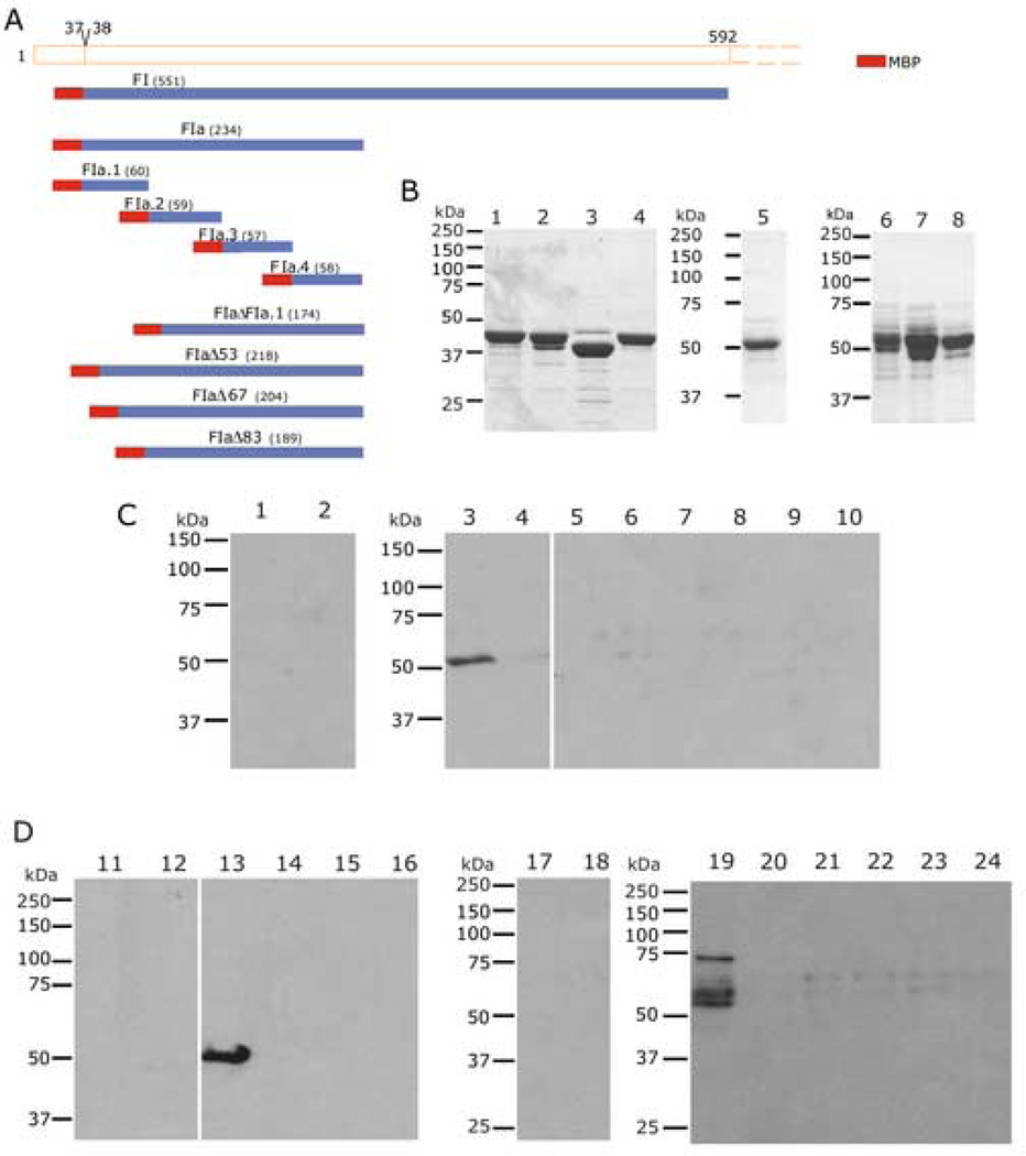
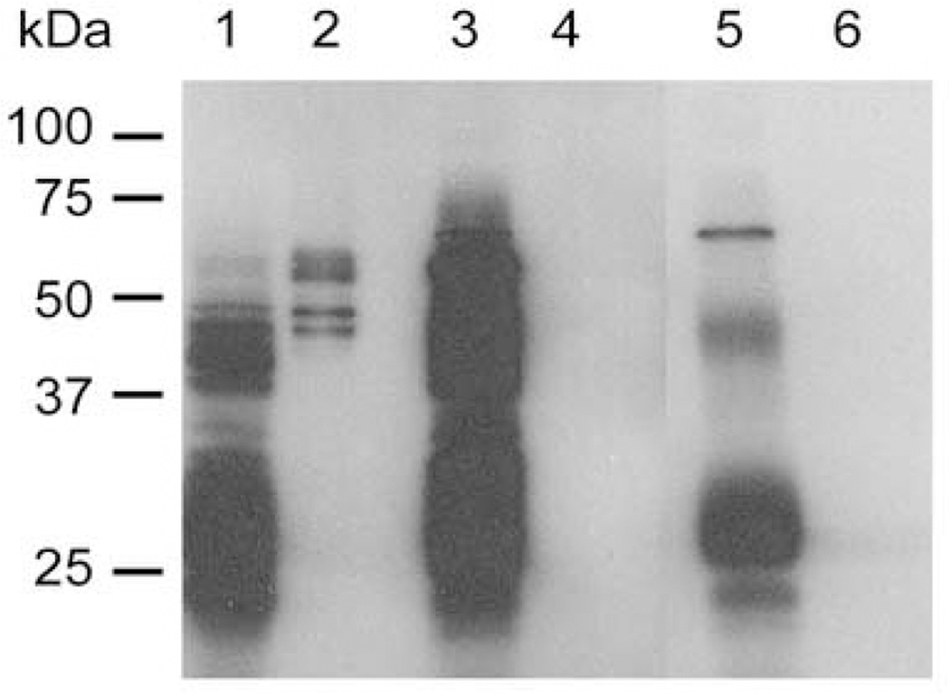
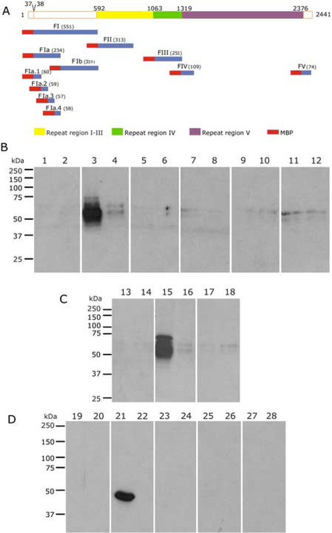
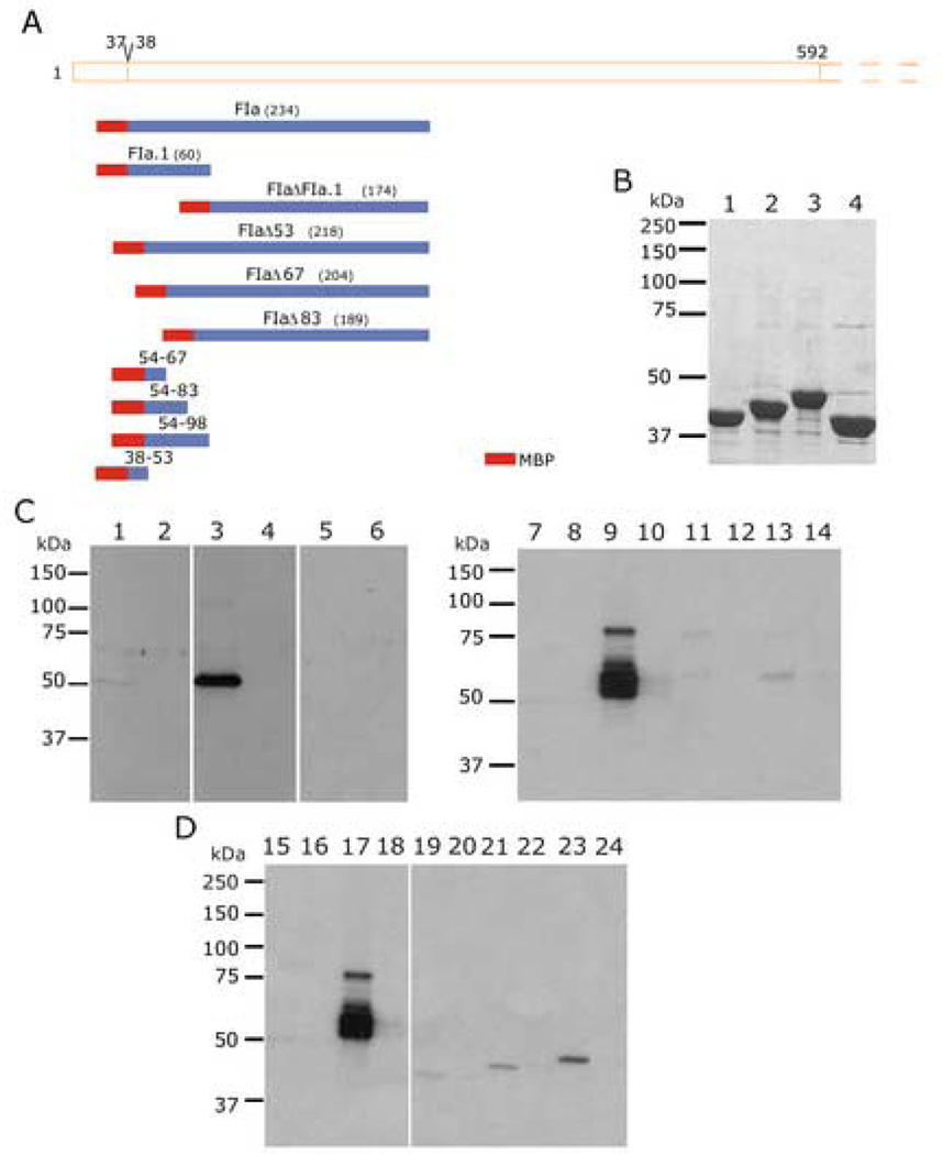
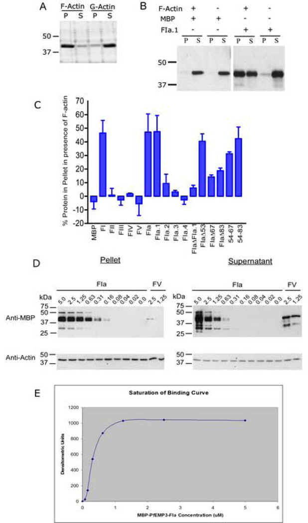
Plasmodium falciparum parasites express and traffick numerous proteins into the red blood cell (RBC), where some associate specifically with the membrane skeleton. Importantly, these interactions underlie the major alterations to the modified structural and functional properties of the parasite-infected RBC. P. falciparum Erythrocyte Membrane Protein 3 (PfEMP3) is one such parasite protein that is found in association with the membrane skeleton. Using recombinant PfEMP3 proteins in vitro, we have identified the region of PfEMP3 that binds to the RBC membrane skeleton, specifically to spectrin and actin. Kinetic studies revealed that residues 38-97 of PfEMP3 bound to purified spectrin with moderately high affinity (K(D(kin))=8.5 x 10(-8) M). Subsequent deletion mapping analysis further defined the binding domain to a 14-residue sequence (IFEIRLKRSLAQVL; K(D(kin))=3.8 x 10(-7) M). Interestingly, this same domain also bound to F-actin in a specific and saturable manner. These interactions are of physiological relevance as evidenced by the binding of this region to the membrane skeleton of inside-out RBCs and when introduced into resealed RBCs. Identification of a 14-residue region of PfEMP3 that binds to both spectrin and actin provides insight into the potential function of PfEMP3 in P. falciparum-infected RBCs.


Figures

Fig. 1
Fig. 1. Mapping the Region of PfEMP3 that Binds to the RBC Skeleton
A. Schematic representation of PfEMP3 and representative sequences located in each distinct peptide repeat region. Repeat regions are shown in solid, shaded boxes, whereas non-repeat regions are shown in open boxes. Residue numbers are indicated above the schematic. The intron splice site, located between the first and second exon of pfemp3 is indicated by the division at residues 37–38 on the schematic. B. Schematic representation of PfEMP3 and the initial sub-fragments used to analyse binding to the RBC skeleton. Residue numbers and fragment lengths are indicated above the schematic and adjacent to the fragment name (shown in brackets), respectively. C. Purified MBP-PfEMP3 fusion proteins. 2µg (total protein) of each purified protein was resolved by SDS-PAGE in 10% polyacrylamide gels, prior to visualisation with Coomassie Brilliant Blue. The protein samples are: MBP (lane 1), -FI (lane 2), -FII (lane 3), -FIII (lane 4), -FIV (lane 5), -FV (lane 6), -FIa (lane 7) and -FIb (lane 8). D. IOV binding assay immunoblots. IOVs (lanes 1, 3, 5, 7, 9, 11, 13, 15 and 17) and BSA (lanes 2, 4, 6, 8, 10, 12, 14, 16 and 18) were coated on the wells of the 96 well plate prior to adding MBP-PfEMP3 fusion proteins or MBP control protein: MBP (lanes 1, 2, 13 and 14), MBP-PfEMP3-FI (lanes 3 and 4), -FII (lanes 5 and 6), -FIII (lanes 7 and 8), FIV (lanes 9 and 10), -FV (lanes 11 and 12), -FIa (lanes 15 and 16) and -Flb (lanes 17 and 18) . -FI, and its sub-fragment –FIa bound to IOVs ( lanes 3 and 15) but not to BSA (lanes 4 and 16). Residual levels of –FV bound to IOVs (lane 11). MBP did not bind to either IOVs (lanes 1 and 13) or BSA (lanes 2 and 14).
Fig. 2
Fig. 2. Finer Mapping of the Region of PfEMP3 that Binds to the Skeleton
A. Schematic representation of PfEMP3-FI region (residues 38–592) and the smaller PfEMP3 sub-fragments and deletion mutant proteins. B. Purified MBP-PfEMP3 fusion proteins. 2µg (total protein) of each purified protein was resolved by SDS-PAGE, prior to visualisation with Coomassie Brilliant Blue. The protein samples are: -FIa.1 (lane 1), -FIa.2 (lane 3), -FIa.3 (lane 3), -FIa.4 (lane 4), -FIaΔFIa.1 (lane 5), -FIaΔ53 (lane 6), -FIaΔ67 (lane 7) and -FIaΔ83 (lane 8). C and D. IOV binding assay immunoblots. IOVs (lanes 1, 3, 5, 7, 9, 11, 13, 15, 17, 19, 21 and 23) and BSA (lanes 2, 4, 6, 8, 10, 12, 14, 16,18, 20, 22 and 24) were coated on the wells of the 96 well plate prior to adding MBP-PfEMP3 fusion proteins or MBP control protein: MBP (lanes 1, 2, 11, 12, 17 and 18), -FIa.1 (lanes 3 and 4), -FIa.2 (lanes 5 and 6), -FIa.3 (lanes 7 and 8), -FIa.4 (lanes 9 and 10), -FIa (lanes 13 and 14), -FIaΔFIa.1 (lanes 15 and 16), -FIaΔ53 (lanes 19 and 20), -FIaΔ67 (lanes 21 and 22) and –FIaΔ83 (lanes 23 and 24). The sub-fragment of –Fla, FIa.1 bound to IOVs (lane 3) and not to BSA (lane 4), thereby mapping the membrane skeleton binding region of PfEMP3 to a 60 residue region. Deletion of these 60 residues from the binding fragment FIa, resulting in the generation –FIaΔFIa.1, abolished binding to IOVs (lane 15). Finer deletion analysis was performed to dissect the 60 residue region, using -FIaΔ53, -FIaΔ67 and -FIaΔ83, showed binding of -FIaΔ53 to IOVs (lane 19) but not to BSA (lane 20). Both -FIaΔ67 and -FIaΔ83 did not bind to IOVs (lanes 21 and 23, respectively) or BSA (lanes 22 and 24, respectively).
Fig. 3
Fig. 3. Membrane Skeleton Association of PfEMP3 FI
TX100 detergent extractions were performed on lysed and resealed RBCs that had been incubated with 5 µM purified MBP-PfEMP3-FI or –FV. TX100 soluble and insoluble fractions were resolved by SDS-PAGE and immuno-blotted with anti-MBP antiserum. MBP-PfEMP3-FI was detected in both the TX100 soluble and insoluble fractions (lanes 1 and 2, respectively). –FV, a region of PfEMP3 that does not bind to the membrane skeleton, was detected only in the TX100 soluble fraction (lane 3) but not the TX100 insoluble fraction (lane 4). The control TX100 soluble and insoluble fractions of normal lysed and resealed (no MBP fusion protein) RBCs are shown in lanes 5 and 6, respectively.
Fig. 4
Fig. 4. Mapping the Region of PfEMP3 that Binds to Spectrin
A. Schematic representation of PfEMP3 and its sub-fragment proteins. B, C and D. Spectrin binding assay immunoblots. Spectrin (lanes 1, 3, 5, 7, 9, 11, 13, 15, 17, 19, 21, 23, 25 and 27) and BSA (lanes 2, 4, 6, 8, 10, 12, 14, 16,18, 20, 22, 24, 26 and 28) were coated on the wells of the 96 well plate prior to adding MBP-PfEMP3 fusion proteins or MBP control protein: MBP (lanes 1, 2, 13, 14, 19 and 20), -FI (lanes 3 and 4), -FII (lanes 5 and 6), -FIII (lanes 7 and 8), -FIV (lanes 9 and 10), -FV (lanes 11 and 12), -FIa (lanes 15 and 16), -FIb (lanes 17 and 18), -FIa.1 (lanes 21 and 22), –FIa.2 (lanes 23 and 24), -Fla.3 (lanes 25 and 26) and – Fla.4 (lanes 27 and 28). Fragment –FI and its successively smaller sub-fragments – FIa and –Fla.1 all bound to IOVs (lanes 3, 15 and 21, respectively) but not to BSA (lanes 4, 16 and 22, respectively). These data map the region of PfEMP3 that binds to spectrin to the same 60 residues that bind to IOVs.
Fig. 5
Fig. 5. Deletion Mapping of PfEMP3 Binding to Spectrin
A. Schematic representation of PfEMP3-FI region (residues 38–592) and the smaller PfEMP3 sub-fragments and deletion fragments used to map the spectrin binding domain. B. Additional MBP-PfEMP3 fusion proteins that were used in these analyses are shown. 2µg (total protein) of each purified protein was resolved by SDS-PAGE and stained with Coomassie Brilliant Blue. The protein samples are: −5438–592) and the smaller PfEMP3 sub-fragments and deletion fragments used to map 67 (lane 1), −5438–592) and the smaller PfEMP3 sub-fragments and deletion fragments used to map 83 (lane 2), −5438–592) and the smaller PfEMP3 sub-fragments and deletion fragments used to map 98 (lane 3) and −38–53 (lane 4). C and D. Spectrin binding assay immunoblots. Spectrin (lanes 1, 3, 5, 7, 9, 11, 13, 15, 17, 19, 21 and 23) and BSA (lanes 2, 4, 6, 8, 10, 12, 14, 16,18, 20, 22 and 24) were coated on the wells of the 96 well plate prior to adding MBP-PfEMP3 fusion proteins or MBP control protein: MBP (lanes 1, 2, 7, 8, 15 and 16), MBP-PfEMP3-FIa (lanes 3 and 4), -FIaΔFIa.1 (lanes 5 and 6), -FIaΔ53 (lanes 9 and 10), -FIaΔ67 (lanes 11 and 12), -FIaΔ83 (lanes 13 and 14), -FIaΔ53 (lanes 17 and 18), −54–67 (lanes 19 and 20), −54–83 (lanes 21 and 22) and –54–98 (lanes 23 and 24). PfEMP3-FIa (lane 3) and its deletion mutant -FIaΔ53 (lane 9 and 17) and −54–67, −54–83 and −54–98 (lanes 19, 21 and 23, respectively) each bound to spectrin only. Taken together, these data map the minimal binding region of PfEMP3 to spectrin to the 14 residue region represented on the protein fragment −54–67.
Fig. 6
Fig. 6. The Region of PfEMP3 that Binds to Actin
A. Preliminary co-sedimentation assays were performed using polymerised F-actin and monomeric G-actin. Samples of F-actin and G-actin were then centrifuged at 25 °C for 10 min at 85,000 rpm (313,000 × g). Pellet and supernatant samples from each reaction were then resolved by SDS-PAGE in 10 % (w/v) polyacrylamide gels and stained with Coomassie Brilliant Blue. Nearly all the polymerised F-actin was found in the pellet fraction (P), whereas most of the monomeric G-actin was found in the supernatant fraction (S). Only residual levels of actin were observed in the other fractions. B. Eqi-molar solutions (5 µM) of F-actin and recombinant MBP-PfEMP3 fusion proteins were interacted and co-sedimented. Immunoblot analysis of resultant supernatant and pellet fractions was performed. Representative immunoblots show MBP alone is unable to bind F-actin, whereas the MBP-PfEMP3-F1a.1 fusion protein co-sedimented in the presence of F-actin, (although a proportion of the recombinant protein also remained in solution). C. Eqi-molar solutions (5 µM) of F-actin and purified MBP-PfEMP3 fusion proteins were interacted and then co-sedimented. The pellet and supernatant fractions were resolved by SDS-PAGE and the immunoblot data analysed by densitometry. The graphed data show pelleting of F-actin with -FI, and its successively smaller sub-fragments, -FIa, -FIa.1. Deletion mutagenesis further defined the binding region to 14 residues (residues 54–67 of PfEMP3), as evidenced by -FIaΔ53 pelleting with F-actin, whereas –FIaΔ67 did not. The presence of only these 14 residues in the −54–67 fragment also resulted in pelleting with F-actin. D. Binding of F-actin to -FIa was demonstrated to be specific and saturable. Constant 5 µM actin was incubated with titrated serial dilutions of –FIa and centrifuged. Pellet and supernatant fractions were analysed by immunoblot, using anti-MBP and anti-actin antibodies and subsequent densitometry. E. Saturation of binding curve constructed from densitometric analyses of titrated – FIa co-sedimented with constant 5 µM actin (Fig. 5D). The graphed data are from a representative assay (mean ± standard deviation).

References

    1. Snow RW, Guerra CA, Noor AM, Myint HY, Hay SI. The global distribution of clinical episodes of Plasmodium falciparum malaria. Nature. 2005;434:214–217. - PMC - PubMed
    1. Cooke BM, Mohandas N, Coppel RL. The malaria-infected red blood cell: structural and functional changes. Adv Parasitol. 2001;50:1–86. - PMC - PubMed
    1. Cooke BM, Mohandas N, Coppel RL. Malaria and the red blood cell membrane. Semin Hematol. 2004;41:173–188. - PubMed
    1. Speicher DW, Morrow JS, Knowles WJ, Marchesi VT. Identification of proteolytically resistant domains of human erythrocyte spectrin. Proc Natl Acad Sci U S A. 1980;77:5673–5677. - PMC - PubMed
    1. Bennett V, Gilligan DM. The spectrin-based membrane skeleton and micron-scale organization of the plasma membrane. Annual Review of Cell Biology. 1993;9:27–66. - PubMed

Publication types

Substances

LinkOut - more resources