. 2007 Jun;26(6):784-97.
doi: 10.1016/j.immuni.2007.04.015.

Transcellular diapedesis is initiated by invasive podosomes

Affiliations

Transcellular diapedesis is initiated by invasive podosomes

Christopher V Carman et al. Immunity. 2007 Jun.

Abstract

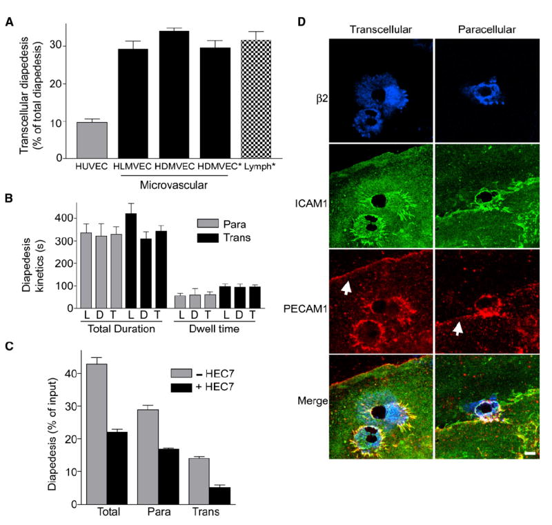
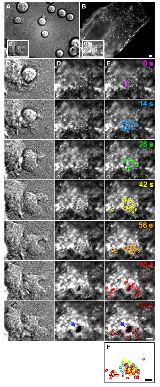
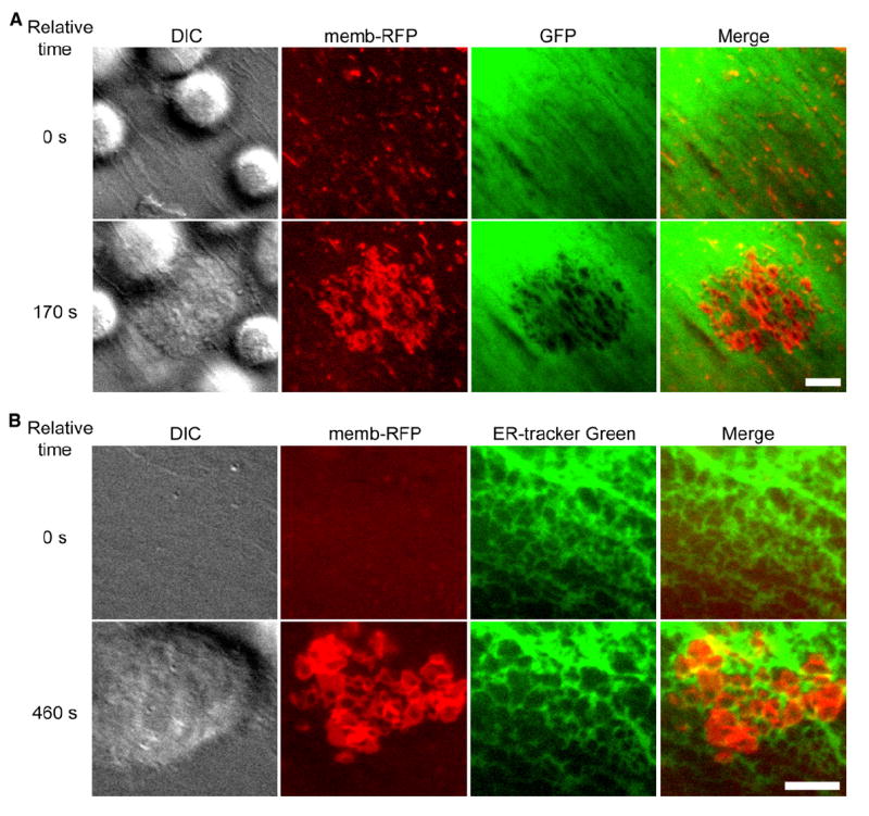
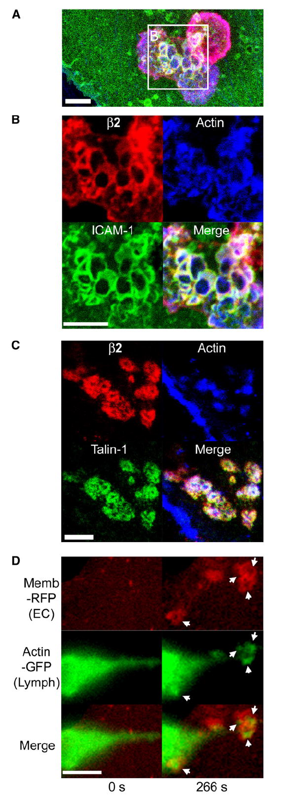
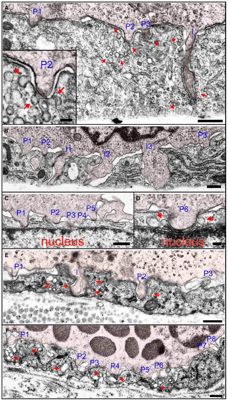
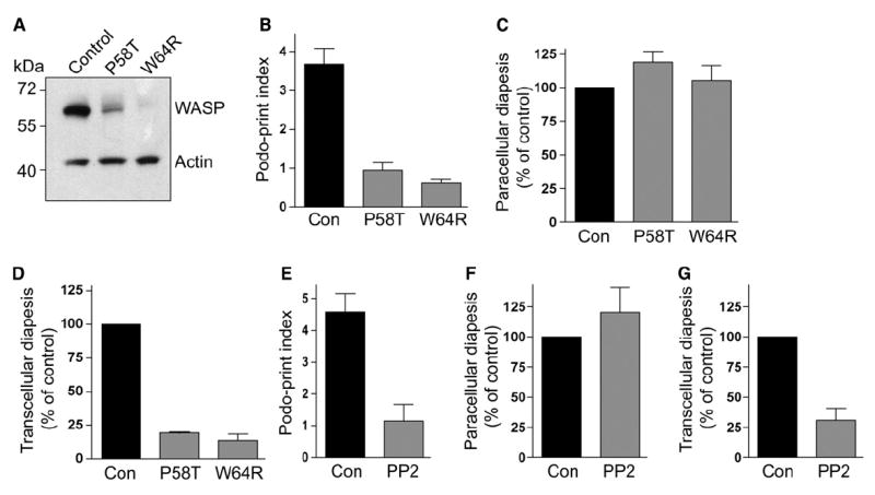
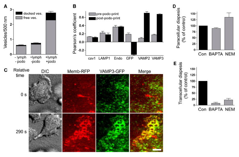
Diapedesis is critical for immune system function and inflammatory responses. This occurs by migration of blood leukocytes either directly through individual microvascular endothelial cells (the "transcellular" route) or between them (the "paracellular" route). Mechanisms for transcellular pore formation in endothelium remain unknown. Here we demonstrate that lymphocytes used podosomes and extended "invasive podosomes" to palpate the surface of, and ultimately form transcellular pores through, the endothelium. In lymphocytes, these structures were dependent on Src kinase and the actin regulatory protein WASP; inhibition of podosome formation selectively blocked the transcellular route of diapedesis. In endothelium, membrane fusion events dependent on the SNARE-containing membrane fusion complex and intracellular calcium were required for efficient transcellular pore formation in response to podosomes. These findings provide insights into basic mechanisms for leukocyte trafficking and the functions of podosomes.


Figures

Figure 1
Figure 1. Transcellular Diapedesis on Microvascular Endothelium
(A) IL-2-cultured lymphocytes were incubated with TNF-α-activated endothelial monolayers for 10 min followed by fixation, staining, and scoring as described in Experimental Procedures. Endothelial cells included HDMVEC and HLMVEC from Cambrex, as well as HDMVEC (HDMVEC*) and lymphatic ECs (Lymph*) selectively isolated from fresh neonatal foreskins. y axis represents the percentage of the total diapedesis that was transcellular. Values are mean ± SEM for at least three separate experiments. (B) Videos from live-cell imaging of lymphocyte diapedesis on HDMVEC and HLMVEC (described in Figure 2, below) were analyzed to determine the total duration of diapedesis and the amount of time lymphocytes resided at a location before initiating diapedesis (dwell time) for transcellular (black bars) or paracellular (gray bars) events. Data for the HLMVECs (L) and HDMVECs (D) were analyzed both separately and together (T). Values are the mean ± SEM for at least seven measurements. (C) Lymphocytes were incubated with activated HLMVEC as in (A) above, in the absence (gray bars) and presence (black bars) of PECAM-1 function-blocking mAb HEC7 (40 μg/ml). Graph depicts total diapedesis, as well as the separate the para- and transcellular diapedesis components, each as a percentage of total cells. Values are the mean ± SEM of three separate experiments. 40 or 120 μg/ml isotype control antibody had no effect on diapedesis (not shown). (D) Lymphocytes were incubated with activated HLMVEC for 10 min followed by fixation and staining with fluorescent antibodies to integrin β2, ICAM-1, and PECAM-1 and subjected to confocal microscopy. Images are Z-stack projections of representative transcellular (left) and paracellular (right) diapedesis events. Note that under one of the two lymphocytes on the left, two transcellular pores have formed. Arrows indicate the PECAM-1-enriched interendothelial junctions. Scale bar represents 5 μm.
Figure 2
Figure 2. Transcellular Diapedesis Is Preceded by Clusters of Micron-Scale ICAM-1-Rich Rings on the Endothelium
TNF-α-activated HDMVEC transiently transfected with ICAM-1-GFP were subjected to live-cell dynamic imaging upon addition of lymphocytes as described in Experimental Procedures. (A) and (C) are DIC, and (B), (D), and (E) are fluorescence images of ICAM-1-GFP. (A) and (B) depict a wide field of view in which most of a single GFP-positive endothelial cell (in the context of a confluent endothelial monolayer) is present. Boxed regions in (A) and (B) depict a location distant from endothelial intercellular junctions. Columns (C), (D), and (E) depict expanded sequential views (at 14 s intervals) of these boxed regions, in which the right-most lymphocyte initiates transcellular diapedesis. The micrographs shown in column (E) are identical to those shown in column (D), but individual ICAM-1-GFP-rich ring-shaped structures have been highlighted, with distinct colors correlating to each time point. (F) represents the overlay of these lines. Note that formation of a transcellular pore (see arrows in [D] and [E] at the 84 s time point), through which the lymphocyte subsequently completes transcellular diapedesis (shown in Movie S1), emerges from the center of two of these rings, which appear to fuse. Scale bars represent 5 μm.
Figure 3
Figure 3. “Ring Structures” Represent Endothelial Cell-Surface Invaginations
TNF-α-activated HLMVEC were subjected to live-cell dynamic imaging upon addition of lymphocytes. (A) HLMVEC were transiently cotransfected with memb-RFP and unmodified GFP. Panels depict DIC, memb-RFP, GFP, and merge of memb-RFP and GFP. Upper panels show a time point shortly after lymphocytes have settled on the endothelium, but before formation of memb-RFP ring structures (relative time = 0 s). Lower panels show a time point after lymphocyte spreading and formation of rings (relative time 170 s). Note that for each memb-RFP ring, a circular region of diminished GFP signal is formed. See corresponding Movie S4. (B) HDMVEC were transiently transfected with memb-RFP and prestained with ER-tracker green. Panels depict DIC, memb-RFP, ER-tracker green, and Merge of memb-RFP and ER-tracker green. Upper panels show a time point before (relative time = 0 s) and lower panels show a time point after (relative time 460 s) the formation of memb-RFP ring structures. Note that memb-RFP ring structures seem to form within the individual reticula of the ER often distorting and expanding these structures. These features can be more readily appreciated in the corresponding Movie S5. Scale bars represent 5 μm.
Figure 4
Figure 4. Endothelial Invaginations Result from Podosome-like Protrusive Structures
(A–C) Confocal imaging of lymphocyte-endothelial interactions. Lymphocytes were incubated with TNF-α-activated HDMVEC for 5 min followed by fixation, staining for F-actin (blue), leukocyte β2 integrin (red), endothelial ICAM-1 (green in [A] and [B]), or talin-1 (green in [C]), and confocal microscopy described in Experimental Procedures. Images are representative Z-stack projections of confocal sections near the plane of the leukocyte-endothelial interaction interface. (A) A merged image of β2 integrin, F-actin, and ICAM-1 is shown in which two adjacent lymphocytes, one spread and somewhat dumbbell-shaped (lower left) and one rounded (upper right), adhere to the surface of the endothelium. (B) An expanded view of the boxed region in (A) is shown as both separate and merged fluorescent channels. (C) In a separate experiment, samples were stained for β2 integrin, F-actin, and talin-1. (D) Live-cell imaging of lymphocyte actin. Lymphocytes transiently expressing GFP-actin were incubated with activated HDMVEC expressing memb-RFP and subjected to live-cell imaging. Left and right panels depict a lymphocyte at time points before (relative time point = 0 s) and after (relative time point = 266 s), respectively, endothelial podoprint formation. Memb-RFP, GFP-actin, and merged images are as indicated. Arrows indicate the appearance of podosome-like (Evans et al., 2003) actin puncta in lymphocytes that are centered within the memb-RFP rings of the endothelial podoprints. Note the distinctly green areas (actin) evident at the center of each ring. See corresponding Movie S8. Scale bars represent 5 μm.
Figure 5
Figure 5. In Vitro and In Vivo Ultrastructure of Lymphocyte Podosomes and Invasive Podosomes
(A–D) Podosomes in vitro. Lymphocytes migrating on TNF-α-activated HDMVEC were fixed after a 5 min coincubation and processed for transmission EM as described in Experimental Procedures. (A) A lymphocyte extends three shallow (~0.3 μm) podosomes (“P1–P3”) and one deep (~1.5 μm) invasive podosome (“I”) into endothelial invaginations. Inset is an expanded view of podosome “P2.” (B) A lymphocyte extends three shallow (~0.2–0.5 μm) podosomes (“P1–P3”) and three deep (~1.5 μm) invasive podosomes (“I1–I3”) that span to nearly the basal surface. (C and D) Shallow lymphocyte podosomes “P1–P6” that have formed directly over the endothelial cell nucleus. “P1” shows the apical plasma membrane nearly in contact with the nuclear envelope and “P6” shows it mildly indenting, but not invaginating, the nucleus. (E and F) Podosomes in vivo. A guinea pig model of dermatitis was prepared and processed for TEM as described in Experimental Procedures. The representative lymphocyte (E) and basophil (F) are from an extensive analysis, including four different experiments and examination of at least 100 distinct lymphocytes and at least 100 distinct basophils. More than half of all both lymphocytes and basophils exhibited at least one podosome in any given section. (E) Lymphocyte podosomes (“P1–P3;” ~0.2–0.5 μm) and an invasive podosome (“I;” ~0.8 μm) project into the endothelial surface of an inflamed vessel. Note that “I” has traversed the entire depth of the endothelial cell and placed the luminal and abluminal membranes into extremely close apposition. (F) Multiple basophil podosomes (“P1–P8;” ~0.2–0.5 μm) protrude into the endothelial surface. In all panels, leukocytes are indicated by a 5% opacity red overlay. In all panels, arrows indicate endothelial vesicles and VVOs, both apparently free in the cytoplasm and fused or docked to the plasma membrane, enriched near leukocyte protrusions. Scale bars represent 500 nm in (A)–(C), (E), and (F) and 100 nm in insert in (A) and (D).
Figure 6
Figure 6. Podosomes and Transcellular Diapedesis Are Dependent on Lymphocyte Src and WASP Activity
(A) Lysates from IL-2-cultured control and WASP-deficient (P58A and W64R) lymphocytes were immunoblotted for WASP and actin as described (Jin et al., 2004). (B) Podosome formation by WASP-deficient lymphocytes. Control, P58A, and W64R lymphocytes were incubated with TNF-α-activated HLMVEC transiently transfected with memb-YFP and subjected to live-cell dynamic imaging. As a readout for podosome formation, podoprint indices were quantified as described in Experimental Procedures. Values are mean ± SEM of measurements obtained from at least 40 lymphocytes. See corresponding Movie S10. (C and D) Para- and transcellular diapedesis in WAS lymphocytes. Control, P58A, and W64R lymphocytes were incubated with activated HLMVEC for 5 min and then fixed, stained, and scored for para- and transcellular diapedesis. (C) Paracellular diapedesis by P58A and W64R lymphocytes were expressed as a percentage of the control value (36.5% ± 1.7% of total cells). (D) Transcellular diapedesis by P58A and W64R lymphocytes were expressed as a percentage of the control value (22.5% ± 1.6% of total cells). Values are mean of normalized values ± SEM for at least three separate experiments. (E) Podoprint indices for lymphocytes pretreated with the Src inhibitor PP2 (1 μM, 60 min) or DMSO (control) were assessed as in (B). (F and G) Para- and transcellular diapedesis in lymphocytes pretreated with PP2 (1 μM, 60 min) or DMSO (control) was as described in (C) and (D), respectively. Control values for para- and transcellular diapedesis were 21.5% ± 2.6% (F) and 12.9% ± 1.6% (G) of total cells, respectively.
Figure 7
Figure 7. Endothelial Calcium- and SNARE-Mediated Membrane Fusion Is Required for Efficient Transcellular Diapedesis
(A) The density of both free (gray) and plasma-membrane-fused or docked (black) vesicles in HDMVEC was quantified in samples without (−) or with (+) lymphocyte incubation and without (−) or with (+) podosomes, as described in Experimental Procedures. Values are mean ± SEM for at least 74 linear segments, each 500 nm long. (B) Pearson’s correlation for the distribution of caveolin-1-GFP (cav1), LAMP1-GFP (LAMP1), pACGFP-Endo (Endo), GFP, VAMP2-GFP (VAMP2), and VAMP3-GFP (VAMP3) relative to memb-RFP, both prior to (gray bars) and during (black bars) podoprint formation, was quantified from live-cell imaging experiments as described in Experimental Procedures. Values are mean ± SEM for at least five measurements. (C) DIC, memb-RFP, VAMP3-GFP, and merged memb-RFP and VAMP3-GFP images are as indicated. Upper panels show a time point before (relative time = 0 s) and lower panels show a time point after (relative time 290 s) the formation of memb-RFP podoprints. See corresponding Movie S12. Scale bars represent 5 μm. (D and E) Activated HLMVEC were pretreated with BAPTA-AM (20 μM, 60 min), NEM (300 μM, 5 min), or DMSO (control) and then incubated with lymphocytes for 5 min followed by fixation, staining, and scoring. Control values for para- and transcellular diapedesis were 29% ± 1.2% (D) and 14.9% ± 1.8% (E) of total cells, respectively.

Comment in

References

    1. Adamson P, Etienne S, Couraud PO, Calder V, Greenwood J. Lymphocyte migration through brain endothelial cell monolayers involves signaling through endothelial ICAM-1 via a rhodependent pathway. J Immunol. 1999;162:2964–2973. - PubMed
    1. Barreiro O, Yanez-Mo M, Serrador JM, Montoya MC, Vicente-Manzanares M, Tejedor R, Furthmayr H, Sanchez-Madrid F. Dynamic interaction of VCAM-1 and ICAM-1 with moesin and ezrin in a novel endothelial docking structure for adherent leukocytes. J Cell Biol. 2002;157:1233–1245. - PMC - PubMed
    1. Buccione R, Orth JD, McNiven MA. Foot and mouth: podosomes, invadopodia and circular dorsal ruffles. Nat Rev Mol Cell Biol. 2004;5:647–657. - PubMed
    1. Calle Y, Chou HC, Thrasher AJ, Jones GE. Wiskott-Aldrich syndrome protein and the cytoskeletal dynamics of dendritic cells. J Pathol. 2004;204:460–469. - PubMed
    1. Carman CV, Springer TA. A transmigratory cup in leukocyte diapedesis both through individual vascular endothelial cells and between them. J Cell Biol. 2004;167:377–388. - PMC - PubMed

Publication types