

Reliability and validity of needle biopsy evaluation of breast-abnormalities using the B-categorization--design and objectives of the Diagnosis Optimisation Study (DIOS)
- PMID: 17570833
- PMCID: PMC1913923
- DOI: 10.1186/1471-2407-7-100
Reliability and validity of needle biopsy evaluation of breast-abnormalities using the B-categorization--design and objectives of the Diagnosis Optimisation Study (DIOS)
Abstract
Background: The planned nationwide implementation of mammography screening 2007 in Germany will increase the occurrence of mammographically detected breast abnormalities. These abnormalities are normally evaluated by minimal invasive core biopsy. To minimize false positive and false negative histological findings, quality assurance of the pathological evaluation of the biopsies is essential. Various guidelines for quality assurance in breast cancer diagnosis recommend applying the B-classification for histopathological categorization. However, to date there are only few studies that reported results about reliability and validity of B-classification. Therefore, objectives of our study are to determine the inter- and intraobserver variability (reliability study) and construct and predictive validity (validity study) of core biopsy evaluation of breast abnormalities. This paper describes the design and objectives of the DIOS Study.
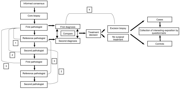
Methods/design: All consecutive asymptomatic and symptomatic women with breast imaging abnormalities who are referred to the University Hospital of Halle for core breast biopsy over a period of 24 months are eligible. According to the sample size calculation we need 800 women for the study. All patients in the study population underwent clinical and radiological examination. Core biopsy is performed by stereotactic-, ultrasound- or magnetic resonance (MR) guided automated gun method or vacuum assisted method. The histopathologic agreement (intra- and interobserver) of pathologists and the histopathologic validity will be evaluated. Two reference standards are implemented, a reference pathologist and in case of suspicious or malignant findings the histopathologic result of excision biopsy. Furthermore, a self administrated questionnaire which contains questions about potential risk factors of breast cancer, is sent to the participants approximately two weeks after core biopsy. This enables us to run a case-control-analysis (woman with breast cancer histological verified after excision are defined as cases, woman without malignant breast lesions are defined as controls) to investigate the predictive values of various risk factors on breast cancer risk.
Conclusion: The analysis of reliability and validity of the histopathological evaluation of core biopsy specimens of breast abnormalities is intended to provide important information needed for a high quality in breast cancer diagnostic and for planning of treatment strategies.
Figures
References
-
- Royal College of Pathologists. NHS Cancer Screening Programes . Guidelines for non-operative diagnostic procedures and reporting in breast cancer screening. Sheffield, NHSBSP publication, no. 50.; 2001.
-
- EC Working Group on Breast Screening Pathology . Quality assurance guidelines for pathology. In: Perry N, Broeders M, de Wolf C, Törnberg S, Holland R and von Karsa L, editor. European guidelines for quality assurance in cancer screening and diagnosis. 4. Vol. 6. European Union; 2006. pp. 219–312. - PubMed
-
- Ibrahim AE, Bateman AC, Theaker JM, Low JL, Addis B, Tidbury P, Rubin C, Briley M, Royle GT. The role and histological classification of needle core biopsy in comparison with fine needle aspiration cytology in the preoperative assessment of impalpable breast lesions. J Clin Pathol. 2001;54:121–125. doi: 10.1136/jcp.54.2.121. - DOI - PMC - PubMed
-
- Collins LC, Connolly JL, Page DL, Goulart RA, Pisano ED, Fajardo LL, Berg WA, Caudry DJ, McNeil BJ, Schnitt SJ. Diagnostic agreement in the evaluation of image-guided breast core needle biopsies: results from a randomized clinical trial. Am J Surg Pathol. 2004;28:126–131. doi: 10.1097/00000478-200401000-00015. - DOI - PubMed
Publication types
MeSH terms
LinkOut - more resources
Full Text Sources
Medical
