

Vasoactive intestinal peptide and the mammalian circadian system
- PMID: 17572414
- PMCID: PMC1994114
- DOI: 10.1016/j.ygcen.2007.04.018
Vasoactive intestinal peptide and the mammalian circadian system
Abstract
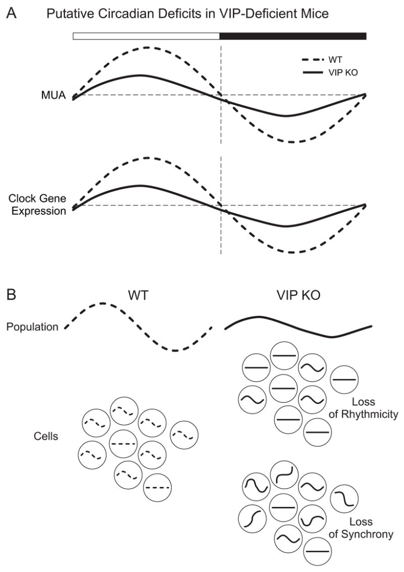
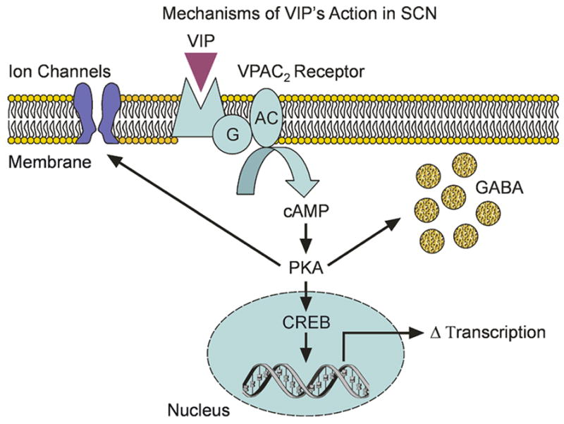
In mammals, the circadian oscillators that drive daily behavioral and endocrine rhythms are located in the hypothalamic suprachiasmatic nucleus (SCN). While the SCN is anatomically well-situated to receive and transmit temporal cues to the rest of the brain and periphery, there are many holes in our understanding of how this temporal regulation occurs. Unanswered questions include how cell autonomous circadian oscillations within the SCN remain synchronized to each other as well as communicate temporal information to downstream targets. In recent years, it has become clear that neuropeptides are critically involved in circadian timekeeping. One such neuropeptide, vasoactive intestinal peptide (VIP), defines a cell population within the SCN and is likely used as a signaling molecule by these neurons. Converging lines of evidence suggest that the loss of VIP or its receptor has a major influence on the ability of the SCN neurons to generate circadian oscillations as well as synchronize these cellular oscillations. VIP, acting through the VPAC(2) receptor, exerts these effects in the SCN by activating intracellular signaling pathways and, consequently, modulating synaptic transmission and intrinsic membrane currents. Anatomical evidence suggests that these VIP expressing neurons connect both directly and indirectly to endocrine and other output targets. Striking similarities exist between the role of VIP in mammals and the role of Pigment Dispersing Factor (PDF), a functionally related neuropeptide, in the Drosophila circadian system. Work in both mammals and insects suggests that further research into neuropeptide function is necessary to understand how circadian oscillators work as a coordinated system to impose a temporal structure on physiological processes within the organism.
Figures


Similar articles
-
Ontogeny of Circadian Rhythms and Synchrony in the Suprachiasmatic Nucleus.J Neurosci. 2018 Feb 7;38(6):1326-1334. doi: 10.1523/JNEUROSCI.2006-17.2017. Epub 2017 Oct 20. J Neurosci. 2018. PMID: 29054877 Free PMC article.
-
Synchronization and maintenance of timekeeping in suprachiasmatic circadian clock cells by neuropeptidergic signaling.Curr Biol. 2006 Mar 21;16(6):599-605. doi: 10.1016/j.cub.2006.02.023. Curr Biol. 2006. PMID: 16546085
-
Vasoactive intestinal polypeptide (VIP) phase-shifts the rat suprachiasmatic nucleus clock in vitro.Eur J Neurosci. 2001 Feb;13(4):839-43. doi: 10.1046/j.0953-816x.2000.01437.x. Eur J Neurosci. 2001. PMID: 11207820
-
The roles of vasoactive intestinal polypeptide in the mammalian circadian clock.J Endocrinol. 2003 Apr;177(1):7-15. doi: 10.1677/joe.0.1770007. J Endocrinol. 2003. PMID: 12697032 Review.
-
An essential role for peptidergic signalling in the control of circadian rhythms in the suprachiasmatic nuclei.J Neuroendocrinol. 2003 Apr;15(4):335-8. doi: 10.1046/j.1365-2826.2003.01005.x. J Neuroendocrinol. 2003. PMID: 12622830 Review.
Cited by
-
Collective timekeeping among cells of the master circadian clock.J Endocrinol. 2016 Jul;230(1):R27-49. doi: 10.1530/JOE-16-0054. Epub 2016 May 6. J Endocrinol. 2016. PMID: 27154335 Free PMC article. Review.
-
How does healthy aging impact on the circadian clock?J Neural Transm (Vienna). 2017 Feb;124(Suppl 1):89-97. doi: 10.1007/s00702-015-1424-2. Epub 2015 Jul 15. J Neural Transm (Vienna). 2017. PMID: 26175004 Review.
-
Endogenous peptide discovery of the rat circadian clock: a focused study of the suprachiasmatic nucleus by ultrahigh performance tandem mass spectrometry.Mol Cell Proteomics. 2010 Feb;9(2):285-97. doi: 10.1074/mcp.M900362-MCP200. Epub 2009 Nov 10. Mol Cell Proteomics. 2010. PMID: 19955084 Free PMC article.
-
Adult Neurogenesis under Control of the Circadian System.Cells. 2022 Feb 22;11(5):764. doi: 10.3390/cells11050764. Cells. 2022. PMID: 35269386 Free PMC article. Review.
-
Reduced VIP Expression Affects Circadian Clock Function in VIP-IRES-CRE Mice (JAX 010908).J Biol Rhythms. 2020 Aug;35(4):340-352. doi: 10.1177/0748730420925573. Epub 2020 May 28. J Biol Rhythms. 2020. PMID: 32460660 Free PMC article.
