. 2007 Aug;87(8):807-17.
doi: 10.1038/labinvest.3700595. Epub 2007 Jun 18.

Rho activation regulates CXCL12 chemokine stimulated actin rearrangement and restitution in model intestinal epithelia

Affiliations

Rho activation regulates CXCL12 chemokine stimulated actin rearrangement and restitution in model intestinal epithelia

Rebecca A Moyer et al. Lab Invest. 2007 Aug.

Abstract

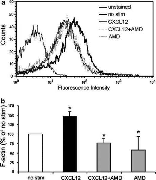
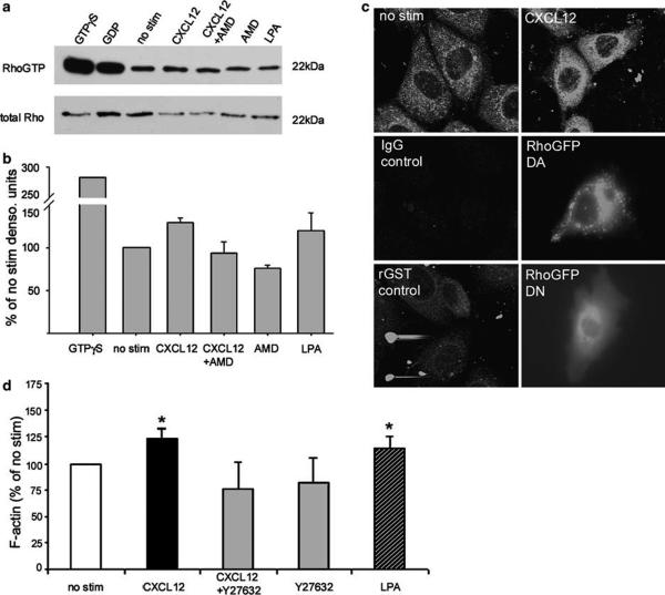
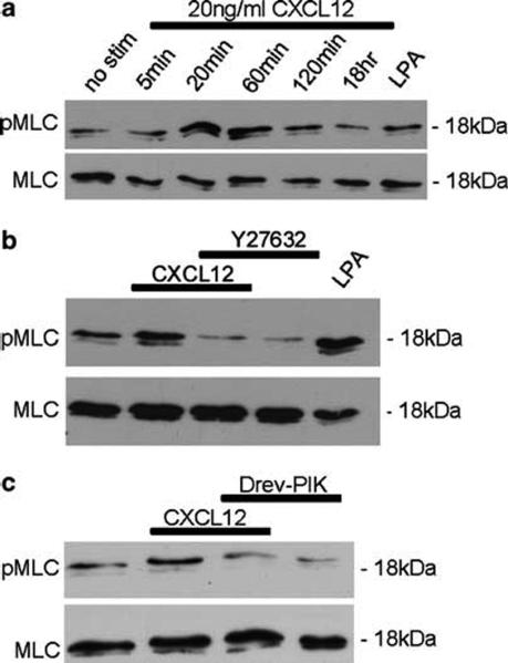
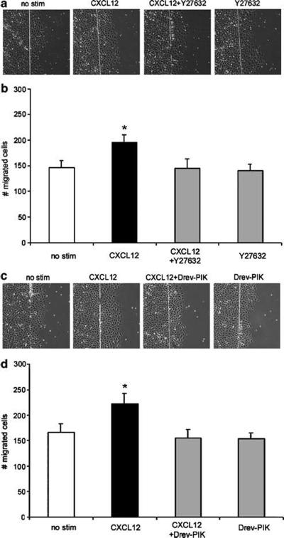
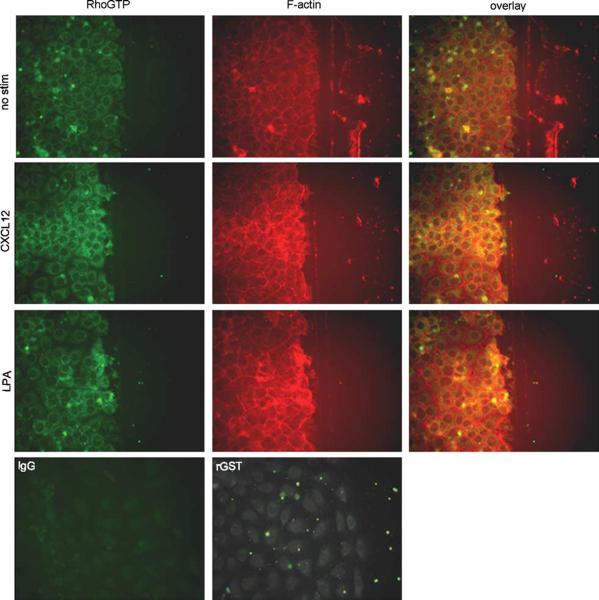
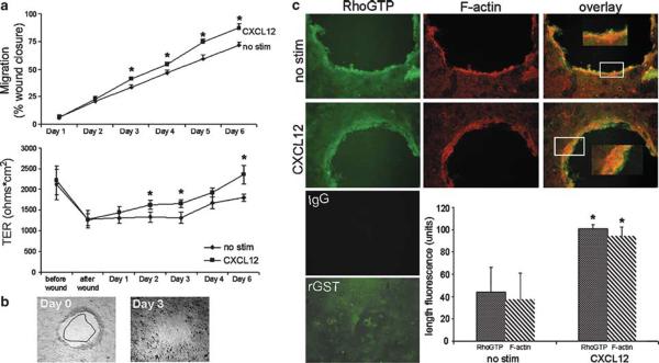
Chemokines are critical regulatory factors that direct migration, proliferation and maturation of receptor expressing target cells within gut mucosa. The aim of the present study was to define the cellular mechanisms whereby engagement of the essential chemokine CXCL12 to CXCR4 regulates restitutive epithelial cell migration. Non-transformed IEC-6 cells or polarized T84 epithelial monolayers were wounded and F-actin accumulation assessed using fluorescence microscopy and flow cytometry. Immunoblot analysis, pull-down assays, fluorescence microscopy and wound healing assays defined activation of Rho, Rho-kinase (ROCK), and myosin light chain (MLC) and the role for those Rho effectors in CXCL12-regulated epithelial restitution. CXCL12 increased RhoGTP and F-actin localization to the leading edge of wounded IEC-6 and T84 monolayers. CXCL12 congruently stimulated an increase in active MLC that was inhibited by blockade of ROCK and myosin light chain kinase and regulated epithelial migration. Our data in model intestinal epithelia suggest CXCR4 and CXCL12 may function as an autocrine and paracrine mucosal signaling network regulating the competency of the epithelial barrier to withstand injury and mediate repair following damage.


Figures

Figure 1
Figure 1
CXCL12 binding to CXCR4 increases F-actin accumulation. (a) Flow cytometric analyses of IEC-6 cells treated with 20 ng/ml CXCL12 (dark line) showed an increase in FITC-phalloidin staining representative of F-actin formation. Increased F-actin was inhibited upon treatment with 5 μg/ml of the specific CXCR4 antagonist AMD3100 (AMD; dotted line). Histogram data are representative of 3-5 experiments. (b) Quantification of F-actin as a percent of unstimulated IEC-6 cells (% no stim) indicated that AMD3100 completely blocked CXCL12-stimulated increases in F-actin. Cells were treated with AMD3100 alone as a control. Values are mean fluorescence intensity units normalized to untreated cells (no stim) for 3-5 independent experiments. Asterisks denote statistically significant difference (P ≤ 0.05) from untreated cells.
Figure 2
Figure 2
F-actin accumulation and distribution correlates to increased migration in CXCL12-treated epithelial cells. (a) FITC-phalloidin fluorescence microscopy of untreated (no stim) or CXCL12 (20 ng/ml)-treated IEC-6 wounded monolayers indicated increased F-actin accumulation at the wound edge 20 h after chemokine stimulation. (b) Enumeration of migrating cells in CXCL12-treated and FITC-phalloidin stained monolayers. Data are representative of three independent experiments. Asterisk denotes statistically significant difference between CXCL12-stimulated and control monolayers (P ≤ 0.05).
Figure 3
Figure 3
CXCR4 signals activation of Rho in IEC-6 cells. (a) RhoGTP pull-down assay with Rhotekin-GST and immunoblot analysis indicated an increase in active Rho in IEC-6 cells stimulated with 20 ng/ml CXCL12, or 1 μg/ml LPA control. CXCL12-stimulated RhoGTP formation was blocked in the presence (5 μg/ml) of the CXCR4 antagonist AMD3100 (AMD). Immunoblots of total Rho were assessed as a loading control. (b) Gel densitometry of active RhoGTP normalized to total Rho protein verified that CXCL12 activated Rho in a CXCR4-dependent manner. Relative densitometry unit data are representative of three independent experiments. (c) Confocal fluorescence microscopy of IEC-6 cells treated with 20 ng/ml CXCL12 confirmed increased active RhoGTP formation. CXCL12-stimulated RhoGTP immunoreactivity was similar to cells transfected with dominant-active (DA)-RhoGFP and distinct from that observed in dominant-negative (DN)-Rho transfectants or the rGST controls. (d) Flow cytometric analysis of IEC-6 cells showed that the downstream Rho effector kinase (ROCK) contributes to CXCL12-mediated F-actin accumulation. CXCL12-stimulated cells were incubated with or without the ROCK inhibitor Y27632, permeablized and stained with FITC-phalloidin and analyzed by flow cytometry. IEC-6 cells stimulated with LPA (1 μg/ml) and Y27632 alone (10 μM) were analyzed as controls. Values are mean fluorescence intensity units normalized to untreated cells (no stim) for 3-5 independent experiments. Asterisks denote statistically significant difference from untreated cells (P ≤ 0.05).
Figure 4
Figure 4
CXCL12 stimulates phosphorylation of MLC via ROCK and MLCK. (a) Immunoblot analysis of phospho-MLC (pMLC) and total MLC in IEC-6 cells stimulated with 20 ng/ml CXCL12 for 5, 20, 60, 120 min, or 18 h showed CXCL12 increased phosphorylation of MLC maximally at 20 and 60 min above unstimulated cells (no stim). (b and c) Inhibition of ROCK with Y27632 (10 μM) or MLCK with Drev-PIK (300 μM) abolished CXCL12-mediated phosphorylation of MLC. Cells treated with Y27632 (10 μM), or Drev-PIK (300 μM) alone or with 1 μg/ml LPA were controls. Data are representative of three independent experiments.
Figure 5
Figure 5
CXCL12-mediated restitution involves ROCK and MLCK. IEC-6 cells were wounded and restitution quantified by enumerating cells that migrated into the denuded ulcer in 3-5 fields of 2-3 wounds per experiment (n = 3 independent experiments). Wounded IEC-6 monolayers treated with 20 ng/ml CXCL12 demonstrated increased restitution relative to unstimulated cells (no stim). Treatment with the ROCK inhibitor Y27632 (10 μM) (a and b) or the MLCK inhibitor Drev-PIK (300 μM) (c and d) impeded CXCL12 restitution. Asterisks denote statistically significant difference from unstimulated monolayers (P ≤ 0.05).
Figure 6
Figure 6
CXCL12 stimulates accumulation of F-actin and RhoGTP at the leading edge of migrating IEC-6 epithelial sheets. IEC-6 monolayers wounded with a razor blade were incubated 20 min with or without 20 ng/ml CXCL12, or 1 μg/ml LPA as a control, fixed, permeablized, and stained for RhoGTP using Rhotekin-GST and an anti-GST antibody, or F-actin with Alexa-fluor-595-phalloidin. Photomicrographs indicated the CXCL12 stimulated accumulation of F-actin and RhoGTP at the leading edge of new wounds greater than that of unstimulated cells (no stim). Cells were examined using fluorescence microscopy at × 400. Data are representative of three independent experiments.
Figure 7
Figure 7
CXCL12 mediates wound closure and induces the accumulation of RhoGTP and F-actin in polarized human model epithelium. CXCL12-treated monolayers demonstrated increased closure and increased TER relative to untreated monolayers. (a, top panel) Wounded polarized T84 monolayers were stimulated with 20 ng/ml CXCL12 and photomicrographs taken directly after wounding (b, 0 h) and daily after that for 4-6 days. Relative area of the wounds was calculated using MetaMorph software and wound closure (a, top) was monitored over time by normalizing wound area to 0 h and shown as a percentage of the initial wound area. (a, bottom panel) TER was measured on the same wounded T84 monolayers as measured in the top panel and demonstrated the parallel increase in barrier integrity with wound closure. (b) Representative T84 monolayer wounds at days 0 and 3 of CXCL12 treatment. Wound closure observed on day 3 outlined and superimposed upon the day 0 wound demonstrated the consistent restitution of CXCL12 treated T84 monolayers. Data are representative of 6-17 independent wounds. (c) Photomicrographs demonstrate the significantly increased accumulation of F-actin and RhoGTP at the leading edge of wounds in CXCL12-treated relative to unstimulated cells (no stim). The length of F-actin and RhoGTP fluorescence from the wound edge was quantified using MetaMorph software as described in Materials and Methods. Cells were examined using fluorescence microscopy at × 100 (insets × 200). Immunofluorescence data are representative of three independent monolayers. Values in (a and c) are the mean±s.d. of three independent monolayers. Asterisks denote statistically significant difference from unstimulated (no stim) monolayers (P≤0.05).

References

    1. Booth C, Brady G, Potten CS. Crowd control in the crypt. Nat Med. 2002;8:1360–1361. - PubMed
    1. Mammen JM, Matthews JB. Mucosal repair in the gastrointestinal tract. Crit Care Med. 2003;31:S532–S537. - PubMed
    1. Dignass AU. Mechanisms and modulation of intestinal epithelial repair. Inflamm Bowel Dis. 2001;7:68–77. - PubMed
    1. Jacinto A, Martinez-Arias A, Martin P. Mechanisms of epithelial fusion and repair. Nat Cell Biol. 2001;3:E117–E123. - PubMed
    1. Zlotnik A, Yoshie O. Chemokines: a new classification system and their role in immunity. Immunity. 2000;12:121–127. - PubMed

Publication types

MeSH terms