. 2007 Oct 15;93(8):2767-80.
doi: 10.1529/biophysj.107.107813. Epub 2007 Jun 15.

Flavonoids affect actin functions in cytoplasm and nucleus

Affiliations

Flavonoids affect actin functions in cytoplasm and nucleus

Markus Böhl et al. Biophys J. .

Abstract

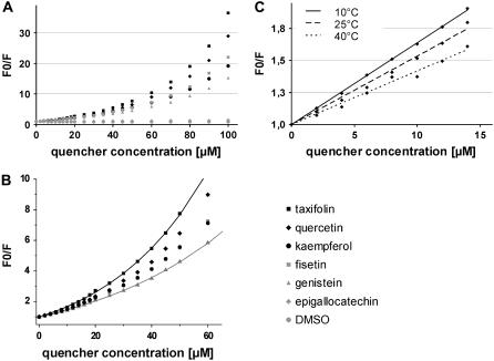
Based on the identification of actin as a target protein for the flavonol quercetin, the binding affinities of quercetin and structurally related flavonoids were determined by flavonoid-dependent quenching of tryptophan fluorescence from actin. Irrespective of differences in the hydroxyl pattern, similar Kd values in the 20 microM range were observed for six flavonoids encompassing members of the flavonol, isoflavone, flavanone, and flavane group. The potential biological relevance of the flavonoid/actin interaction in the cytoplasm and the nucleus was addressed using an actin polymerization and a transcription assay, respectively. In contrast to the similar binding affinities, the flavonoids exert distinct and partially opposing biological effects: although flavonols inhibit actin functions, the structurally related flavane epigallocatechin promotes actin activity in both test systems. Infrared spectroscopic evidence reveals flavonoid-specific conformational changes in actin which may mediate the different biological effects. Docking studies provide models of flavonoid binding to the known small molecule-binding sites in actin. Among these, the mostly hydrophobic tetramethylrhodamine-binding site is a prime candidate for flavonoid binding and rationalizes the high efficiency of quenching of the two closely located fluorescent tryptophans. The experimental and theoretical data consistently indicate the importance of hydrophobic, rather than H-bond-mediated, actin-flavonoid interactions. Depending on the rigidity of the flavonoid structures, different functionally relevant conformational changes are evoked through an induced fit.


Figures

FIGURE 1
FIGURE 1
Structures of flavonoids used in this study. (A) Flavonoid generic molecular formula. (B) Chemical structures of flavonoids used in this study. The flavonols kaempferol, quercetin, and fisetin show a different hydroxyl group substitution pattern and they share the positions 3, 7, 4′ (boxed). The isoflavone genistein, the flavane epigallocatechin, and the flavanone taxifolin represent common flavonoids from other subclasses.
FIGURE 2
FIGURE 2
Effect of spectral properties of flavonoids on the intensity of tryptophan emission. (A) Quenching effect of flavonoids used in this study (Ex295 nm/Em345 nm). (B) Actin emission spectra (Ex295 nm) in the presence of rising quercetin concentration. (C) Actin emission spectra (Ex295 nm) in the presence of rising taxifolin concentration. (D) Flavonoids used in this study show an overlap in their absorption spectrum with the actin emission spectrum (Ex295 nm).
FIGURE 3
FIGURE 3
Actin tryptophan-dependent fluorescence is quenched by flavonoids. (A) SV analysis of the quenching data according to Eq. 1 (see Materials and Methods). (B) Analysis of the SV plots according to Eq. 2 (see Materials and Methods) can explain the upward curvature of the plots as exemplified here for taxifolin (solid black line) and genistein (solid gray line). The corresponding Kd values are displayed in Table 2. (C) SV analysis on the temperature dependence of quercetin quenching according to Eq. 1.
FIGURE 4
FIGURE 4
Evaluation of flavonoid-dependent actin functions. (A) In vitro polymerization of rabbit skeletal muscle actin in the presence of 5, 10, and 25 μM of the respective flavonoid. (B) In vitro transcription activity of HeLa cell nuclear extract in the presence of different flavonoids (5 and 25 μM). Columns represent the mean value of duplicates or triplicates; error bars indicate the standard deviation.
FIGURE 5
FIGURE 5
ATR-FTIR spectra in the amide I and II spectral range of free and flavonoid-bound actin. (A) Actin in the absence of flavonoid. (B) Actin in the presence of 50 μM epigallocatechin. (C) Actin in the presence of 50 μM quercetin. The amide I contour was fitted by Gaussian/Lorentzian bands as described (Materials and Methods) and as summarized in Table 2. Experiments were done at 23 °C, in 5× buffer G.
FIGURE 6
FIGURE 6
Structural flexibility of actin and binding of low molecular weight ligands. Superimposed are the crystallographically determined structures of actin bound to ADP/tetramethylrhodamine (magenta; PDB ID 1J6Z), ATP/swinholide (white; PDB ID 1YXQ), latrunculin (blue; PDB ID: 2A5X), and reidispongiolide (yellow; PDB ID 2ASM). The original ligands for the studied binding sites are depicted in sticks: ATP (color coded) latrunculin (blue), reidispongiolide (yellow), and tetramethylrhodamine (magenta). The tryptophans responsible for 90% of actin fluorescence, i.e., W-340 (center) and W-356 (lower right corner), are shown in space fill mode. Their closeness to the tetramethylrhodamin-binding site, which we propose to also accommodate the flavonoids, agrees with the strong quenching of tryptophan emission in the flavonoid-bound state. This figure was prepared with Chimera.
FIGURE 7
FIGURE 7
Docking poses of quercetin in the different low molecular weight ligand-binding sites of actin. (From top to bottom) Latrunculin-, reidispongiolide-, and tetramethylrhodamine-binding sites. On the left, the original ligand from the PDB structure is shown in thin blue sticks and the docked quercetin in thick colored sticks. In the reidispongiolide-binding site, we omitted the original ligand for clarity. On the right, the predicted poses for the six different ligands are shown to visualize the variability in the docking results. Except for a single H-bond to the backbone carbonyl of F-352, the tetramethylrhodamine-binding site (bottom panels) provides hydrophobic interactions with the quercetin rings, and the hydroxyls of the flavonoid are exposed. Clustering of two binding modes in the tetramethylrhodamine-binding site may be relevant to shifting the actin conformation in a flavonoid structure-dependent manner (see text for details).

References

    1. Middleton, E., C. Kandaswami, and T. C. Theoharides. 2000. The effects of plant flavonoids on mammalian cells: implications for inflammation, heart disease, and cancer. Pharmacol. Rev. 52:673–751. - PubMed
    1. Knekt, P., J. Kumpulainen, R. Jarvinen, H. Rissanen, M. Heliovaara, A. Reunanen, T. Hakulinen, and A. Aromaa. 2002. Flavonoid intake and risk of chronic diseases. Am. J. Clin. Nutr. 76:560–568. - PubMed
    1. Manach, C., A. Scalbert, C. Morand, C. Remesy, and L. Jimenez. 2004. Polyphenols: food sources and bioavailability. Am. J. Clin. Nutr. 79:727–747. - PubMed
    1. Nichenametla, S. N., T. G. Taruscio, D. L. Barney, and J. H. Exon. 2006. A review of the effects and mechanisms of polyphenolics in cancer. Crit. Rev. Food Sci. Nutr. 46:161–183. - PubMed
    1. Chen, D., K. G. Daniel, M. S. Chen, D. J. Kuhn, K. R. Landis-Piwowar, and Q. P. Dou. 2005. Dietary flavonoids as proteasome inhibitors and apoptosis inducers in human leukemia cells. Biochem. Pharmacol. 69:1421–1432. - PubMed

Publication types

LinkOut - more resources