Increased O-GlcNAc levels during reperfusion lead to improved functional recovery and reduced calpain proteolysis
- PMID: 17573462
- PMCID: PMC2850209
- DOI: 10.1152/ajpheart.00285.2007
Increased O-GlcNAc levels during reperfusion lead to improved functional recovery and reduced calpain proteolysis
Abstract
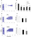
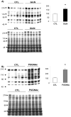
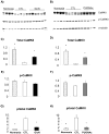
We have previously shown that preischemic treatment with glucosamine improved cardiac functional recovery following ischemia-reperfusion, and this was mediated, at least in part, via enhanced flux through the hexosamine biosynthesis pathway and subsequently elevated O-linked N-acetylglucosamine (O-GlcNAc) protein levels. However, preischemic treatment is typically impractical in a clinical setting; therefore, the goal of this study was to investigate whether increasing protein O-GlcNAc levels only during reperfusion also improved recovery. Isolated perfused rat hearts were subjected to 20 min of global, no-flow ischemia followed by 60 min of reperfusion. Administration of glucosamine (10 mM) or an inhibitor of O-GlcNAcase, O-(2-acetamido-2-deoxy-D-glucopyranosylidene)amino-N-phenylcarbamate (PUGNAc; 200 microM), during the first 20 min of reperfusion significantly improved cardiac functional recovery and reduced troponin release during reperfusion compared with untreated control. Both interventions also significantly increased the levels of protein O-GlcNAc and ATP levels. We also found that both glucosamine and PUGNAc attenuated calpain-mediated proteolysis of alpha-fodrin as well as Ca(2+)/calmodulin-dependent protein kinase II during reperfusion. Thus two independent strategies for increasing protein O-GlcNAc levels in the heart during reperfusion significantly improved recovery, and this was correlated with attenuation of calcium-mediated proteolysis. These data provide further support for the concept that increasing cardiac O-GlcNAc levels may be a clinically relevant cardioprotective strategy and suggest that this protection could be due, at least in part, to inhibition of calcium-mediated stress responses.
Figures


Comment in
-
The sweet nature of cardioprotection.Am J Physiol Heart Circ Physiol. 2007 Sep;293(3):H1324-6. doi: 10.1152/ajpheart.00697.2007. Epub 2007 Jun 22. Am J Physiol Heart Circ Physiol. 2007. PMID: 17586610 No abstract available.
References
-
- Bartoli M, Bourg N, Stockholm D, Raynaud F, Delevacque A, Han Y, Borel P, Seddik K, Armande N, Richard I. A Mouse Model for Monitoring Calpain Activity under Physiological and Pathological Conditions. J Biol Chem. 2006;281:39672–39680. - PubMed
-
- Benter IF, Juggi JS, Khan I, Yousif MH, Canatan H, Akhtar S. Signal transduction mechanisms involved in cardiac preconditioning: role of Ras-GTPase, Ca2+/calmodulin-dependent protein kinase II and epidermal growth factor receptor. Mol Cell Biochem. 2005;268:175–183. - PubMed
-
- Bertinchant JP, Polge A, Robert E, Sabbah N, Fabbro-Peray P, Poirey S, Laprade M, Pau B, Juan JM, Bali JP, de la Coussaye JE, Dauzat M. Time-course of cardiac troponin I release from isolated perfused rat hearts during hypoxia/reoxygenation and ischemia/reperfusion. Clin Chim Acta. 1999;283:43–56. - PubMed
-
- Bolli R, Becker L, Gross G, Mentzer R, Jr, Balshaw D, Lathrop DA. Myocardial protection at a crossroads: the need for translation into clinical therapy. Circ Res. 2004;95:125–134. - PubMed
-
- Champattanachai V, Chatham JC, Marchase RB. Glucosamine protects neonatal cardiomyocytes from ischemia-reperfusion injury via increased protein-associated O-GlcNAc. Am J Physiol Cell Physiol. 2006 In Press. - PubMed
Publication types
MeSH terms
Substances
Grants and funding
LinkOut - more resources
Full Text Sources
Molecular Biology Databases
Miscellaneous

