. 2007 Sep;189(17):6351-8.
doi: 10.1128/JB.00509-07. Epub 2007 Jun 15.

The N-terminal domain of OmpATb is required for membrane translocation and pore-forming activity in mycobacteria

Affiliations

The N-terminal domain of OmpATb is required for membrane translocation and pore-forming activity in mycobacteria

Anuradha Alahari et al. J Bacteriol. 2007 Sep.

Abstract

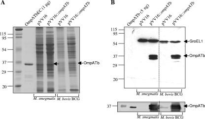
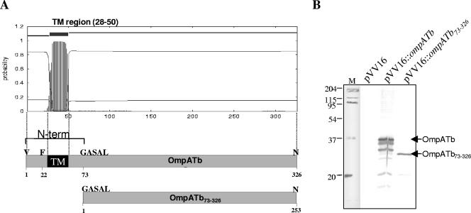
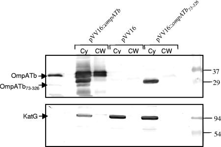
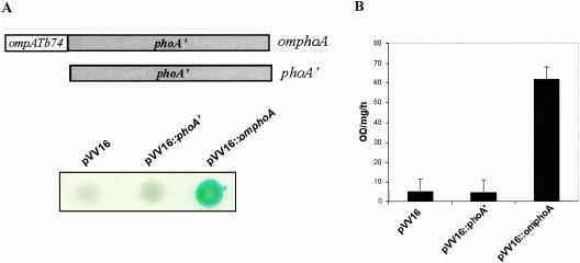
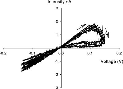
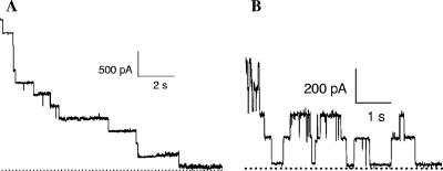
OmpATb is the prototype of a new family of porins in Mycobacterium tuberculosis and Mycobacterium bovis BCG. Although the pore-forming activity of this protein has been clearly established by using recombinant protein produced in Escherichia coli, characterization of the native porin has been hampered by the scarce amount of protein present in the M. tuberculosis detergent extracts. To this aim, we have developed a protocol to overproduce and obtain high yields of OmpATb in both Mycobacterium smegmatis and M. bovis BCG. The protein could be extracted and purified from the cell wall fraction and subsequently used for analysis of the pore-forming activity in multichannel and single-channel conductance experiments. Our results indicate that OmpATb produced in mycobacteria presents an average conductance value of 1,600+/-100 pS, slightly higher than that of OmpATb produced in E. coli, suggesting the occurrence of OmpATb in a highly ordered organization within the mycobacterial cell wall. In contrast to OmpATb, a truncated form lacking the first 72 amino acids (OmpATb73-326) was essentially found in the cytosol and was not active in planar lipid bilayers. This suggested that the N-terminal domain of OmpATb could participate in targeting of OmpATb to the cell wall. This was further confirmed by analyzing M. smegmatis clones expressing a chimeric protein consisting of a fusion between the N-terminal domain of OmpATb and the E. coli PhoA reporter. The present study shows for the first time that the N terminus of OmpATb is required for targeting the porin to the cell wall and also appears to be essential for its pore-forming activity.


Figures

FIG. 1.
FIG. 1.
Overexpression of OmpATb in mycobacteria. M. smegmatis and M. bovis BCG were transformed with either the empty pVV16 or pVV16::ompATb that permits production of OmpATb as a C-terminal His-tagged fusion protein. Recombinant mycobacteria were disrupted, and lysates equivalent to 20 μg of total protein were separated by SDS-PAGE. (A) Coomassie blue-stained gel showing overexpression of OmpATb in crude cell lysates of mycobacterial strains carrying plasmids as indicated. Purified recombinant OmpATb from E. coli was used for comparison of molecular size. The numbers on the left indicate sizes of the molecular weight markers. (B) Corresponding immunoblots using either a His-Probe-HRP along with a chemiluminescent substrate that reveals the presence of His-tagged proteins (upper panel) or anti-OmpATb antiserum (lower panel). The GroEL1 is detected in the crude lysates by the virtue of the naturally occurring C-terminal His residues (22).
FIG. 2.
FIG. 2.
Overexpression of OmpATb73-326 in M. smegmatis. (A) Graphical representation of OmpATb as predicted by TMHMM 2.0 software (http://www.cbs.dtu.dk/services/TMHMM-2.0/). One transmembrane helix (TM) was predicted to be present in the N terminus of the protein (residues 28 to 50) (upper panel). The N terminus of OmpATb, lacking in OmpATb73-326, comprises the first 72 amino acids and includes the TM region (lower panel). (B) Western blot analysis of crude lysates of recombinant M. smegmatis strains overexpressing either OmpATb or OmpATb73-326. Equal amounts (20 μg) of total lysates were loaded in each lane, resolved on SDS-PAGE, transferred to a nitrocellulose membrane and probed with anti-OmpATb antiserum.
FIG. 3.
FIG. 3.
Subcellular localization of OmpATb and OmpATb73-326 in M. smegmatis. Recombinant M. smegmatis strains overproducing either OmpATb or OmpATb73-326 were lysed and fractionated to separate the cytoplasm (Cy) from the cell wall (CW). The fractions were resuspended into the same volume of buffer and 20 μl of each fraction were subjected to SDS-PAGE, electroblotted onto a nitrocellulose membrane, and probed with anti-OmpATb antiserum (upper panel) or monoclonal anti-KatG (lower panel) antibodies.
FIG. 4.
FIG. 4.
Purification of OmpATb and OmpATb73-326 produced in mycobacteria and E. coli. (A) Coomassie blue-stained gel showing OmpATb73-326BCG (lane 1), OmpATb73-326SMEG (lane 2), OmpATbEC (lane 3), OmpATbBCG (lane 5), and OmpATbSMEG (lane 6). Proteins (15 μg) were separated by SDS-12% PAGE. The molecular mass standards used were, from top to bottom, 97, 66, 45, and 30 kDa. (B) Immunoreactivity of the purified proteins. Lane 1, Coomassie blue-stained gel of OmpATbBCG; lane 2, immunoblot of OmpATbBCG probed with anti-OmpATb antibodies. The molecular mass standards used were from top to bottom: 97, 66, 45, 30, 20.1 kDa.
FIG. 5.
FIG. 5.
Expression and translocation of PhoA across the cell membrane in M. smegmatis. (A, top) Representation of the omphoA fusion construct. The phoA gene encoding the E. coli alkaline phosphatase has been fused to the DNA sequence corresponding to the first 74 amino acids of OmpATb and designated omphoA. phoA′ corresponds to the phoA gene missing its signal sequence. (A, bottom) PhoA activity in M. smegmatis harboring pVV16::omphoA. Liquid cultures of M. smegmatis harboring either pVV16, pVV16::phoA′, or pVV16::omphoA were spotted onto indicator LB plates containing 40 μg of BCIP/ml, generating a blue-colored bacteria when expressing an active PhoA phosphatase. (B) Alkaline phosphatase activity assayed on whole M. smegmatis cells. PhoA activities were measured on intact cells and are expressed as OD410/ml of culture/min. Error bars represent the standard deviation from triplicate analyses.
FIG. 6.
FIG. 6.
Macroscopic current-voltage (I/V) curves of OmpATb SMEG in azolectin membranes. I/V curves between −200 and +200 mV at a ramp sweep of 10 mV/s in 1 M KCl and 10 mM HEPES (pH 5) at room temperature. A total of 30 ng of protein was added in the cis-side compartment.
FIG. 7.
FIG. 7.
Single-channel traces for OmpATbs in azolectin bilayers. (A) OmpATbSMEG at +166 mV and pH 7. (B) OmpATbBCG at +100 mV and pH 6. The electrolyte solution was composed of 1 M KCl and 10 mM HEPES. A total of 5 ng of protein was added in the cis-side compartment. The dotted lines represent zero current.

References

    1. Boyd, D., C. Manoil, and J. Beckwith. 1987. Determinants of membrane protein topology. Proc. Natl. Acad. Sci. USA 84:8525-8529. - PMC - PubMed
    1. Braunstein, M., A. M. Brown, S. Kurtz, and W. R. Jacobs, Jr. 2001. Two nonredundant SecA homologues function in mycobacteria. J. Bacteriol. 183:6979-6990. - PMC - PubMed
    1. Brennan, P. J., and H. Nikaido. 1995. The envelope of mycobacteria. Annu. Rev. Biochem. 64:29-63. - PubMed
    1. Faller, M., M. Niederweis, and G. E. Schulz. 2004. The structure of a mycobacterial outer-membrane channel. Science 303:1189-1192. - PubMed
    1. Heinz, C., H. Engelhardt, and M. Niederweis. 2003. The core of the tetrameric mycobacterial porin MspA is an extremely stable beta-sheet domain. J. Biol. Chem. 278:8678-8685. - PubMed

Publication types

MeSH terms

LinkOut - more resources