. 2007 Aug;144(4):1863-77.
doi: 10.1104/pp.107.099226. Epub 2007 Jun 15.

Resistance to Botrytis cinerea in sitiens, an abscisic acid-deficient tomato mutant, involves timely production of hydrogen peroxide and cell wall modifications in the epidermis

Affiliations

Resistance to Botrytis cinerea in sitiens, an abscisic acid-deficient tomato mutant, involves timely production of hydrogen peroxide and cell wall modifications in the epidermis

Bob Asselbergh et al. Plant Physiol. 2007 Aug.

Abstract

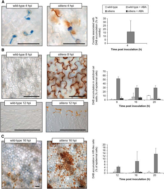
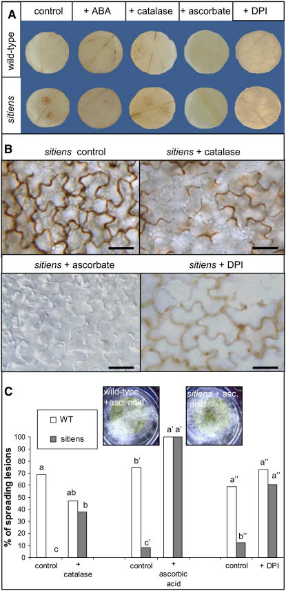
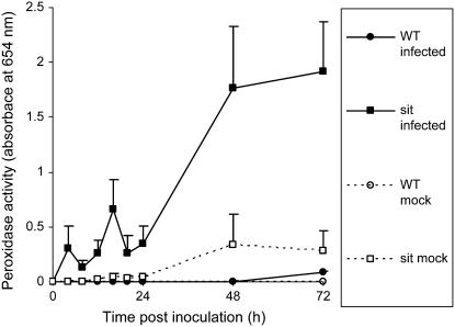
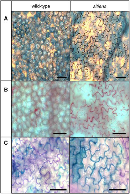
Plant defense mechanisms against necrotrophic pathogens, such as Botrytis cinerea, are considered to be complex and to differ from those that are effective against biotrophs. In the abscisic acid-deficient sitiens tomato (Solanum lycopersicum) mutant, which is highly resistant to B. cinerea, accumulation of hydrogen peroxide (H(2)O(2)) was earlier and stronger than in the susceptible wild type at the site of infection. In sitiens, H(2)O(2) accumulation was observed from 4 h postinoculation (hpi), specifically in the leaf epidermal cell walls, where it caused modification by protein cross-linking and incorporation of phenolic compounds. In wild-type tomato plants, H(2)O(2) started to accumulate 24 hpi in the mesophyll layer and was associated with spreading cell death. Transcript-profiling analysis using TOM1 microarrays revealed that defense-related transcript accumulation prior to infection was higher in sitiens than in wild type. Moreover, further elevation of sitiens defense gene expression was stronger than in wild type 8 hpi both in number of genes and in their expression levels and confirmed a role for cell wall modification in the resistant reaction. Although, in general, plant defense-related reactive oxygen species formation facilitates necrotrophic colonization, these data indicate that timely hyperinduction of H(2)O(2)-dependent defenses in the epidermal cell wall can effectively block early development of B. cinerea.


Figures

Figure 1.
Figure 1.
Disease symptoms on wild-type and sitiens tomato leaf discs after infection with two 5-μL drops of a B. cinerea conidial suspension. A, Macroscopic disease symptoms. In wild-type leaf discs, spreading gray disintegration of the host tissue is visible and in sitiens discs necrotic spots at the site of the infection droplet are observed. B, Close up of macroscopic disease symptoms. In sitiens, primary necrotic lesions contain a high number of distinct dark spots at 48 and 72 hpi. In wild type, primary necrotic spots at 48 hpi are typically larger, less numerous, have a paler shade of brown, and more often develop into spreading water-soaked lesions.
Figure 2.
Figure 2.
Temporal evolution of H2O2 accumulation in wild-type and sitiens tomato after infection with B. cinerea. DAB staining of leaf discs infected with two 5-μL drops of a conidial suspension was performed at different time points post inoculation (4, 8, 12, 16, 20, 24, 48, and 72 hpi). One representative disc out of three replicates is shown for each time point. [See online article for color version of this figure.]
Figure 3.
Figure 3.
Effect of ABA on H2O2 accumulation in epidermal cells after infection with B. cinerea. A, Association of DAB accumulation with B. cinerea conidia. In sitiens, H2O2 was located at the site of penetration and in some parts of the anticlinal wall of the penetrated cell, whereas in wild type and in sitiens supplemented with ABA, no H2O2 accumulation was detected. Germinating conidia were classified in two groups based on the presence or absence of associated DAB accumulation in the epidermal cells, whose percentage is shown. B, DAB accumulation in epidermal cell walls. In sitiens, DAB staining was general in the entire outline of the anticlinal cell wall, whereas in wild type and sitiens supplemented with ABA, no H2O2 accumulation was detected at 8 hpi (top). The restriction of sitiens DAB accumulation to the epidermal layer was confirmed on cross sections (bottom). Epidermal cells were classified in two groups based on the presence or absence of DAB accumulation in the anticlinal walls, whose percentage is shown in the anticlinal walls at 8, 16, and 20 hpi. C, Intracellular DAB accumulation in epidermal cells showing an HR-like reaction. At 16 hpi, wild-type and ABA-supplemented sitiens epidermal cells did not accumulate intracellular DAB, whereas in sitiens, groups of HR-like cells with intracellular DAB accumulation were present near the site of fungal penetration. Epidermal cells were classified in two groups based on the presence or absence of intracellular DAB accumulation and the percentage is shown at 12, 16, and 20 hpi. In all graphs, bars represent the means and the sds of data from six inoculation droplets originating from three plants. In each inoculation droplet, at least 50 conidia (A) or 300 epidermal cells (B and C) originating from representative zones within each inoculation droplet were counted. Data from one experiment is presented. The experiment was repeated with similar results. Scale bar = 50 μm.
Figure 4.
Figure 4.
HR-like reaction of sitiens epidermal cells at 12 hpi. Cells near the site of fungal penetration with this reaction showed intracellular DAB accumulation (A), cytoplasmic aggregation (B), and green to yellow autofluorescence after UV excitation (C). Scale bar = 50 μm. [See online article for color version of this figure.]
Figure 5.
Figure 5.
Effect of ABA, ascorbate, catalase, and DPI treatment on H2O2 accumulation and symptom development in wild-type and sitiens tomato leaf discs after inoculation with two 5-μL droplets of B. cinerea conidial suspension. A, DAB staining at 20 hpi. One representative disc out of three replicates is presented. B, Micrograph of sitiens H2O2 accumulation at 8 hpi. C, B. cinerea symptom development. For each treatment, at least 18 leaf discs from at least five plants were infected and the number of spreading lesions was evaluated at 4 dpi. Data were statistically analyzed using binary logistic regression. Bars with different letters are significantly different at P < 0.05. Profuse pathogen development is visible on discs treated with ascorbic acid at 4 dpi. All experiments were repeated with similar results.
Figure 6.
Figure 6.
Extracellular peroxidase activity in wild-type and sitiens leaf discs infected with B. cinerea. Leaf discs were inoculated with two 5-μL droplets of B. cinerea (infected) or with the control (mock) solution and fixed in ethanol at the different time points. Peroxidase activity was measured at 654 nm after addition of TMB and 0.03% H2O2. The mean and se of the absorbance of the incubation solution from three discs of different plants are presented.
Figure 7.
Figure 7.
Cell wall modifications in wild-type and sitiens tomato inoculated with B. cinerea. In sitiens, cell wall modifications were first detected at the sites of H2O2 accumulation and were generally present from 8 hpi beneath the inoculation droplet in the epidermal anticlinal cell walls, with an increase in staining intensity at subsequent time points. In wild-type tomato, only few cells had limited cell wall modifications. Cell wall modifications were visualized with Coomassie Blue staining after SDS denaturation to detect protein cross-linking (dark blue; A) with safranin-O (red-pink; B) and with toluidine blue to detect phenolic compounds (turquoise), whereas pectic fragments stain purple (C). For each time point and stain, infection sites on at least three leaf discs from independent sitiens and wild-type plants were examined and gave the same pattern of cell wall fortification. Scale bar = 50 μm.
Figure 8.
Figure 8.
Effect of ascorbate and catalase treatment on epidermal cell wall modification in sitiens tomato inoculated with B. cinerea. The sitiens leaf discs that were floated in a solution of ascorbate or catalase and infected with B. cinerea were fixed in ethanol at 16 hpi and stained with safranin-O. Ascorbate treatment completely removed the cell wall staining, whereas catalase treatment resulted in fainter accumulation of the stain compared to the control treatment. For each treatment, at least three discs were examined and gave the same pattern of cell wall modification. Scale bar = 50 μm.
Figure 9.
Figure 9.
Schematic model of the location and sequence of fungal growth, H2O2 accumulation, and cell wall modification in wild-type and sitiens leaf tissue infected with B. cinerea. Wild-type tissue displays only very few zones with H2O2 accumulation or cell wall modification until 24 hpi. Between 24 and 48 hpi, strong H2O2 accumulation appears mainly in the mesophyll cell layer and is associated with cell death and fungal spreading. In sitiens, H2O2 accumulation and modifications coincide in a first phase (around 8 hpi) mainly in the anticlinal walls of epidermal cells. At later time points (24 and 48 hpi), fungal growth inside sitiens is blocked by these modifications and is thereby limited to the epidermal outer periclinal wall. Intracellular H2O2 is mainly restricted to HR-like epidermal cells. co, Conidium; cu, cuticula; ep, epidermal cell layer; me, mesophyll cell layer; pe, point of penetration.

References

    1. AbuQamar S, Chen X, Dhawan R, Bluhm B, Salmeron J, Lam S, Dietrich RA, Mengiste T (2006) Expression profiling and mutant analysis reveals complex regulatory networks involved in Arabidopsis response to Botrytis infection. Plant J 48 28–44 - PubMed
    1. Achuo EA, Audenaert K, Meziane H, Höfte M (2004) The salicylic acid-dependent defence pathway is effective against different pathogens in tomato and tobacco. Plant Pathol 53 65–72 - PubMed
    1. Achuo EA, Prinsen E, Höfte M (2006) Influence of drought, salt stress and abscisic acid on the resistance of tomato to Botrytis cinerea and Oidium neolycopersici. Plant Pathol 55 178–186
    1. Alba R, Fei Z, Payton P, Liu Y, Moore SL, Debbie P, Cohn J, D'Ascenzo M, Gordon JS, Rose JKC, et al (2004) ESTs, cDNA microarrays, and gene expression profiling: tools for dissecting plant physiology and development. Plant J 39 697–714 - PubMed
    1. Anderson JP, Badruzsaufari E, Schenk PM, Manners JM, Desmond OJ, Ehlert C, Maclean DJ, Ebert PR, Kazan K (2004) Antagonistic interaction between abscisic acid and jasmonate-ethylene signaling pathways modulates defense gene expression and disease resistance in Arabidopsis. Plant Cell 16 3460–3479 - PMC - PubMed

Publication types

LinkOut - more resources