Review
. 2007 Aug;211(2):250-60.
doi: 10.1111/j.1469-7580.2007.00764.x. Epub 2007 Jun 15.

Topographical and laminar distribution of cortical input to the monkey entorhinal cortex

Affiliations
Review

Topographical and laminar distribution of cortical input to the monkey entorhinal cortex

A Mohedano-Moriano et al. J Anat. 2007 Aug.

Abstract

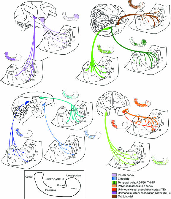
Hippocampal formation plays a prominent role in episodic memory formation and consolidation. It is likely that episodic memory representations are constructed from cortical information that is mostly funnelled through the entorhinal cortex to the hippocampus. The entorhinal cortex returns processed information to the neocortex. Retrograde tracing studies have shown that neocortical afferents to the entorhinal cortex originate almost exclusively in polymodal association cortical areas. However, the use of retrograde studies does not address the question of the laminar and topographical distribution of cortical projections within the entorhinal cortex. We examined material from 60 Macaca fascicularis monkeys in which cortical deposits of either (3)H-amino acids or biotinylated dextran-amine as anterograde tracers were made into different cortical areas (the frontal, cingulate, temporal and parietal cortices). The various cortical inputs to the entorhinal cortex present a heterogeneous topographical distribution. Some projections terminate throughout the entorhinal cortex (afferents from medial area 13 and posterior parahippocampal cortex), while others have more limited termination, with emphasis either rostrally (lateral orbitofrontal cortex, agranular insular cortex, anterior cingulate cortex, perirhinal cortex, unimodal visual association cortex), intermediate (upper bank of the superior temporal sulcus, unimodal auditory association cortex) or caudally (parietal and retrosplenial cortices). Many of these inputs overlap, particularly within the rostrolateral portion of the entorhinal cortex. Some projections were directed mainly to superficial layers (I-III) while others were heavier to deep layers (V-VI) although areas of dense projections typically spanned all layers. A primary report will provide a detailed analysis of the regional and laminar organization of these projections. Here we provide a general overview of these projections in relation to the known neuroanatomy of the entorhinal cortex.


Figures

Fig. 1
Fig. 1
Schematic representation of the ventromedial surface of the monkey brain (A). The entorhinal cortex and the constituting entorhinal cortex subfields are indicated. In (B) a photomicrograph of a histological section of the intermediate subfield of the entorhinal cortex is presented. Note the different appearance of the subfields indicated, and the laminar organization. (C) Human brain with the same orientation as A. Note the similar location of the main cortical fields, as well as the subfield composition of the human entorhinal cortex. (D) Photomicrograph of histological section of the medial temporal lobe at the level of the hippocampal head and amygdala. Note the laminar organization of the entorhinal cortex, comparable with that in the monkey, in particular medial intermediate subfield (Brodmann's area 34). Scale bars in A–C, 1 cm; B, 2 mm; D, 1 mm.
Fig. 2
Fig. 2
Dark-field photomicrograph of a coronal section through the caudal part of the monkey entorhinal cortex. A deposit of 1% WGA-HRP was made in the posterior part of the body of the hippocampus. Labelled neurons occupy mainly layers II and III in the entorhinal cortex and stop at the border with the adjacent perirhinal cortex. Scale bar equals 350 µm.
Fig. 3
Fig. 3
Schematic representation of different views of the cynomolgous monkey brain on which are plotted the locations of cortical areas that showed labelling in the retrograde studies and that were injected with anterograde tracers. The agranular insular and the representation of unimodal auditory association cortex located in the lateral aspect of the superior temporal sulcus are depicted in A. Orbitofrontal cortex and the cortical belt surrounding the entorhinal cortex (that includes temporopolar, perirhinal (area 35–36) and posterior parahippocampal cortex, here shown as a single band) are shown in B. Medial frontal (area 25) and cingulate cortices (areas 24, 23 and 29–30) are presented in C. Temporopolar cortex on the left-hand side, and polymodal association cortex at the upper bank of the superior temporal sulcus, as well as unimodal visual association cortex (area TE) at the lower bank of the superior temporal sulcus, are represented in the frontal view in D.
Fig. 5
Fig. 5
Bright-field photomicrographs of the labelling extending on both banks of the fundus of the rhinal sulcus after a BDA deposit in polymodal association cortex. Section has been counterstained with thionin. At low power magnification (A) a very dense patch of termination can be appreciated in area 36 of the perirhinal cortex, as well as in area 35. The patch in area 35 extends to the lateral rostral subfield of the entorhinal cortex, and is present at higher magnification in B in a different case with a similar deposit. Note the density of labelling in layer V at the lateral part of the lateral rostral subfield. Scale bar in A equals 1 mm, and 320 µm in B.
Fig. 4
Fig. 4
The series of histograms of this and subsequent figures depict the afferent cortical input from selected areas of the brain in which anterograde tracer deposits were placed to study the direct projection to the entorhinal cortex. Each histogram depicts the percentage of the area covered by the projection in the entorhinal cortex subfields, irrespective of the density of the projection. The percentage extent of the projection (i.e., 100% is the total extent of each subfield) is presented along the ordinate, while the abscissa represents the entorhinal cortex subfields. White columns are for superficial (layers I–III) of the entorhinal cortex, while black columns are for deep layers (V–VI). Overall, note that the largest contribution in percentage and number of subfields corresponds to the cortical belt surrounding the entorhinal cortex. Polymodal association cortex (presented at two different rostrocaudal levels) and unimodal association cortices such as visual (TE) and auditive (parabelt region) present a lower percentage of projection in the entorhinal cortex subfields.
Fig. 6
Fig. 6
Histograms showing the projection from cortical areas such as the orbitofrontal cortex, lateral (A) and medial (B). Note the projection to deep layers in all subfields of the entorhinal cortex. Agranular insular cortex (C) shows a preferential projection to olfactory subfield of entorhinal cortex. Area 25 of the medial frontal cortex (D) also presents a preferential projection to the deep layers of entorhinal cortex. Anterior cingulate cortex (E) distributes widespread afferents to the entorhinal cortex, while retrosplenial cortex (F) is directed specifically to the caudal subfields EC and ECL.
Fig. 7
Fig. 7
Histograms depicting the percentage of the area covered in each subfield of the entorhinal cortex by the afferent projection of the different cortical areas providing cortical input to the entorhinal cortex. Note that the largest cortical input falls in the lateral entorhinal cortex, lateral rostral and lateral caudal subfields. Rostral subfield follows in extent of the projection, while the remaining subfields present more limited cortical projection.
Fig. 8
Fig. 8
Summary diagram of the projection of cortical input to the entorhinal cortex subfields. The thickness of the arrows relates to the extent of the projection. The inset presents a schematic view of the monkey hippocampus with its various portions. Each cortical area projecting to the entorhinal cortex is presented as a particular colour, and the presumed corresponding termination in the hippocampus is likewise indicated.

Similar articles

Cited by

References

    1. Amaral DG, Cowan WM. Subcortical afferents to the hippocampal formation in the monkey. J Comp Neurol. 1980;189:573–591. - PubMed
    1. Amaral DG, Insausti R, Cowan WM. Evidence for a direct projection from the superior temporal gyrus to the entorhinal cortex in the monkey. Brain Res. 1983;275:263–277. - PubMed
    1. Amaral DG, Insausti R, Cowan WM. The commissural connections of the monkey hippocampal formation. J Comp Neurol. 1984;224:307–336. - PubMed
    1. Amaral DG, Insausti R, Cowan WM. The entorhinal cortex of the monkey, I. Cytoarchitectonic organization. J Comp Neurol. 1987;264:326–355. - PubMed
    1. von Bonin G, Bailey P. The Neocortex of Macaca mulatta. Urbana, IL: University of Illinois Press; 1947.

Publication types

MeSH terms

LinkOut - more resources