. 2007 Aug 1;308(1):144-57.
doi: 10.1016/j.ydbio.2007.05.018. Epub 2007 May 24.

mef2ca is required in cranial neural crest to effect Endothelin1 signaling in zebrafish

Affiliations

mef2ca is required in cranial neural crest to effect Endothelin1 signaling in zebrafish

Craig T Miller et al. Dev Biol. .

Abstract

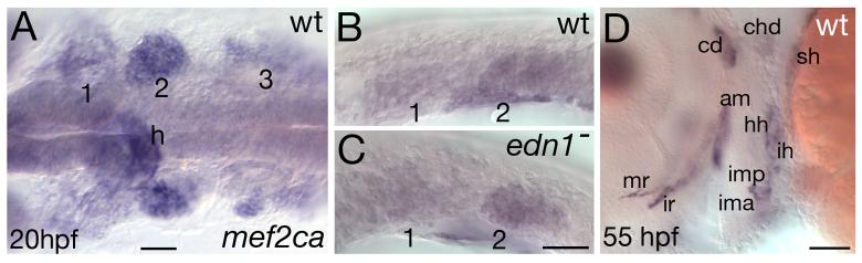
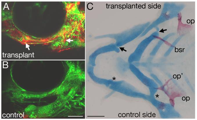
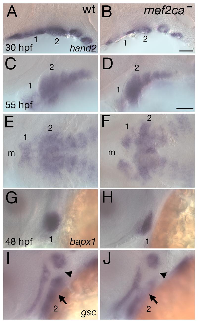
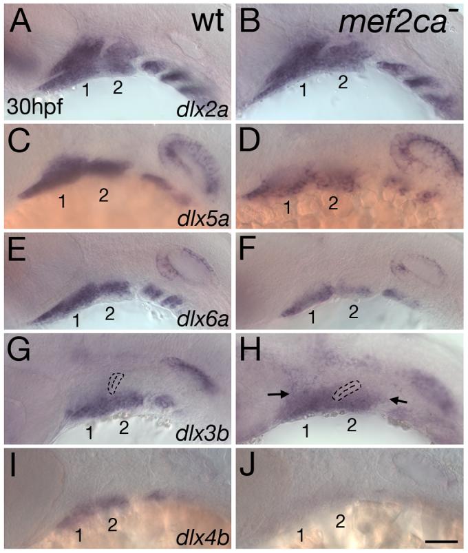
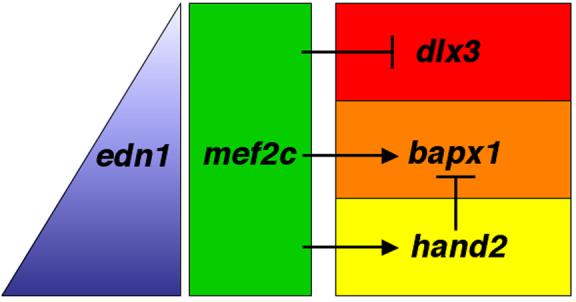
Mef2 genes encode highly conserved transcription factors involved in somitic and cardiac mesoderm development in diverse bilaterians. Vertebrates have multiple mef2 genes. In mice, mef2c is required for heart and vascular development. We show that a zebrafish mef2c gene (mef2ca) is required in cranial neural crest (CNC) for proper head skeletal patterning. mef2ca mutants have head skeletal phenotypes resembling those seen upon partial loss-of-function of endothelin1 (edn1). Furthermore, mef2ca interacts genetically with edn1, arguing that mef2ca functions within the edn1 pathway. mef2ca is expressed in CNC and this expression does not require edn1 signaling. Mosaic analyses reveal that mef2ca is required in CNC for pharyngeal skeletal morphogenesis. Proper expression of many edn1-dependent target genes including hand2, bapx1, and gsc, depends upon mef2ca function. mef2ca plays a critical role in establishing the proper nested expression patterns of dlx genes. dlx5a and dlx6a, known Edn1 targets, are downregulated in mef2ca mutant pharyngeal arch CNC. Surprisingly, dlx4b and dlx3b are oppositely affected in mef2ca mutants. dlx4b expression is abolished while the edn1-dependent dlx3b is ectopically expressed in more dorsal CNC. Together our results support a model in which CNC cells require mef2ca downstream of edn1 signaling for proper craniofacial development.


Figures

Fig. 1
Fig. 1
Molecular identification of the lesions in hoover(mef2ca) mutants. (A) Schematic showing the map position of hoover, which is non-recombinant with mef2ca in 1008 meioses. (B) mef2ca is mutated in all three hoover alleles. In b631 mutants, an A-to-C mutation is predicted to change the initiator methionine to leucine. In b1086 mutants, an A-to-T mutation creates an early stop in exon two, predicted to create a truncated protein lacking a MEF2 domain. In tn213 mutants, a T-to-C mutation alters the conserved second nucleotide of the intron 7 splice donor.
Fig. 2
Fig. 2
Reduction of mef2ca function causes facial deformities which phenocopy partial edn1 loss-of-function. (A-B) Live phenotypes of mef2ca mutants at 5 dpf. mef2ca mutants have malformed faces, open mouths, and ventrally displaced jaws. (C-D) Cartilage and bone phenotypes in the pharyngeal arches of mef2ca mutants. Dorsal/ventral joints (arrows) are missing in mef2ca mutants (asterisks). Mutants also have ectopic cartilage nodules (arrowhead), distinctive ectopic medial processes emanating from the upper jaw cartilage (see Table 2) and homeotic transformations of hyoid dermal bone identity. In mef2ca mutants, a ventral hyoid bone, the branchiostegal ray, is enlarged, assumes the shape of and fuses to the dorsal hyoid bone, the opercle. (E-F) mef2ca-MO injection phenocopies mef2ca mutations. Lateral views of wholemount 4.5 dpf wild-type larvae, uninjected (E) and injected with 5 ng of mef2ca morpholino (F). mef2ca-MO injected fish display characteristic mef2ca mutant cartilage phenotypes. Joints are lost (asterisks) and ectopic cartilage nodules (arrowhead) are seen near the basihyal. (G-I) mef2ca mutation phenocopies gradual reduction in edn1 function. Ventral views of 4.5 dpf Alcian stained wild-type (G), mef2cab631 mutant (H), and low level edn1-MO injected (I) larvae. mef2ca mutants and low level edn1-MO morphants display joint loss (black asterisks), ectopic medially-projecting processes on the upper jaw cartilage, the palatoquadrate (white asterisks), and ectopic cartilage nodules near the basihyal (arrowheads). bh, basihyal; bsr, branchiostegal ray; ch, ceratohyal; hs, hyosymplectic; M, Meckel's; op, opercle; pq, palatoquadrate.
Fig. 3
Fig. 3
Complex genetic interactions between mef2ca and edn1. Flat-mounted pharyngeal skeletons of 5 dpf fish stained for cartilage in blue and bone in red for fish of different mef2ca and edn1 genotypes. (A) In the wild-type second arch, a prominent fan-shaped opercle bone (op) articulates with the hyosymplectic (hs). Its serial homolog, the branchiostegal ray (bsr) appears saber-shaped and articulates with the ceratohyal (ch). We typically detect no phenotypes in edn1 or mef2ca single heterozygous classes (B and D), although rarely very mild phenotypes are seen in edn1 mutant heterozygotes (Table 2). (C) In mef2ca homozygous mutants, the opercle is enlarged, and the ventral branchiostegal ray is enlarged and transformed towards opercle morphology. The dorsal/ventral joint is lost (asterisk showing joint loss in the hyoid arch). The hyoid ventral cartilage is reduced. (E) Fish heterozygous for both mef2ca and edn1 (mef2ca+/−; edn1+/−) display mef2ca mutant phenotypes such as joint loss (asterisk) and enlarged opercles fused to malformed branchiostegal rays. (F) Heterozygosity of edn1 enhances the mef2ca cartilage and bone phenotypes (compare to C, and see Table 2). (G-I) Heterozygosity for mef2ca mutation (H) partially and subtly rescues cartilage and bone phenotypes in edn1−/− homozygous mutants (G), while homozygosity for mef2ca mutation more significantly partially rescues hyoid cartilage and bone phenotypes of edn1 homozygous mutants (I).
Fig. 4
Fig. 4
Epistatic interactions between mef2ca and edn1. Hyoid bone (opercle, OP) (A-C) and cartilage (ceratohyal, CH) (D-F) phenotypes as a function of: edn1+ alleles in homozygous mef2ca mutants (A,D), mef2ca+ alleles in homozygous edn1 mutants (B, E), and genotypes at both mef2ca and edn1 (C,F). In C and F, edn1 genotype is denoted by the number of edn1+ alleles: open box = 2 edn1+ alleles; “X” = 1 edn1+ allele, “+” = 0 edn1+ alleles. Phenotypes were scored on individual sides of PCR-genotyped larvae (see Methods). OP bone scoring scale: strong edn1 loss-of-function phenotype (loss of bone) = 0, weak edn1 loss-of-function phenotype (expanded bone) = 1, wild-type = 2. CH cartilage scoring scale: strong edn1 loss-of-function phenotype (complete loss) = 0, weak edn1 loss-of-function phenotype (partial loss) = 1, wild-type = 2. See Kimmel et al. (2003) and Miller and Kimmel (2001) for strong and weak edn1 loss-of-function bone and cartilage mutant phenotypes. Phenotypes for each genotypic category are shown as least square means (LS means). Error bars represent 95% confidence intervals from ANOVA. (G) Genetic model for zebrafish hyoid cartilage and dermal bone development. mef2ca represses bone and cartilage formation. edn1 represses mef2ca, but also activates a mef2ca-independent pathway for ventral cartilage development.
Fig. 5
Fig. 5
mef2ca expression in early CNC and late head muscles. In situ hybridization showing mef2ca expression in wild-type (A,B,D) and edn1 mutant (C) embryos at 20 hpf (A-C) and 55 hpf (D). (A-B) Dorsal (A) and lateral (B) views of mef2ca expression in all three migrating streams of CNC at 20 hpf. (C) CNC expression in edn1 mutants appears unaffected. (D) Lateral view of mef2ca expression in head muscles at 55 hpf. For A-C, arches are numbered. Pharyngeal pouches are outlined in B-C. For description of the larval head muscle pattern see (Lin et al., 2006; Schilling and Kimmel, 1997). am adductor mandibulae; cd, constrictor dorsalis; chd, constrictor hyoideus dorsalis; hh, hyohyoideus; ih, interhyoideus; ima, intermandibularis anterior; imp, intermandibularis posterior; ir, inferior rectus; mr, medial rectus; sh, sternohyoideus.
Fig. 6
Fig. 6
mef2ca is autonomously required in cranial neural crest for skeletal patterning. Wild-type rhodamine-labeled fli1:EGFP CNC cells were unilaterally transplanted into mef2ca mutant hosts. (A-B) Confocal micrographs of mef2ca mutant with wild-type CNC mosaic fish from the tranplanted side (A) and control side (B). On the transplanted side, lineage tracer is detected throughout dorsal and ventral cartilages, as well as joint regions (arrows). On the control side, only mef2ca mutant host fli1:GFP CNC derivatives are seen. (C) Flatmounted pharyngeal skeleton, double stained for cartilage in blue and bone in red. Dorsal/ventral cartilage joints have been rescued on the transplanted side (arrows) but not on the control side (asterisks). Opercle and branchiostegal ray morphology is also completely rescued on the transplanted side, but not the control side. bsr, branchiostegal ray; op, opercle.
Fig. 7
Fig. 7
mef2ca is required for proper expression of hand2, bapx1, and gsc in postmigratory CNC. Lateral (A-D, G-J) and ventral (E,F) views of expression of hand2 (A-F) at 30 hpf (A,B) and 55 hpf (C-F), and bapx1 (G,H) and gsc (I,J) at 48 hpf in wild-type (A,C,E,G,I) and mef2ca mutants (B,D,F,H,J). (A-F) Ventrally restricted expression of hand2 is reduced in mef2ca mutants at 30 hpf, but recovers by 55 hpf. (G,H) bapx1 expression prefiguring the jaw joint is reduced in mef2ca mutants. (I,J) Ventral hyoid expression of gsc (arrow) is downregulated in mef2ca mutants. The hyoid joint region (arrowhead) remains devoid of gsc expression. Arches are numbered. m, mouth.
Fig. 8
Fig. 8
mef2ca activates dlx5a, dlx6a, and dlx4b but represses dlx3b. Lateral views of expression of dlx2a (A,B), dlx5a (C,D), dlx6a (E,F), dlx3b (G,H) and dlx4b (I,J) in wild-type (A,C,E,G,I) and mef2ca mutant (B,D,F,H,J) heads of 30 hpf zebrafish embryos. (A,B) mef2ca mutants have no detectable alteration in dlx2a expression, which is expressed throughout pharyngeal arch CNC. (C-F) Expression of both dlx5a and dlx6a, in ventral and intermediate CNC in wild-types, is reduced in mef2ca mutants. (G,H) dlx3b expression is ectopically expressed in dorsal arch CNC of mef2ca mutants (arrows). (I,J) In contrast, dlx4b expression, restricted to ventral arch CNC, is largely undetectable in mef2ca mutants. The first two arches are numbered.
Fig. 9
Fig. 9
The edn1-effector mef2c regulates dorsal/ventral patterning in pharyngeal arch CNC. Edn1 secreted from ventral pharyngeal epithelia and mesoderm forms a gradient of Edn1 (blue triangle), with high levels ventrally (bottom of triangle) and low levels dorsally (top of triangle). mef2c, broadly expressed in early arch postmigratory CNC (green rectangle) exerts differential effects on downstream target genes to help specify domains along the dorsal-ventral axis. In the dorsal arch (red), mef2c represses dlx3 expression. In the intermediate (orange) and ventral (yellow) domains, mef2c activates transcription of bapx1 and hand2, two effectors of intermediate and ventral skeletal fates, respectively. hand2 helps delimit the intermediate domain by repressing bapx1 expression from the ventral domain (Miller et al., 2003). The red, orange, and yellow domains are in part defined by the nested expression patterns of dlx genes and hand2. The dorsal red domain expresses dlx2a, but not dlx3b or dlx6a. The orange and yellow domains express dlx2a, dlx3b, dlx5a, and dlx6a. The expression of hand2 defines the ventral yellow compartment. See Walker et al. (2006a) for a more detailed spatiotemporal model of dlx and hand2 regulation by Edn1.

Similar articles

Cited by

References

    1. Akimenko MA, et al. Combinatorial expression of three zebrafish genes related to distal-less: part of a homeobox gene code for the head. J Neurosci. 1994;14:3475–86. - PMC - PubMed
    1. Angelo S, et al. Conservation of sequence and expression of Xenopus and zebrafish dHAND during cardiac, branchial arch and lateral mesoderm development. Mech Dev. 2000;95:231–7. - PubMed
    1. Bi W, et al. The transcription factor MEF2C-null mouse exhibits complex vascular malformations and reduced cardiac expression of angiopoietin 1 and VEGF. Dev Biol. 1999;211:255–67. - PubMed
    1. Black BL, Olson EN. Transcriptional control of muscle development by myocyte enhancer factor-2 (MEF2) proteins. Annu Rev Cell Dev Biol. 1998;14:167–96. - PubMed
    1. Chang S, et al. An expression screen reveals modulators of class II histone deacetylase phosphorylation. Proc Natl Acad Sci U S A. 2005;102:8120–5. - PMC - PubMed

Publication types

MeSH terms