. 2007 Jul 15;313(12):2597-610.
doi: 10.1016/j.yexcr.2007.05.009. Epub 2007 May 18.

Cholesterol depletion alters detergent-specific solubility profiles of selected tight junction proteins and the phosphorylation of occludin

Affiliations

Cholesterol depletion alters detergent-specific solubility profiles of selected tight junction proteins and the phosphorylation of occludin

Robert D Lynch et al. Exp Cell Res. .

Abstract

Differential centrifugation of Triton X-100 or CHAPS lysates from control and cholesterol (CH)-depleted MDCK II cells, segregated integral tight junction (TJ) proteins associated with detergent-resistant membranes (DRMs) into two groups. Group A proteins (occludin, claudin-2 and -3) were detected in large, intermediate and small aggregates in both detergents, whereas group B proteins (claudin-1, -4 and -7) were observed in small aggregates in TX-100 and in intermediate and small aggregates in CHAPS. Depletion of CH altered the distribution of group A and B proteins among the three size categories in a detergent-specific manner. In lysates produced with octyl glucoside, a detergent that selectively extracts proteins from DRMs, group A proteins were undetectable in large aggregates and CH depletion did not alter the distribution of either group A or B proteins in intermediate or small aggregates. Neither occludin (group A) nor claudin-1 (group B) was in intimate enough contact with CH to be cross-linked to [(3)H]-photo-cholesterol. However, antibodies to either TJ protein co-immunoprecipitated caveolin-1, a CH-binding protein. Unlike claudins, occludin's presence in TJs and DRMs did not require palmitoylation. Equilibrium density centrifugation on discontinuous OptiPrep gradients revealed detergent-related differences in the densities of TJ-bearing DRMs. There was little or no change in those densities after CH depletion. Removing CH from the plasma membrane increased tyrosine and threonine phosphorylation of occludin, and transepithelial electrical resistance (TER) within 30 min. After 2 h of CH efflux, phospho-occludin levels and TER fell below control values. We conclude that the association of integral TJ proteins with DRMS, pelleted at low speeds, is partially CH-dependent. However, the buoyant density of TJ-associated DRMs is a function of the detergent used and is insensitive to decreases in CH.


Figures

Figure 1
Figure 1
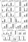
Western blot analysis of MDCK II cell monolayers incubated with 10 mM MBCD for 0, 0.5 or 2 h. Monolayers were lysed either in TX-100 or CHAPS at 4°C and centrifuged at 1000 g for 30 min. Low speed pellets (LSP) and aliquots of the low speed supernatants (LSS) were retrieved. The remaining low speed supernatants were centrifuged at 4°C for 18 h at 107,600 g. High-speed supernatants (HSS) and high-speed pellets (HSP) were harvested. All supernatant and pellet fractions were processed for Western blotting and probed for occludin, claudin-1, -2, -3, -4, -7, caveolin-1, annexin-2, ZO-1 and actin. The data shown are representative of those obtained from a minimum of two independent experiments using TX-100 and CHAPS.
Figure 2
Figure 2
Densitometric data derived from control MDCK II cells (black bar), treated for 30 min (light gray bars) or 2 h (dark gray bars) with 10 mM MBCD and lysed in either TX-100 or CHAPS at 4°C. The large aggregates are derived from low-speed pellets (LSP), the medium size aggregates are from high-speed pellets (HSP) and the small aggregates are obtained from high-speed supernatants (HSS). Densitometric measurements are derived from experiments described in the Fig. 1 legend. Values on the ordinate represent the percent of the protein in each fraction. Densitometric data from the two groups of integral TJ proteins (A and B), the lipid binding proteins (C) and TJ associated cytoplasmic proteins (D). Densitometric data from cell monolayers incubated with MBCD for 30 min and extracted with either 3.25% TX-100 or 4% CHAPS (E). Densitometric data of transferrin receptor from cells treated as in (E) and extracted with either of two concentrations of TX-100 or CHAPS (F). In (D) and (E) only data from control cultures and those incubated with MBCD for 30 min are shown.
Figure 3
Figure 3
Densitometric data derived from control MDCK II cells (black bar), treated for 30 min (light gray bars) with 10 mM MBCD and lysed in 1.75% OG. In contrast to TX-100 and CHAPS lysates, virtually no group A TJ proteins are detected as large aggregates in the LSP, while the distribution of group B TJ proteins is similar to that observed in TX-100 and CHAPS lysates. Furthermore, in contrast to TX-100 and CHAPS preparations, incubation with 10 mM MBCD did not alter the distribution of these proteins in the different fractions.
Figure 4
Figure 4
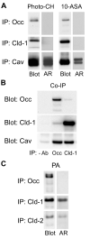
A. MDCK II cell monolayers were incubated with either [3H]-photo-CH or [3H]-choline and 10-ASA as described in Materials and Methods. Following photo-activation, occludin, claudin-1 and caveolin-1 were sequentially immunoprecipitated (IP) in RIPA buffer. Dried PVDF membranes were subjected to autoradiography (AR) and probed for the respective immunoprecipitated protein (Blot). Whereas [3H]-photo-CH and [3H]-photo-PC prominently labeled caveolin-1, neither occludin nor claudin-1 were labeled. B. Occludin or claudin-1 was immunoprecipitated from lysates in 1% CHAPS, 0.05% SDS and the blots were probed with anti-occludin, anti-claudin-1 or anti-caveolin-1. Caveolin-1 is co-immunoprecipitated by both anti-occludin and anti-claudin-1. C. MDCK II cell monolayers were metabolically labeled with [3H]-palmitic acid (PA) as described in Materials and Methods. Occludin, claudin-1 and claudin-2 were sequentially immunoprecipitated. Dried PVDF membranes were subjected to autoradiography (AR) and probed for the respective immunoprecipitated protein (Blot). [3H]-palmitate labels claudin-1 and -2 but not occludin.
Figure 5
Figure 5
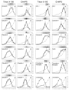
Densitometric data of 10 fractions each, obtained from discontinuous OptiPrep step gradients of control MDCK II cells (black circles) or MDCK II cells treated with 10 mM MBCD for 2h (white circles). The cells were lysed in either 1% TX-100 or 1.2% CHAPS. Lysates were prepared and OptiPrep gradients were centrifuged as described in Materials and Methods. Fractions, 0.5 ml, were collected beginning from the top of the gradient and prepared for Western blotting as described in Materials and Methods. Parallel gradients were run for CH determinations and to determine the refractive index for each fraction (white circles and dotted lines). Density in g/ml is indicated on the right hand Y-axis, while fraction number is indicated on the X-axis. The percent of a particular protein in each fraction is plotted on the left Y-axis and was estimated by dividing the pixel intensity of that protein in that fraction by the sum of its pixel intensities in all 10 fractions. All data obtained from TX-100 extracts are the mean and range bars of three independent experiments, whereas the data from the CHAPS extracts are derived from two independent experiments. Data from the transferrin receptor, a non-DRM associated protein, are included for comparison.
Figure 6
Figure 6
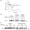
A. Sequential TER measurements of MDCK II cell monolayers that were treated with 10 mM MBCD for 2 h (white circles). The control monolayers (black circles) underwent medium changes at the same time that MBCD was either added or removed from the experimental monolayers. TER falls to near zero within 2 h of adding MBCD. Within 24 h of removing MBCD, TER returns to control levels. B. Occludin was immunoprecipitated from untreated monolayers or from monolayers incubated for 0.5 or 2 h with 10 mM MBCD and the immunoprecipitates were subjected to Western blotting. The Western blots were probed with anti-occludin, anti-phosphotyrosine or anti-phosphothreonine antibodies. C. Monolayers of MDCK II cells were treated with 10 mM MBCD without or with 100 μM Genistein (G) for 0, 0.5 or 2 h. Occludin was then immunoprecipitated and Western blots were probed using either anti-occludin (loading control) or anti-phosphotyrosine antibodies. TER data are the mean +/- SD of triplicate inserts. Data in Fig. 6A and B are representative of three independent experiments and the data in Fig 6C are from a single experiment.

Similar articles

Cited by

References

    1. Furuse M, Hirase T, Itoh M, Nagafuchi A, Yonemura S, Tsukita S, Tsukita S. Occludin: A novel integral membrane protein localizing at tight junctions. Journal of Cell Biology. 1993;123:1777–1788. - PMC - PubMed
    1. Furuse M, Fujita K, Fujimoto K, Tsukita S. Claudin 1 and 2: Novel integral membrane proteins localizing at tight junctions with no sequence similarity to occludin. Journal of Cell Biology. 1998;141:1539–1550. - PMC - PubMed
    1. Tsukita S, Furuse M, Itoh M. Multifunctional strands in tight junctions. Nature Reviews Molecular Cell Biology. 2001;2:285–293. - PubMed
    1. Staehelin LA. Further observations on the fine structure of freeze-cleaved tight junctions. Journal of Cell Science. 1973;13:763–786. - PubMed
    1. Vogelmann R, Nelson WJ. Fractionation of the epithelial apical junctional complex: Reassessment of protein distributions in different substructures. Molecular Biology of the Cell. 2005;16:701–716. - PMC - PubMed

Publication types

MeSH terms