. 2007 Jul 15;313(12):2667-79.
doi: 10.1016/j.yexcr.2007.05.013. Epub 2007 May 18.

RACK1 regulates Src activity and modulates paxillin dynamics during cell migration

Affiliations

RACK1 regulates Src activity and modulates paxillin dynamics during cell migration

Ashley T Doan et al. Exp Cell Res. .

Abstract

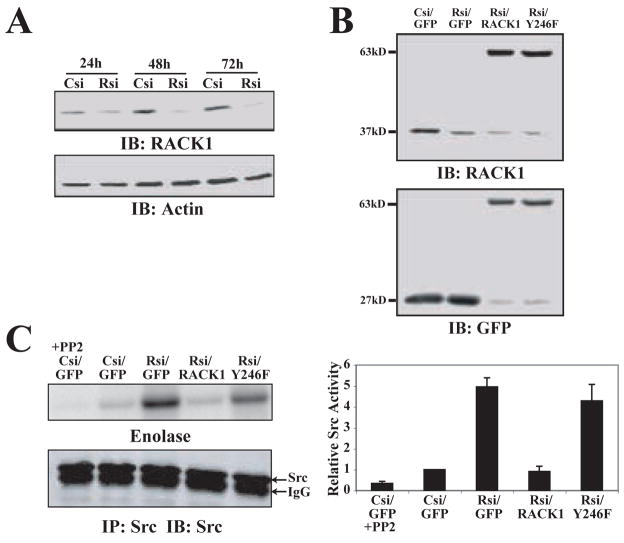
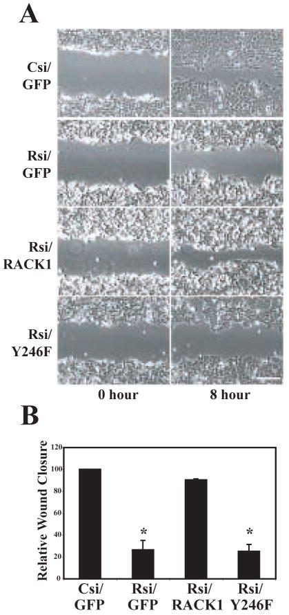
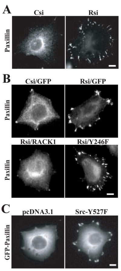
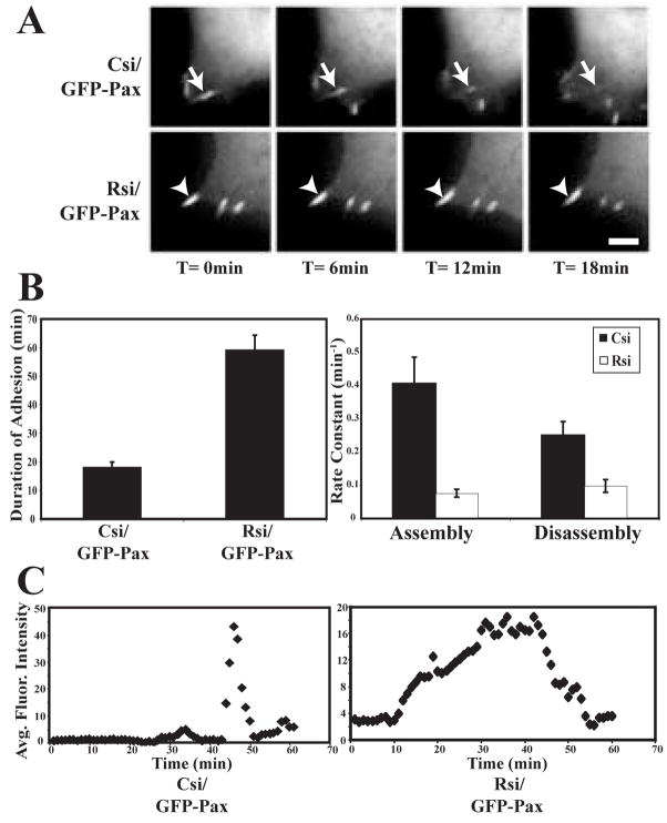
Receptor for Activated C Kinase, RACK1, is an adaptor protein that regulates signaling via Src and PKC-dependent pathways, and has been implicated in cell migration. In this study we demonstrate novel functions for RACK1 in regulating adhesion dynamics during cell migration. We report that cells lacking RACK1 are less motile and show reduced dynamics of paxillin and talin at focal complexes. To investigate the role of the RACK1/Src interactions in adhesion dynamics, we used RACK1 in which the putative Src binding site has been mutated (RACK Y246F). RACK1-deficient cells showed enhanced c-Src activity that was rescued by expression of wild type RACK1, but not by RACK Y246F. Expression of wild type RACK1, but not RACK Y246F, was also able to rescue the adhesion and migration defects observed in the RACK1-deficient cells. Furthermore, our findings indicate that RACK1 functions to regulate paxillin phosphorylation and that its effects on paxillin dynamics require the Src-mediated phosphorylation of tyrosine 31/118 on paxillin. Taken together, these findings support a novel role for RACK1 as a key regulator of cell migration and adhesion dynamics through the regulation of Src activity, and the modulation of paxillin phosphorylation at early adhesions.


Figures

Figure 1
Figure 1
RACK1 is a negative regulator of endogenous c-Src activity. (A) HEK cells were transiently transfected with pooled control siRNA (Csi) or pooled RACK1 siRNA (Rsi) for 24 h, 48 h and 72 h as indicated. Representative blot shows expression of RACK1. Actin was used as a loading control. (B) HEK cells were transiently transfected with single target control siRNA (Csi) or RACK1 siRNA (Rsi) co-expressed with GFP, GFP-RACK1 (RACK1) or GFP-RACK Y246F (Y246F) as described in Materials and Methods. Representative blot shows knockdown of endogenous RACK1 and expression of GFP constructs as indicated by immunoblotting with α-RACK1 or α-GFP. (C) HEK cells were transiently transfected with control siRNA (Csi) or RACK1 siRNA (Rsi) co-expressed with GFP, GFP-RACK1 (RACK1) or GFP-RACK Y246F (Y246F). Immunoprecipitation was performed on lysates using a Src-specific antibody. Kinase activity assays were performed with or without Src inhibitor PP2 as described in Materials and Methods and the autoradiograph is shown. Quantification of Src activity from three separate experiments is shown as the mean ± SEM.
Figure 2
Figure 2
RACK1 modulates cell migration through its regulation of Src activity. (A) HEK cells were co-transfected with control siRNA (Csi) or RACK1 siRNA (Rsi) with GFP, GFP-RACK1 (RACK1) or GFP-RACK Y246F (Y246F) as indicated. Wound assays were performed as described in Materials and Methods. Representative images from three separate experiments are shown at time 0 h and time 8 h after wounding. See supplemental movies 1–4. Bar, 50 μm. (B) Quantification of wound assay was performed by measuring the change in wound area from 0 h to 8 h post-wound relative to control cells as described in Materials and Methods. Data represent the average from three separate experiments, with error bars representing as the mean ± SEM. *p<0.01 in a Student’s t test in comparison to control.
Figure 3
Figure 3
RACK1 regulates paxillin organization at focal complexes. (A) HEK cells were plated for 2 h on coverslips coated with 3 μg/ml fibronectin as described in the Materials and Methods. Representative images from more than four separate experiments show the distribution of paxillin in cells that express pooled control siRNA (Csi) or pooled RACK1 siRNA (Rsi). Bar, 10 μm. (B) HEK cells were co-transfected with single target control siRNA (Csi) or RACK1 siRNA (Rsi) with GFP, GFP-RACK1 (RACK1) or GFP-RACK Y246F (Y246F) as indicated. Representative images are shown from more than four separate experiments. Bar, 10 μm. (C) HEK cells were co-transfected with pcDNA3.1 or Src-Y527F with GFP-paxillin as indicated. Representative images are from three separate experiments. Bar, 10 μm.
Figure 4
Figure 4
Turnover of paxillin at focal complexes is reduced in the absence of RACK1. (A) GFP-paxillin was co-transfected with pooled control (Csi) or RACK1 (Rsi) siRNA. in HEK cells. Cells were plated on 3 μg/ml fibronectin and imaged by time-lapse fluorescence microscopy. Representative frames are shown from time-lapse sequences used to quantify duration and assembly/disassembly rates. See supplemental movies 5 and 6. For each sequence, T= 0 min is the first frame in which the adhesion marked by the arrow was observed. Bar, 3 μm. (B) Quantification of the persistence of GFP-paxillin in focal complexes demonstrates that paxillin remains at focal complexes for longer durations in the absence of RACK1. Duration measurements were made by counting the amount of time elapsed between the first and last frames in which an individual adhesion was observed in HEK cells that co-express GFP-paxillin and control siRNA (Csi) or RACK1 siRNA (Rsi). Rate constants for assembly and disassembly kinetics were calculated as described in the Materials and Methods. Quantification shows the mean ± SEM from three separate experiments (total of 45 adhesions quantified for each condition). (C) Representative fluorescence intensity graphs for HEK cells that express control (Csi) and RACK1 (Rsi) siRNA over time (min).
Figure 5
Figure 5
RACK1 regulates the dynamics of both paxillin and talin at focal complexes. Cells were plated on 3 μg/ml fibronectin and imaged by time-lapse fluorescence microscopy as described in Materials and Methods. Representative frames are shown from time-lapse sequences collected every 2 min. See supplemental movies 7 and 8. Dual imaging of GFP-talin and dsRed-paxillin in HEK cells that expressed pooled control (Csi) and RACK1 (Rsi) siRNA. Bar, 10 μm.
Figure 6
Figure 6
RACK1 regulates phosphorylation of focal adhesion proteins through a Src-dependent pathway. (A) HEK cells were transiently transfected with pooled control siRNA (Csi) or RACK1 siRNA (Rsi) for 24 h, 48 h and 72with h. Representative blots show paxillin phosphorylation at Y118. Total paxillin was used as a loading control. (B) HEK cells transiently transfected with pooled control (Csi) or RACK1 (Rsi) siRNA were treated with control vehicle (−) or the Src inhibitor, PP2. Cell lysates were collected and immunoblotted for phospho-paxillin (pY118), total paxillin and RACK1 as indicated. Representative blot from three experiments is shown. (C) HEK cells were transiently transfected with single target control siRNA (Csi) or RACK1 siRNA (Rsi) co-expressed with GFP, GFP- GFP-RACK1 (RACK1) or GFP-RACK Y246F (Y246F). Representative blot shows paxillin phosphorylation at Y118 from cells plated on fibronectin as described in Materials and Methods. Total paxillin was used as a loading control. (D) Quantification of paxillin phosphorylation relative to control lysates for conditions described in (C). Data represents the average from five separate experiments, with error bars represented as the mean ± SEM. *p<0.03 using a Student’s t test (in comparison with control). (E) HEK cells were transiently transfected with single target control siRNA (Csi) or RACK1 siRNA (Rsi) co-expressed with GFP, GFP-RACK1 (RACK1) or GFP-RACK Y246F (Y246F). Representative blot shows FAK phosphorylation at the Src-specific site Y576 and paxillin phosphorylation at Y31 from cells plated on fibronectin as described in Materials and Methods. Total FAK and paxillin were used as loading controls.
Figure 7
Figure 7
Paxillin phosphorylation is required for RACK1 effects on paxillin morphology and dynamics. (A) HEK cells were co-transfected with control siRNA or RACK1 siRNA with either wild type GFP-paxillin or GFP-paxillin that can not be phosphorylated at tyrosine 31 or 118 (GFP-paxillin Y31,118F). Representative images show the localization of GFP-paxillin or GFP-paxillin Y31,118F. Bar, 10 μm. (B) Representative frames are shown from time-lapse sequences used to quantify duration of adhesions. For each sequence, T= 0 min is the first frame in which the adhesion marked by the arrow was observed. Bar, 3 μm. (C) Quantification of GFP-paxillin Y31,118F duration at focal complexes in control and RACK1-deficient cells. The graph represents the average from three separate experiments, with error bars represented as the mean ± SEM.
Figure 8
Figure 8
Turnover of phosphoproteins at focal complexes is reduced in the absence of RACK1. (A) GFP-dSH2 was co-transfected with pooled control (Csi) or RACK1 (Rsi) siRNA in HEK cells. Cells were plated on 3 μg/ml fibronectin and imaged by time-lapse fluorescence microscopy. Representative frames are shown from time-lapse sequences used to quantify duration of adhesions. For each sequence, T= 0 min is the first frame in which the adhesion marked by the arrow was observed. Bar, 3 μm. (B) Quantification of the persistence of GFP-dSH2 in focal complexes demonstrates that phosphoproteins remain at focal complexes for longer durations in the absence of RACK1. Duration measurements were made by counting the amount of time elapsed between the first and last frames in which an individual adhesion was observed in HEK cells that express control siRNA (Csi) or RACK1 siRNA (Rsi). The graph represents the average from at least three separate experiments, with error bars represented as the mean ± SEM. (C) HEK cells were transiently transfected with dSH2-GFP and either control (Csi) or RACK1 (Rsi) siRNA for 48 h. Immunoprecipitation for GFP or nonspecific IgG control was performed as described in Materials and Methods and immunoblotted for paxillin. Total cell lysates from control and RACK1-deficient cells were immunoblotted for phosphopaxillin (pY118), total paxillin and RACK1 as indicated. Representative blots from three separate experiments are shown.

References

    1. Jockusch BM, Bubeck P, Giehl K, Kroemker M, Moschner J, Rothkegel M, Rudiger M, Schluter K, Stanke G, Winkler J. The molecular architecture of focal adhesions. Annu Rev Cell Dev Biol. 1995;11:379–416. - PubMed
    1. Hynes RO. Integrins: bidirectional, allosteric signaling machines. Cell. 2002;110:673–87. - PubMed
    1. Burridge K, Fath K, Kelly T, Nuckolls G, Turner C. Focal adhesions: transmembrane junctions between the extracellular matrix and the cytoskeleton. Annu Rev Cell Biol. 1988;4:487–525. - PubMed
    1. Schwartz MA, Schaller MD, Ginsberg MH. Integrins: emerging paradigms of signal transduction. Annu Rev Cell Dev Biol. 1995;11:549–99. - PubMed
    1. Ilic D, Furuta Y, Kanazawa S, Takeda N, Sobue K, Nakatsuji N, Nomura S, Fujimoto J, Okada M, Yamamoto T. Reduced cell motility and enhanced focal adhesion contact formation in cells from FAK-deficient mice. Nature. 1995;377:539–44. - PubMed

Publication types