

Induction of potent local cellular immunity with low dose X4 SHIV(SF33A) vaginal exposure
- PMID: 17574643
- PMCID: PMC2756750
- DOI: 10.1016/j.virol.2007.05.021
Induction of potent local cellular immunity with low dose X4 SHIV(SF33A) vaginal exposure
Abstract
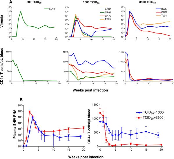
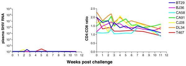
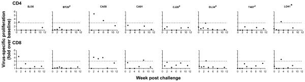
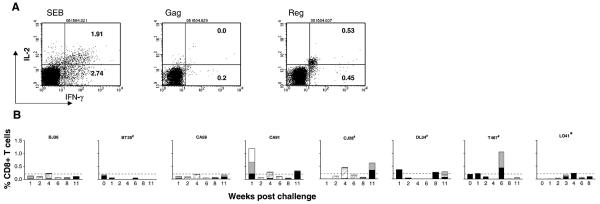
Intravaginal inoculation of rhesus macaques with varying doses of the CXCR4 (X4)-tropic SHIV(SF33A) isolate revealed a threshold inoculum for establishment of systemic virus infection and a dose dependency in overall viral burden and CD4+ T cell depletion. While exposure to inoculum size of 1000 or greater 50% tissue infectious dose (TCID(50)) resulted in high viremia and precipitous CD4+ T cell loss, occult infection was observed in seven of eight macaques exposed to 500 TCID(50) of the same virus. The latter was characterized by intermittent detection of low level virus with no evidence of seroconversion or CD4+ T cell decline, but with signs of an ongoing antiviral T cell immune response. Upon vaginal re-challenge with the same limiting dose 11-12 weeks after the first, classic pathogenic X4 SHIV(SF33A) infection was established in four of the seven previously exposed seronegative macaques, implying enhanced susceptibility to systemic infection with prior exposure. Pre-existing peripheral SIV gag-specific CD4+ T cells were more readily demonstrable in macaques that became systemically infected following re-exposure than those that were not. In contrast, early presence of circulating polyfunctional cytokine secreting CD8+ T cells or strong virus-specific proliferative responses in draining lymph nodes and in the gut associated lymphoid tissue (GALT) following the first exposure was associated with protection from systemic re-infection. These studies identify the gut and lymphoid tissues proximal to the genital tract as sites of robust CD8 T lymphocyte responses that contribute to containment of virus spread following vaginal transmission.
Figures
 ), or both IFN-γ and IL-2 to SIV Gag (
 ) and HIV/SIV Reg (□) of all seven ES macaques. Dashed line denotes a 0.2% response level, the upper limit of cumulative responding cells to viral peptides in six naïve macaques analyzed. Baseline values available for two of the macaques (BT29 and T467) as well as data for L041 are also shown. * indicates systemic infection and # indicates animals with viral blips or intermittent proviral SHIV PCR positivity.
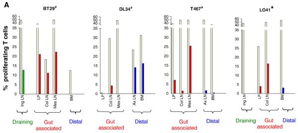
 ), or both IFN-γ and IL-2 to SIV Gag (
 ) and HIV/SIV Reg (□) is shown. For both sets of data, the colors designate three broadly classified tissue sites: green, draining lymph nodes; red, gut associated lymphoid tissue; purple, distal or secondary lymphoid tissues. # indicates animals with viral blips or intermittent proviral SHIV PCR positivity, and − /+ indicates the absence or presence respectively of SHIV specific DNA in the various tissue compartments as determined by semi-nested env PCR.
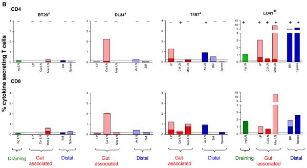 ), or both IFN-γ and IL-2 to SIV Gag () and HIV/SIV Reg (□) is shown. For both sets of data, the colors designate three broadly classified tissue sites: green, draining lymph nodes; red, gut associated lymphoid tissue; purple, distal or secondary lymphoid tissues. # indicates animals with viral blips or intermittent proviral SHIV PCR positivity, and − /+ indicates the absence or presence respectively of SHIV specific DNA in the various tissue compartments as determined by semi-nested env PCR.
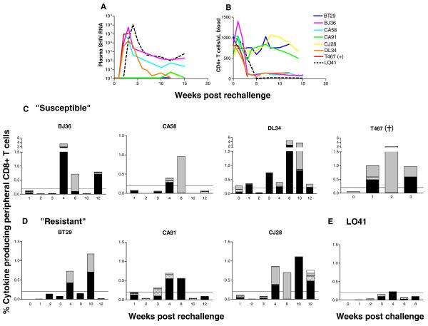
 ), or both IFN-γ and IL-2 to SIV Gag (
 ) and HIV/SIV Reg (□) were monitored at various time points post re-challenge. Virologic (A) and immunologic data (B, E) from L041, the macaque infected following a single exposure to 500 TCID50 of X4 SHIVSF33A were included for comparison. +, indicates death due to euthanasia.
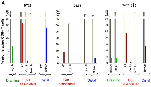
 ), or both IFN-γ and IL-2 to SIV Gag (
 ) and HIV/SIV Reg (□) is shown. Color designations are the same as those outlined in Figure 4. The ratio of total CD4 to CD8 cytokine producing T cells in the various tissue compartments of each macaque was generated and shown for comparison. − /+ indicate the absence or presence respectively of SHIV specific DNA as determined by semi-nested env PCR. +, indicates death due to euthanasia. NA, not applicable.
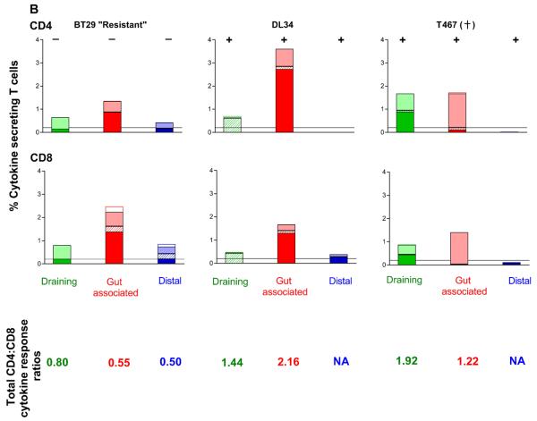 ), or both IFN-γ and IL-2 to SIV Gag () and HIV/SIV Reg (□) is shown. Color designations are the same as those outlined in Figure 4. The ratio of total CD4 to CD8 cytokine producing T cells in the various tissue compartments of each macaque was generated and shown for comparison. − /+ indicate the absence or presence respectively of SHIV specific DNA as determined by semi-nested env PCR. +, indicates death due to euthanasia. NA, not applicable.
References
-
- Beattie T, Rowland-Jones S, Kaul R. HIV-1 and AIDS: what are protective immune responses? J HIV Ther. 2002;7(2):35–9. - PubMed
-
- Bevan MJ. Helping the CD8(+) T-cell response. Nat Rev Immunol. 2004;4(8):595–602. - PubMed
-
- Bogers WM, Cheng-Mayer C, Montelaro RC. Developments in preclinical AIDS vaccine efficacy models. AIDS. 2000;14(Suppl 3):S141–51. - PubMed
-
- Champagne P, Ogg GS, King AS, Knabenhans C, Ellefsen K, Nobile M, Appay V, Rizzardi GP, Fleury S, Lipp M, Forster R, Rowland-Jones S, Sekaly RP, McMichael AJ, Pantaleo G. Skewed maturation of memory HIV-specific CD8 T lymphocytes. Nature. 2001;410(6824):106–11. - PubMed
-
- Clerici M, Shearer GM. Correlates of protection in HIV infection and the progression of HIV infection to AIDS. Immunol Lett. 1996;51(12):69–73. - PubMed
Publication types
MeSH terms
Substances
Grants and funding
LinkOut - more resources
Full Text Sources
Research Materials
