. 2007 Aug;73(15):4984-95.
doi: 10.1128/AEM.00988-07. Epub 2007 Jun 15.

Transposon insertion reveals pRM, a plasmid of Rickettsia monacensis

Affiliations

Transposon insertion reveals pRM, a plasmid of Rickettsia monacensis

Gerald D Baldridge et al. Appl Environ Microbiol. 2007 Aug.

Abstract

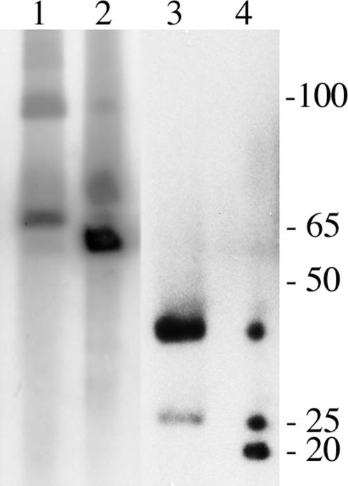
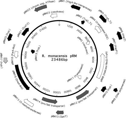
Until the recent discovery of pRF in Rickettsia felis, the obligate intracellular bacteria of the genus Rickettsia (Rickettsiales: Rickettsiaceae) were thought not to possess plasmids. We describe pRM, a plasmid from Rickettsia monacensis, which was detected by pulsed-field gel electrophoresis and Southern blot analyses of DNA from two independent R. monacensis populations transformed by transposon-mediated insertion of coupled green fluorescent protein and chloramphenicol acetyltransferase marker genes into pRM. Two-dimensional electrophoresis showed that pRM was present in rickettsial cells as circular and linear isomers. The 23,486-nucleotide (31.8% G/C) pRM plasmid was cloned from the transformant populations by chloramphenicol marker rescue of restriction enzyme-digested transformant DNA fragments and PCR using primers derived from sequences of overlapping restriction fragments. The plasmid was sequenced. Based on BLAST searches of the GenBank database, pRM contained 23 predicted genes or pseudogenes and was remarkably similar to the larger pRF plasmid. Two of the 23 genes were unique to pRM and pRF among sequenced rickettsial genomes, and 4 of the genes shared by pRM and pRF were otherwise found only on chromosomes of R. felis or the ancestral group rickettsiae R. bellii and R. canadensis. We obtained pulsed-field gel electrophoresis and Southern blot evidence for a plasmid in R. amblyommii isolate WB-8-2 that contained genes conserved between pRM and pRF. The pRM plasmid may provide a basis for the development of a rickettsial transformation vector.


Figures

FIG. 1.
FIG. 1.
GFPuv-fluorescent Rmona658B transformant cells in tick ISE6 cells. Arrows indicate single (bottom right), doublet form (top left), and multicell chain form (bottom center) rickettsiae. Bar, 10 μm. Magnification, ×1,000, with a fluorescein isothiocyanate filter.
FIG. 2.
FIG. 2.
Southern blot analysis of pMODompACAT\GFPuv658 transposon insertions in genomic DNA of R. monacensis transformant populations Rmona658, Rmona658A, and Rmona658B. Untransformed R. monacensis (lanes 1 and 5), Rmona658 (lanes 2 and 6), Rmona658A (lanes 3 and 7), Rmona658B (lanes 4 and 8), pMOD-2<MCS> (lane 9), and pMODompACAT\GFPuv658 (lane 10) DNA was digested with HindIII (lanes 1 to 5) or EcoRI (lanes 6 to 10). The membranes were probed with a GFPuv gene-specific probe (6). Migration positions of linear DNA markers in a 1-kbp ladder are indicated at right (3 to 12 kbp).
FIG. 3.
FIG. 3.
PFGE and Southern blot analyses of R. felis and R. amblyommii isolate WB-8-2 DNA hybridized with the R. felis pRF plasmid probe (32). R. felis DNA (lane 1) digested with PvuI (lane 2) and R. amblyommii DNA (lane 3) digested with PvuI (lane 4). Migration positions of linear DNA markers in a 5-kbp ladder are indicated at right.
FIG. 4.
FIG. 4.
PFGE and Southern blot analyses of R. monacensis pMOD658 transformants. (A) Ethidium bromide-stained PFGE gel with DNA from untransformed R. amblyommii isolate WB-8-2 (lane 1), untransformed R. monacensis (lane 2) digested with HindIII (lane 3), Rmona658 (lane 4) digested with HindIII (lane 5), and Rmona658A (lane 6) digested with HindIII (lane 7). Lanes 8 and 9 contain 5- and 1-kbp DNA ladders, respectively, with sizes indicated at right. (B) Southern blot of gel shown in panel A hybridized with a GFPuv gene probe specific for the pMOD658 transposon (6). (C) Ethidium bromide-stained PFGE gel with DNA from untransformed R. monacensis (lane 1), Rmona658 (lane 2), Rmona658B (lane 3) digested with NcoI (lane 4) or SmaI (lane 5), and Rmona658B QIAGEN plasmid prep DNA (lane 6) digested with NcoI (lane 7) or SmaI (lane 8). Lane 9 contains 1- and 5-kbp DNA marker ladders with sizes indicated at right. (D) Southern blot of gel shown in panel C hybridized with the GFPuv gene probe. Arrowheads indicate positions of the rickettsial plasmid containing the pMOD658 transposon (DNA bands migrating in a range between 23 and 100 kbp).
FIG. 5.
FIG. 5.
Two-dimensional gel electrophoresis and Southern blot analysis of the R. monacensis pRM plasmid. (A) Ethidium bromide-stained PFGE gel with R. monacensis DNA (lane 1) and linear DNA markers (lane 2) in a 5-kbp ladder with sizes indicated at right. The large arrow indicates the negative-to-positive polarity of current. (B) Constant field electrophoresis (CFE) of PFGE gel shown in panel A. The large arrow indicates the negative-to-positive polarity of current. (C) Southern blot analysis of gel shown in panel B with a probe specific for the pRM6 gene. Putative conformational identities of DNA species present in lanes 1 of all panels are indicated at left as chromosomal (chr), mixed circular and linear dimeric plasmid (c/li), supercoiled plasmid (sc), and linear monomeric plasmid (li).
FIG. 6.
FIG. 6.
The R. monacensis pRM plasmid cloning and sequencing strategy. The plasmid is represented as a circle, with the number of base pairs from an arbitrary origin indicated on the inner surface. Lines with double arrowheads indicate the lengths and relative positions of sequenced restriction fragments obtained by CHL marker rescue of Rmona658A and Rmona658B DNA restriction enzyme digests. Lines with single arrowheads indicate the relative positions and orientations of DNA primers used to PCR amplify portions of the plasmid from Rmona658B DNA. The hatched and filled bars represent, respectively, a sequenced PCR product encompassing that portion of the plasmid not present on the Rmona658A and Rmona658B restriction fragment clones and a sequenced product spanning their overlap. Filled circles indicate the relative locations of the pMOD658 transposon within the plasmid in the Rmona658A and Rmona658B transformants.
FIG. 7.
FIG. 7.
Predicted genes and pseudogenes on pRM. The plasmid is represented as a circle, with the number of base pairs from an arbitrary origin indicated on the inner surface. Black arrows indicate genes identified as BLAST best matches to R. felis pRF plasmid genes. Gray arrows indicate genes (pRM12, pRM13, pRM14, and pRM22) identified as best matches to R. felis or R. bellii chromosomal genes. The hatched arrow indicates a hypothetical protein gene unique to R. monacensis. Unfilled arrows indicate genes identified as BLAST best matches to genes of bacteria other than Rickettsia species.
FIG. 8.
FIG. 8.
Southern blot confirmation of the circular pRM sequence and evidence for the presence of orthologous genes on the plasmid of R. amblyommii. (A) SYBR green-stained PFGE gel with DNA from the Rmona658B transformant of R. monacensis (lanes 2, 4, 6, and 8), untransformed R. monacensis (lane 3), and R. amblyommii isolate WB-8-2 (lanes 5, 7, and 9). Migration positions of linear DNA markers in a 5-kbp ladder (lane 1) are indicated at left. (B) Southern blot analysis of gel shown in panel A hybridized with probes specific for the GFPuv gene (lanes 1, 2, and 3), the R. monacensis pRM16 gene (lanes 4 and 5), the pRM21 gene (lanes 6 and 7), and the pRM6 gene (lanes 8 and 9).

References

    1. Akman, L., R. V. Rio, C. B. Beard, and S. Aksoy. 2001. Genome size determination and coding capacity of Sodalis glossinidus, an enteric symbiont of tsetse flies, as revealed by hybridization to Escherichia coli gene arrays. J. Bacteriol. 183:4517-4525. - PMC - PubMed
    1. Akman, L., A. Yamashita, H. Watanabe, K. Oshima, T. Shiba, M. Hattori, and S. Aksoy. 2002. Genome sequence of the endocellular obligate symbiont of tsetse flies, Wigglesworthia glossinidia. Nat. Genet. 32:402-407. - PubMed
    1. Andersson, S. G., A. Zomorodipour, J. O. Andersson, T. Sicheritz-Ponten, U. C. Alsmark, R. M. Podowski, A. K. Naslund, A. Eriksson, H. H. Winkler, and C. G. Kurland. 1998. The genome sequence of Rickettsia prowazekii and the origin of mitochondria. Nature 396:133-140. - PubMed
    1. Azad, A. F., and C. B. Beard. 1998. Rickettsial pathogens and their arthropod vectors. Emerg. Infect. Dis. 4:179-186. - PMC - PubMed
    1. Baldridge, G. D., N. Burkhardt, M. J. Herron, T. J. Kurtti, and U. G. Munderloh. 2005. Analysis of fluorescent protein expression in transformants of Rickettsia monacensis, an obligate intracellular tick symbiont. Appl. Environ. Microbiol. 71:2095-2105. - PMC - PubMed

Publication types

Substances

Associated data