. 2007 Jun 15;21(12):1519-29.
doi: 10.1101/gad.1547707.

Nucleosome stability mediated by histone variants H3.3 and H2A.Z

Affiliations

Nucleosome stability mediated by histone variants H3.3 and H2A.Z

Chunyuan Jin et al. Genes Dev. .

Abstract

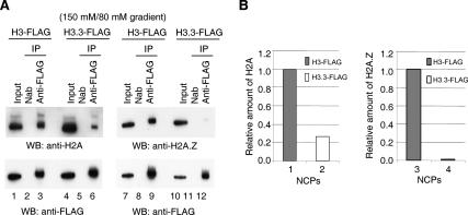
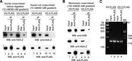
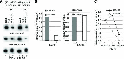
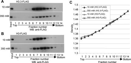
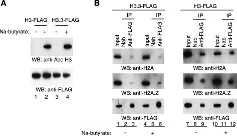
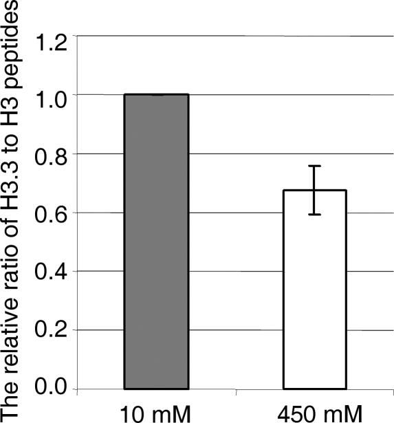
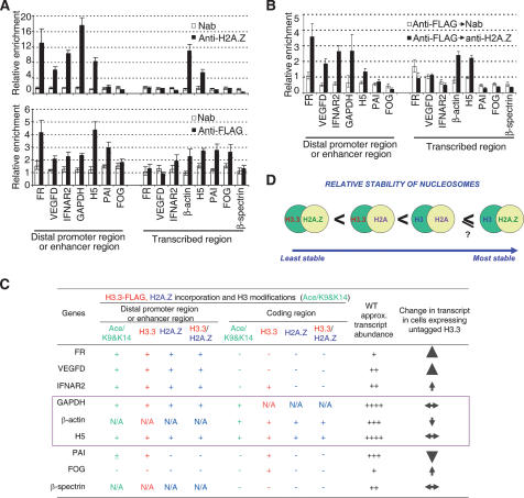
Nucleosomes containing the histone variant H3.3 tend to be clustered in vivo in the neighborhood of transcriptionally active genes and over regulatory elements. It has not been clear, however, whether H3.3-containing nucleosomes possess unique properties that would affect transcription. We report here that H3.3 nucleosomes isolated from vertebrates, regardless of whether they are partnered with H2A or H2A.Z, are unusually sensitive to salt-dependent disruption, losing H2A/H2B or H2A.Z/H2B dimers. Immunoprecipitation studies of nucleosome core particles (NCPs) show that NCPs that contain both H3.3 and H2A.Z are even less stable than NCPs containing H3.3 and H2A. Intriguingly, NCPs containing H3 and H2A.Z are at least as stable as H3/H2A NCPs. These results establish an hierarchy of stabilities for native nucleosomes carrying different complements of variants, and suggest how H2A.Z could play different roles depending on its partners within the NCP. They also are consistent with the idea that H3.3 plays an active role in maintaining accessible chromatin structures in enhancer regions and transcribed regions. Consistent with this idea, promoters and enhancers at transcriptionally active genes and coding regions at highly expressed genes have nucleosomes that simultaneously carry both H3.3 and H2A.Z, and should therefore be extremely sensitive to disruption.


Figures

Figure 1.
Figure 1.
Relative stability of NCPs containing H3 and H3.3. (A) NCPs were prepared from cells expressing either H3-Flag or H3.3-Flag. NCP monomers suspended in a solvent containing 150 mM NaCl were obtained by fractionation on a sucrose gradient (see Materials and Methods) in a solvent containing 80 mM NaCl and buffers. In each case, the sample was immunoprecipitated with antibody to Flag, histones were isolated and fractionated by gel electrophoresis, and Western blots of the samples were probed with antibody to H2A (left) or H2A.Z (right). Input lanes are loaded with an aliquot representing 10% of the starting sample. The label “150 mM/80 mM gradient” indicates the highest NaCl concentration used in NCP preparation and the NaCl concentration in the gradient. (Nab) No antibody control. (B) Comparative recoveries of H2A and H2A.Z from the data in A. The amounts of H2A or H2A.Z in H3-Flag-containing NCPs were set to 1. The relative amount was calculated by comparing the intensity of immunoprecipitated H2A or H2A.Z with that of immunoprecipitated H3-Flag or H3.3-Flag.
Figure 2.
Figure 2.
NCPs containing H3.3 are disrupted during purification. (A) Complete NCPs containing H3.3 and H2A.Z are present in vivo. (Left panel) Nuclei isolated from 6C2 cells in 10 mM NaCl were fixed with formaldehyde (see Materials and Methods) and then digested with micrococcal nuclease. NCPs carrying H3.3Flag or H3-Flag were purified on sucrose gradients containing 80 mM NaCl and analyzed for H2A.Z content on Western blots. (Right panel) Control in which formaldehyde treatment was omitted. (B) Experiments like those in Figure 1A, except NCPs were cross-linked with formaldehyde before immunoprecipitation, and Western blots were probed for H2A.Z and H2B. Input lanes are loaded with an aliquot representing 10% of the starting sample. (C) Length distribution of DNA isolated from NCPs. NCPs from cells carrying H3-Flag or H3.3-Flag were immunoprecipitated with anti-Flag, and the nucleosomal DNA was purified and analyzed by gel electrophoresis.
Figure 3.
Figure 3.
Unusual sensitivity of nucleosomes containing H3.3 to the ionic strength of the solvent. (A) Experiments like those in Figure 1, but NCPs were isolated from a sucrose gradient containing 10 mM NaCl without exposure to any higher-ionic-strength solvents. (B) Comparative recoveries of H2A and H2A.Z from the data in A. The relative amounts of histone H2A and H2A.Z in NCPs were calculated as described in Figure 1B. (C) Dynamic changes of relative amount of H2A or H2A.Z in H3.3-containing NCPs during purification. The relative amount of H2A.Z and H2A in H3.3 NCPs in nuclei (shown as in vivo) (Fig. 2A; data not shown) were measured as described and set to 1. These amounts were then compared with those presented in B (10 mM) and in Figure 1B (150 mM).
Figure 4.
Figure 4.
Sensitivity of purified NCPs to the higher ionic strength of the solvent. NCPs carrying H3.3-Flag (A) or H3-Flag (B) were dialyzed into solvents containing 10 mM or 350 mM NaCl, formaldehyde-fixed, and centrifuged to equilibrium in CsCl density gradients. Fractions were collected, histones were fractionated as in Figure 1, and Western blots were probed with anti-Flag antibody. Density increases with increasing fraction number. (C) Density distribution within the gradient.
Figure 5.
Figure 5.
Loss of histone acetylation does not measurably affect relative stabilities of H3 and H3.3 NCPs. (A) Loss of histone acetylation in the absence of sodium butyrate. NCPs containing either H3-Flag or H3.3-Flag isolated from cells in the absence or presence of sodium butyrate were immunoprecipitated with anti-Flag antibody, and histones were isolated and subjected to Western blot analysis with antibody to acetylated H3. (B) Loss of acetylation does not affect the relative instability of H3.3 NCPs. Immunoprecipitation studies similar to those in Figure 1, in the presence or absence of sodium butyrate.
Figure 6.
Figure 6.
Sensitivity of the NCPs consisting of untagged histones to the ionic strength of the solvent. NCPs from chicken erythrocytes were dialyzed into 10 mM NaCl and 450 mM NaCl, respectively. The octamer fractions were cross-linked with formaldehyde and isolated by CsCl gradient sedimentation, and the ratios of H3.3 to H3 at each ionic strength were determined by mass spectrometry (see Materials and Methods). The results are the average of two independent experiments. The ratio at 10 mM NaCl was set to 1.
Figure 7.
Figure 7.
(A) ChIP analysis of H3.3-Flag and H2A.Z over distal promoter or enhancer regions and transcribed regions of a variety of genes in 6C2 cells expressing H3.3-Flag. (Open bars) No antibody control; (filled bars) anti-Flag or anti-H2A.Z immunoprecipitation. Error bars reflect three separate measurements. (PAI) Plasminogen activator inhibitor; (FOG) friend of GATA. (B) Double ChIP analysis over same regions. First ChIPs by anti-Flag were followed by second ChIPs by anti-H2A.Z antibodies. (C) Summary of ChIP and double ChIP results; level of Ac/H3K9&K14; relative expression level of those genes surveyed in wild-type 6C2 cells and in the cells overexpressing untagged H3.3. The ChIP data are from A and B, while all others are from our previous study (Jin and Felsenfeld 2006). (N/A) Not applicable. (D) Schematic representation of relative stability of nucleosomes containing different histone variants.

References

    1. Abbott D.W., Ivanova V.S., Wang X., Bonner W.M., Ausio J., Ivanova V.S., Wang X., Bonner W.M., Ausio J., Wang X., Bonner W.M., Ausio J., Bonner W.M., Ausio J., Ausio J. Characterization of the stability and folding of H2A.Z chromatin particles: Implications for transcriptional activation. J. Biol. Chem. 2001;276:41945–41949. - PubMed
    1. Ahmad K., Henikoff S., Henikoff S. The histone variant H3.3 marks active chromatin by replication-independent nucleosome assembly. Mol. Cell. 2002;9:1191–1200. - PubMed
    1. Ashe H.L., Monks J., Wijgerde M., Fraser P., Proudfoot N.J., Monks J., Wijgerde M., Fraser P., Proudfoot N.J., Wijgerde M., Fraser P., Proudfoot N.J., Fraser P., Proudfoot N.J., Proudfoot N.J. Intergenic transcription and transinduction of the human β-globin locus. Genes & Dev. 1997;11:2494–2509. - PMC - PubMed
    1. Bruce K., Myers F.A., Mantouvalou E., Lefevre P., Greaves I., Bonifer C., Tremethick D.J., Thorne A.W., Crane-Robinson C., Myers F.A., Mantouvalou E., Lefevre P., Greaves I., Bonifer C., Tremethick D.J., Thorne A.W., Crane-Robinson C., Mantouvalou E., Lefevre P., Greaves I., Bonifer C., Tremethick D.J., Thorne A.W., Crane-Robinson C., Lefevre P., Greaves I., Bonifer C., Tremethick D.J., Thorne A.W., Crane-Robinson C., Greaves I., Bonifer C., Tremethick D.J., Thorne A.W., Crane-Robinson C., Bonifer C., Tremethick D.J., Thorne A.W., Crane-Robinson C., Tremethick D.J., Thorne A.W., Crane-Robinson C., Thorne A.W., Crane-Robinson C., Crane-Robinson C. The replacement histone H2A.Z in a hyperacetylated form is a feature of active genes in the chicken. Nucleic Acids Res. 2005;33:5633–5639. - PMC - PubMed
    1. Burton D.R., Butler M.J., Hyde J.E., Phillips D., Skidmore C.J., Walker I.O., Butler M.J., Hyde J.E., Phillips D., Skidmore C.J., Walker I.O., Hyde J.E., Phillips D., Skidmore C.J., Walker I.O., Phillips D., Skidmore C.J., Walker I.O., Skidmore C.J., Walker I.O., Walker I.O. The interaction of core histones with DNA: Equilibrium binding studies. Nucleic Acids Res. 1978;5:3643–3663. - PMC - PubMed

Publication types