. 2007 Jun 15;21(12):1530-45.
doi: 10.1101/gad.1544207.

RNAi-dependent H3K27 methylation is required for heterochromatin formation and DNA elimination in Tetrahymena

Affiliations

RNAi-dependent H3K27 methylation is required for heterochromatin formation and DNA elimination in Tetrahymena

Yifan Liu et al. Genes Dev. .

Abstract

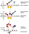
Methylated H3K27 is an important mark for Polycomb group (PcG) protein-mediated transcriptional gene silencing (TGS) in multicellular eukaryotes. Here a Drosophila E(z) homolog, EZL1, is characterized in the ciliated protozoan Tetrahymena thermophila and is shown to be responsible for H3K27 methylation associated with developmentally regulated heterochromatin formation and DNA elimination. Importantly, Ezl1p-catalyzed H3K27 methylation occurs in an RNA interference (RNAi)-dependent manner. H3K27 methylation also regulates H3K9 methylation in these processes. Furthermore, an "effector" of programmed DNA elimination, the chromodomain protein Pdd1p, is shown to bind both K27- and K9-methylated H3. These studies provide a framework for an RNAi-dependent, Polycomb group protein-mediated heterochromatin formation pathway in Tetrahymena and underscore the connection between the two highly conserved machineries in eukaryotes.


Figures
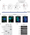
Figure 1.
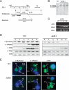
Figure 1.
H3K27me3 is a general mark for heterochromatin in Tetrahymena. (A) Schematic representation of key nuclear events in Tetrahymena conjugation. Electron-dense chromatin bodies (open arrowhead) are dispersed in the somatic macronucleus (Mac) of vegetatively growing (nonmating) cells (0 h). Two Tetrahymena cells of different mating types can pair during conjugation (only one partner is shown in immunofluorescence pictures). During this sexual pathway, the germline micronucleus (Mic) gives rise to two new micronuclei and two developing macronuclei, also referred to as anlagen (AN), supported by transcription from the parental macronuclei (PM) (6 h). As anlagen formation progresses, the old macronucleus (OM) degenerates (10 h). At late conjugation, specialized DNA elimination heterochromatic structures (solid arrowhead) form in anlagen as pairs separate (>12 h). Heterochromatic structures are highlighted (red). (B) Localization of H3K27me3. Wild-type cells were processed for immunofluorescence staining with H3K27me3-specific antibody and counterstained with DAPI. From left to right, typical examples of stained cells are shown as vegetatively growing, micronuclei/anlagen differentiation, and early and late anlagen stages, aligned with their schematic representations in A. (C) Differential usage of H3K27me3 and H3K9me3 in distinct Tetrahymena nuclei. Acid extracts from purified micronuclei and macronuclei in vegetatively growing cells (Veg) or anlagen isolated from 10-h conjugating cells (Cnj) were resolved on 10% SDS-PAGE, blotted, and probed with the indicated antibodies. Note that H3K27me3 was only observed in the fast-migrating form of micronuclear H3 (arrowhead), which corresponds to a mature, proteolytically processed H3 (Allis et al. 1980). Another further truncated form was observed in anlagen. (D) Expression of EZLs mRNA during conjugation. Total RNA samples from wild-type and ΔEZL1 cells from different conjugation time points (0, 4, 8, and 12 h post-mixing) were reverse-transcribed and analyzed by PCR with primers specific for EZL1, EZL2, EZL3, PDD1 (a conjugation-specific chromodomain protein), and HHP1 (a gene encoding a chromodomain protein ubiquitously expressed in vegetative and conjugating cells).
Figure 2.
Figure 2.
EZL1 is required for H3K27me3 and H3K9me3 in conjugating Tetrahymena. (A) Schematic representation of endogenous EZL1 locus and the knockout construct. Full-length EZL1 mRNA is aligned with the endogenous genomic locus. In the knockout construct, 1.2 kb of the endogenous locus, corresponding to the conserved pre-SET and SET domain regions, was replaced by a drug-resistance cassette, neo3. The probe and restriction enzyme sites used in Southern blot analysis (shown in B) was also mapped. (X) XmnI. (B) Southern blot analysis of genomic DNA from wild-type or ΔEZL1 cells was digested with XmnI and hybridized with the EZL1 probe. (C) RT–PCR analysis of ΔEZL1 cells. Total RNA samples of ΔEZL1 cells from different conjugation time points were reverse-transcribed and analyzed by PCR with primers specific for EZL1, PDD1, and HHP1. (D) Changes in H3K27me3 and H3K9me3 levels during the conjugation pathway. Whole-cell extract samples from different conjugation time points of wild-type and ΔEZL1 cells were resolved on 10% SDS-PAGE, blotted, and probed with the indicated antibodies. (E) Localization of H3K27me3 and H3K9me3 in conjugating cells. Wild-type and ΔEZL1 cells from early anlagen stages were processed for immunofluorescence staining with antibodies specific for H3K27me3, H3K9me3, or H4; nuclei were counterstained with DAPI. (Mic) Micronuclei; (AN) macronuclear anlagen; (OM) old macronuclei.
Figure 3.
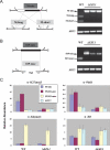
Figure 3.
EZL1 is required for DNA elimination and chromosome breakage in conjugating Tetrahymena. (A) Single-cell PCR assay of DNA elimination in Tetrahymena. (Left) A schematic representation of DNA elimination at M-element, an IES. Boxes indicate micronuclear-limited sequences to be eliminated; lines indicate macronuclear-retained sequences. Note that the micronuclear-specific form of M-element (M-mic) can be processed into either of the two macronuclear-specific forms (M-long and M-short). (Right) Individual conjugation progeny from wild-type and ΔEZL1 strains were isolated at 48 h into conjugation and analyzed by multiplex PCR. The positions of PCR products corresponding to micronuclear- and macronuclear-specific forms are indicated by solid and open arrowheads, respectively. HHT2 locus was examined as a control for DNA integrity. (B) Single-cell PCR assay of chromosome breakage in Tetrahymena. (Left) A schematic representation of CBS 819 locus. Circles indicate telomeres (Tel) added to the newly generated macronuclear chromosomal ends. (Right) PCR results from wild-type and ΔEZL1 strains. (C) Specific association of H3K27me3 with IES. Conjugating wild-type and ΔEZL1 cells (10 h) were processed for ChIP with the indicated antibodies. ChIP data were quantified by real-time PCR. The results shown are from duplicate experiments and are normalized against the input (shown as percentage pulled down). See text for details.
Figure 4.
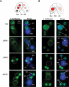
Figure 4.
H3K27me3 in parental macronucleus and macronuclear anlagen in conjugating Tetrahymena is RNAi dependent. Localization of H3K27me3 in anlagen (small white circles) at early anlagen formation (10 h conjugation) (A) and in parental macronuclei (large white circles) at micronucleus/macronucleus differentiation stages (6 h conjugation) (B). Wild-type, ΔEZL1, and RNAi-deficient (ΔTWI1 and ΔDCL1) mutants were processed for immunofluorescence staining with H3K27me3-specific antibody and counterstained with DAPI. Note the abnormal accumulation of H3K27me3 in the new micronuclei instead of macronuclear anlagen in ΔTWI1 and ΔDCL1 cells at both stages. (Mic) Micronuclei; (PM) parental macronuclei; (AN) macronuclear anlagen; (OM) old macronuclei.
Figure 5.
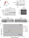
Figure 5.
Relationship between H3K27 methylation and RNAi pathway. (A) Changes in H3K27me3 level in anlagen of RNAi-deficient cells. Acids extract from unit gravity-purified anlagen from 10-h conjugating wild-type, ΔDCL1, and ΔTWI1 cells were resolved on 15% SDS-PAGE, blotted, and probed with the indicated antibodies. (B) Changes in H3K27me3 and H3K9me3 level in ΔTWI1 cells during conjugation. Whole-cell extract samples from different conjugation time points of ΔTWI1 cells were resolved on 10% SDS-PAGE, blotted, and probed with the indicated antibodies. (C) EZL1 mRNA expression in ΔTWI1 cells. Total RNA samples from ΔTWI1 cells from different conjugation time points were reverse-transcribed and analyzed by PCR with primers specific for EZL1, PDD1, and HHP1. (D) Accumulation of M-element-specific small RNAs. Total RNA samples from wild-type and ΔEZL1 conjugating cells were resolved by 12% sequencing gel, blotted, and probed with 32P end-labeled DNA oligos specific for the micronuclear-limited sequence (M-mic) or macronuclear-retained sequence (M-mac), respectively. (E) Accumulation of IES-derived transcripts during conjugation. Total RNA samples from wild-type and DNA elimination-deficient mutants (ΔTWI1, ΔEZL1, and ΔPDD1) at different conjugation time points (0, 2, 4, 6, 8, 10, 12, and 24 h into conjugation, respectively) were reverse-transcribed and quantified by real-time PCR with primers specific for micronuclear-limited sequence of M-element (M-mic) or the control PGM1 locus (PGM1-mac). Expression levels were normalized against total RNA input (OD260) and plotted relative to the level before the initiation of conjugation (0 h).
Figure 6.
Figure 6.
Pdd1p is a “dual-binder” for H3K27me3 and H3K9me3. (A) Localization of H3K27me3, H3K9me3, Pdd1p, and Pdd3p in the DNA elimination heterochromatic structures. Wild-type conjugating cells at late anlagen stages were processed for sequential double-immunofluorescence staining (red and green) with the indicated antibodies and counterstained with DAPI. (B) Binding specificity of Pdd1p and Pdd3p chromodomains for H3K27me3 and H3K9me3 peptides. The chromodomains (GST-tagged) were purified and incubated with the indicated FITC-labeled peptides. The interaction was quantified by fluorescence anisotropy. A representative set of binding data is shown with fitted binding curves. (C) The binding affinity data (KD) calculated from triplicate experiments in C. (D) Pdd1p localization in wild-type and H3K27me3-deficient (ΔEZL1, ΔTWI1, and ΔDCL1) cells at early anlagen stages (10 h). Wild-type cells at late anlagen stages are also shown. The nuclei were counterstained with DAPI. (Mic) micronuclei; (AN) macronuclear anlagen; (OM) old macronuclei. (E) PCR assay of DNA elimination efficiency. Wild-type, S10E, and S28E strains were completely assorted to have only M-long form in macronuclei. The reappearance of M-short form during conjugation can be used as an indicator for the processing efficiency of M-element (Liu et al. 2004). Mass mating cell samples were taken at 24 h and 48 h into conjugation and analyzed by PCR with primers flanking M-element. (F) Immunofluorescence staining of H3 S10E and H3 S28E conjugating cells with antibodies specific for H3K27me3, H3K9me3, and Pdd1p, respectively. The nuclei were also stained with DAPI. Note the abnormal aggregation of H3K9me3 and Pdd1p in macronuclear anlagen of S28E cells (arrowheads).
Figure 7.
Figure 7.
Revised model of RNAi-dependent heterochromatin formation pathway in Tetrahymena. Ezl1p is recruited to IES regions by homologous siRNAs in association with Twi1p, leading to H3K27 methylation. This is followed by H3K9 methylation, possibly also deposited by Ezl1p. H3K27me3 and H3K9me3 recruit Pdd1p and Pdd3p. Chromatin condensation ensues, and eventually IES is eliminated from the macronuclear genome. For details, see text and previous proposals (Mochizuki et al. 2002; Mochizuki and Gorovsky 2004b) from which this model is derived.

References

    1. Allis C.D., Bowen J.K., Abraham G.N., Glover C.V., Gorovsky M.A., Bowen J.K., Abraham G.N., Glover C.V., Gorovsky M.A., Abraham G.N., Glover C.V., Gorovsky M.A., Glover C.V., Gorovsky M.A., Gorovsky M.A. Proteolytic processing of histone H3 in chromatin: A physiologically regulated event in Tetrahymena micronuclei. Cell. 1980;20:55–64. - PubMed
    1. Baldauf S.L., Roger A.J., Wenk-Siefert I., Doolittle W.F., Roger A.J., Wenk-Siefert I., Doolittle W.F., Wenk-Siefert I., Doolittle W.F., Doolittle W.F. A kingdom-level phylogeny of eukaryotes based on combined protein data. Science. 2000;290:972–977. - PubMed
    1. Bannister A.J., Zegerman P., Partridge J.F., Miska E.A., Thomas J.O., Allshire R.C., Kouzarides T., Zegerman P., Partridge J.F., Miska E.A., Thomas J.O., Allshire R.C., Kouzarides T., Partridge J.F., Miska E.A., Thomas J.O., Allshire R.C., Kouzarides T., Miska E.A., Thomas J.O., Allshire R.C., Kouzarides T., Thomas J.O., Allshire R.C., Kouzarides T., Allshire R.C., Kouzarides T., Kouzarides T. Selective recognition of methylated lysine 9 on histone H3 by the HP1 chromo domain. Nature. 2001;410:120–124. - PubMed
    1. Bastow R., Mylne J.S., Lister C., Lippman Z., Martienssen R.A., Dean C., Mylne J.S., Lister C., Lippman Z., Martienssen R.A., Dean C., Lister C., Lippman Z., Martienssen R.A., Dean C., Lippman Z., Martienssen R.A., Dean C., Martienssen R.A., Dean C., Dean C. Vernalization requires epigenetic silencing of FLC by histone methylation. Nature. 2004;427:164–167. - PubMed
    1. Bender L.B., Cao R., Zhang Y., Strome S., Cao R., Zhang Y., Strome S., Zhang Y., Strome S., Strome S. The MES-2/MES-3/MES-6 complex and regulation of histone H3 methylation in C. elegans. Curr. Biol. 2004;14:1639–1643. - PubMed

Publication types

MeSH terms