. 2007 Jun 15;21(12):1546-58.
doi: 10.1101/gad.436307.

Bone marrow-derived endothelial progenitor cells are a major determinant of nascent tumor neovascularization

Affiliations

Bone marrow-derived endothelial progenitor cells are a major determinant of nascent tumor neovascularization

Daniel J Nolan et al. Genes Dev. .

Abstract

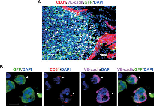
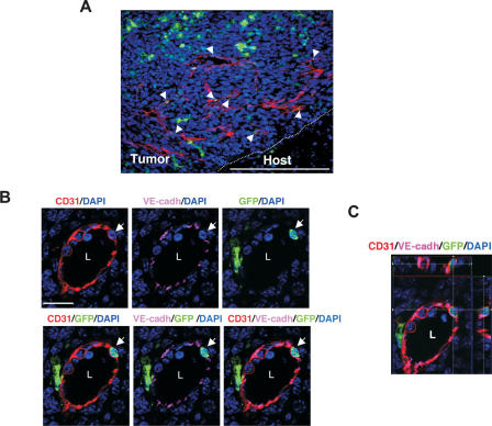
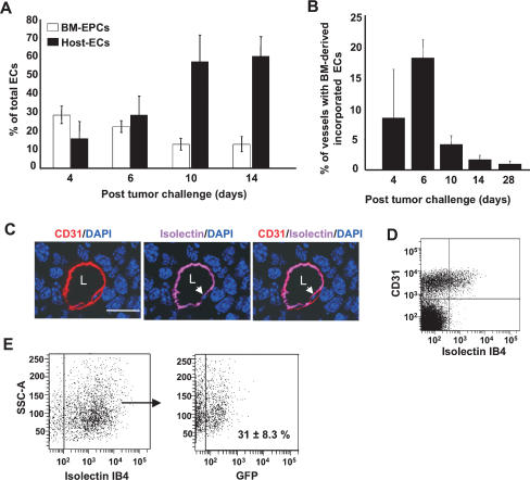
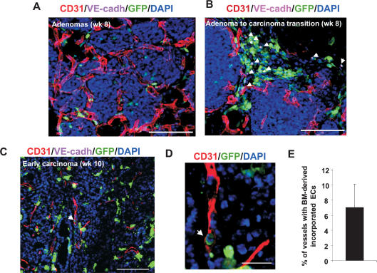
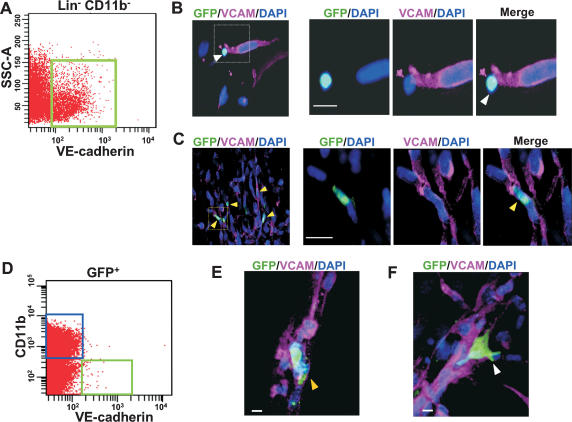
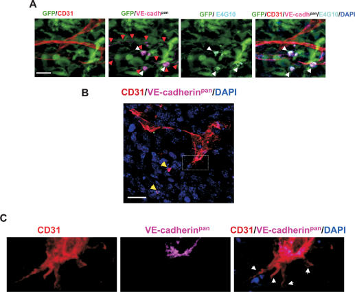
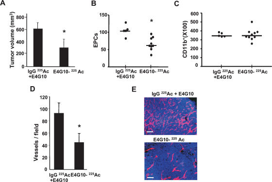
Tumors build vessels by cooption of pre-existing vasculature and de novo recruitment of bone marrow (BM)-derived endothelial progenitor cells (EPCs). However, the contribution and the functional role of EPCs in tumor neoangiogenesis are controversial. Therefore, by using genetically marked BM progenitor cells, we demonstrate the precise spatial and temporal contribution of EPCs to the neovascularization of three transplanted and one spontaneous breast tumor in vivo using high-resolution microscopy and flow cytometry. We show that early tumors recruit BM-derived EPCs that differentiate into mature BM-derived endothelial cells (ECs) and luminally incorporate into a subset of sprouting tumor neovessels. Notably, in later tumors, these BM-derived vessels are diluted with non-BM-derived vessels from the periphery, which accounts for purported differences in previously published reports. Furthermore, we show that specific ablation of BM-derived EPCs with alpha-particle-emitting anti-VE-cadherin antibody markedly impaired tumor growth associated with reduced vascularization. Our results demonstrate that BM-derived EPCs are critical components of the earliest phases of tumor neoangiogenesis.


Figures

Figure 1.
Figure 1.
BM-derived EPCs are recruited at the periphery of early tumors. (A) A representative fluorescent image showing recruitment of BM-derived GFP+ VE-cadherin+ EPCs (arrows) at the periphery of early nonvascularized LLC tumors (days 4–6; n = 15). CD31+ mature vessels are observed in the host tissue but not in the tumors. The dotted line separates the host tissue from the tumor. Bar, 100 μm. (B) High-resolution image showing GFP+ VE-cadherin+ CD31low EPCs (arrows) at the periphery of LLC tumors (day 4). DAPI was used to stain the nucleus of all cells. Bar, 5 μm.
Figure 2.
Figure 2.
BM-derived ECs are luminally incorporated in tumor neovasculature. (A) Representative fluorescent image showing CD31+ mature vessels in LLC tumors (days 6–8; n = 15). Arrows depict incorporated BM-derived ECs in these vessels. Bar, 200 μm. The dotted line separates the host tissue from the tumor. (B) High-resolution image of a representative blood vessel showing an incorporated mature BM-derived GFP+ CD31+ VE-cadherin+-coexpressing cell (arrow). The lumen of the vessel (L) and VE-cadherin staining the adherens junctions between ECs are shown. Bar, 20 μm. (C) Optical sectioning (Z-stack resolution, 0.275 μm) of the BM-derived ECs showing that GFP and CD31 signals are localized within the same individual cell (XZ- and YZ-axes are represented by the top and side panels).
Figure 3.
Figure 3.
Contribution of BM-derived EPCs and ECs as a function of tumor progression. (A) Flow cytometry analysis of LLC tumors (days 4–14; n = 5 per group), showing relative contribution of BM-derived EPCs (GFP+ VE-cadherin+ CD31low, CD11b), and host-derived ECs (GFP VE-cadherin+ CD31+, CD11b). Total number of cells analyzed was 2 × 10⁵ per animal, except for day 4, due to smaller tumor size. Each analysis was performed in duplicate. Error bars represent standard deviations. This experiment was repeated three times, and identical trends were observed. (B) Quantification of vessels in LLC tumors (days 4–28) with incorporated BM-derived ECs (GFP+, CD31+, VE-cadherin+), a minimum of 400 vessels counted per time point from six nonsequential sections, and 20 images per tumor (Z-stacks evaluated for each section). Error bars represent standard deviations. (C) A representative image showing a tumor vessel from an animal perfused with isolectin GS-IB4 and stained with CD31. Arrows indicate isolectin IB4 staining the luminal surface of the EC in the vessel. (L) Lumen. Bar, 20 μm. (D) Flow cytometric analysis of LLC tumors (days 6–8) showing that fluorescent isolectin specifically stains a population of luminally incorporated CD31+ ECs. (E, left) Flow cytometric analysis showing that fluorescent isolectin stains a population of luminally incorporated ECs (CD31+ CD11b) derived from LLC tumors. (Right) Of these, 31% ± 8.3% are BM derived (GFP+ CD31+ Isolectin+ CD11b). The averages and standard deviation were determined by analyzing 1 × 10⁵ cells per animal (n = 5). (SSC-A) Side scatter values.
Figure 4.
Figure 4.
Contribution of BM-derived EPCs in spontaneous breast tumors. (A) Primary adenoma lesions in mammary gland sections from a PyMT mouse at 8 wk of age. Pre-existing CD31+ vessels are observed surrounding the adenomas. Bar, 100 μm. (B) Recruitment of BM-derived GFP+ VE-cadherin+ EPCs (arrows) at the periphery of the avascular adenoma–carcinoma progression is shown. Bar, 100 μm. (C) An early carcinoma showing recruited CD31+ mature vessels in the tumor mass (10 wk of age). Arrow depicts incorporated BM-derived ECs in a vessel. Bar, 100 μm. (D) High-resolution image of a representative blood vessel (box in C) showing an incorporated mature BM-derived GFP+ CD31+-coexpressing cell (arrow). Bar, 20 μm. DAPI was used to stain the nucleus of all cells. (E) Quantification of vessels in breast tumors (10 wk old) with incorporated BM-derived ECs (GFP+ CD31+ VE-cadherin+). A minimum of 250 vessels were counted from nonsequential sections from four animals, and Z-stacks were evaluated for each section. Error bars represent standard deviations.
Figure 5.
Figure 5.
BM-derived EPCs differentiate into mature ECs and incorporate into vascular networks. (A) Scatter plot showing VE-cadherin-expressing EPCs in the BM-derived Lin CD11b fraction (green box). (B) Immunostaining of cocultured BM-derived GFP+ EPCs and mature ECs (non-GFP) in 3D matrigel at 12 h. (Right panels) A high-resolution image showing an unincorporated, BM-derived GFP+ VCAM EPC (white arrow). (C) Multiple BM-derived ECs (GFP+ VCAM+, yellow arrows) that have incorporated into vascular tubes 48 h following coculture. (Right panels) A high-resolution image showing an incorporated BM-derived GFP+ EC (yellow arrow). (D) Scatter plot showing GFP+ cells from day 4 LLC tumors. GFP+ VE-cadherin+ CD11b EPCs (green box) and GFP+ VE-cadherin CD11b+ hematopoietic cells (blue box) were live-sorted. (E) Immunostaining of the cocultured BM-derived GFP+ EPCs sorted in D and mature ECs (non-GFP) in 3D matrigel at 48 h. High-resolution microscopy showing an incorporated BM-derived GFP+ VCAM+ EC (yellow arrow). (F) Immunostaining of the cocultured GFP+ CD11b+ hematopoietic cells sorted in D and mature ECs (non-GFP) in 3D matrigel at 48 h. High-resolution microscopy showing an unincorporated BM-derived GFP+ VCAM cell (white arrow). Bars, 10 μm. (SSC-A) Side scatter values.
Figure 6.
Figure 6.
VE-cadherin monoclonal antibody E4G10 specifically recognizes EPCs and not mature ECs. (A) Five-color immunostaining of early LLC tumors (day 6) with VE-cadherin antibody E4G10 (white arrows), pan-VE-cadherin antibody 11D4.1 (VE-cadhpan, red arrows), and CD31. E4G10 exclusively staining EPCs is shown (double red and white arrows). Bar, 20 μm. (B) Immunostaining of early tumors (day 6) with CD31 and a pan-VE-cadherin antibody. Low-magnification image showing endothelial projections of the invading tumor vasculature. Yellow arrows depict VE-cadherin+ EPCs in the vicinity. Bar, 20 μm. GFP staining is not included for precise visualization. (C) Higher magnification of the area denoted by a rectangle in B, showing that the endothelial projections are devoid of VE-cadherin (white arrows).
Figure 7.
Figure 7.
Selective ablation of EPCs results in delayed tumor growth in vivo. (A) LLC tumor volume at day 14 in animals administered with isotype control 225Act-IgG and unlabeled E4G10 or 225Act-E4G10 antibody (n = 5, 10 per group, respectively). (B) Number of BM-derived EPCs (GFP+ VE-cadherin+ CD31low CD11b) cells in early tumors (day 6) in animals administered with control or 225Act-E4G10 antibody at day 3 and 5 post-tumor innoculation (n = 5, 10, respectively; 1 × 10⁵ events counted per n). Bars denote averages. (C) BM-derived GFP+ CD11b+ cells in tumors from animals treated as in B (n = 5, 10, respectively; 1 × 10⁵ events counted per n). Bars denote averages. (D) Vessel density in tumors in animals treated with control and test antibody (day 14; n = 5, 7 per group, respectively) (E) CD31 immunostaining of tumor sections (day 14) isolated from E4G10-treated and control-treated animals. Bar, 50 μm. Error bars represent standard deviations. (*) Significant by t-test; P < 0.05.

References

    1. Aozuka Y., Koizumi K., Saitoh Y., Ueda Y., Sakurai H., Saiki I., Koizumi K., Saitoh Y., Ueda Y., Sakurai H., Saiki I., Saitoh Y., Ueda Y., Sakurai H., Saiki I., Ueda Y., Sakurai H., Saiki I., Sakurai H., Saiki I., Saiki I. Anti-tumor angiogenesis effect of aminopeptidase inhibitor bestatin against B16–BL6 melanoma cells orthotopically implanted into syngeneic mice. Cancer Lett. 2004;216:35–42. - PubMed
    1. Asahara T., Murohara T., Sullivan A., Silver M., van der Zee R., Li T., Witzenbichler B., Schatteman G., Isner J.M., Murohara T., Sullivan A., Silver M., van der Zee R., Li T., Witzenbichler B., Schatteman G., Isner J.M., Sullivan A., Silver M., van der Zee R., Li T., Witzenbichler B., Schatteman G., Isner J.M., Silver M., van der Zee R., Li T., Witzenbichler B., Schatteman G., Isner J.M., van der Zee R., Li T., Witzenbichler B., Schatteman G., Isner J.M., Li T., Witzenbichler B., Schatteman G., Isner J.M., Witzenbichler B., Schatteman G., Isner J.M., Schatteman G., Isner J.M., Isner J.M. Isolation of putative progenitor endothelial cells for angiogenesis. Science. 1997;275:964–967. - PubMed
    1. Asahara T., Takahashi T., Masuda H., Kalka C., Chen D., Iwaguro H., Inai Y., Silver M., Isner J.M., Takahashi T., Masuda H., Kalka C., Chen D., Iwaguro H., Inai Y., Silver M., Isner J.M., Masuda H., Kalka C., Chen D., Iwaguro H., Inai Y., Silver M., Isner J.M., Kalka C., Chen D., Iwaguro H., Inai Y., Silver M., Isner J.M., Chen D., Iwaguro H., Inai Y., Silver M., Isner J.M., Iwaguro H., Inai Y., Silver M., Isner J.M., Inai Y., Silver M., Isner J.M., Silver M., Isner J.M., Isner J.M. VEGF contributes to postnatal neovascularization by mobilizing bone marrow-derived endothelial progenitor cells. EMBO J. 1999;18:3964–3972. - PMC - PubMed
    1. Baumann C.I., Bailey A.S., Li W., Ferkowicz M.J., Yoder M.C., Fleming W.H., Bailey A.S., Li W., Ferkowicz M.J., Yoder M.C., Fleming W.H., Li W., Ferkowicz M.J., Yoder M.C., Fleming W.H., Ferkowicz M.J., Yoder M.C., Fleming W.H., Yoder M.C., Fleming W.H., Fleming W.H. PECAM-1 is expressed on hematopoietic stem cells throughout ontogeny and identifies a population of erythroid progenitors. Blood. 2004;104:1010–1016. - PubMed
    1. Bertolini F., Shaked Y., Mancuso P., Kerbel R.S., Shaked Y., Mancuso P., Kerbel R.S., Mancuso P., Kerbel R.S., Kerbel R.S. The multifaceted circulating endothelial cell in cancer: Towards marker and target identification. Nat. Rev. Cancer. 2006;6:835–845. - PubMed

Publication types