. 2007 Jun 15;21(12):1559-71.
doi: 10.1101/gad.1535007.

Gravin regulates mesodermal cell behavior changes required for axis elongation during zebrafish gastrulation

Affiliations

Gravin regulates mesodermal cell behavior changes required for axis elongation during zebrafish gastrulation

Douglas C Weiser et al. Genes Dev. .

Abstract

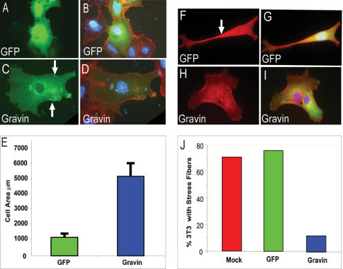
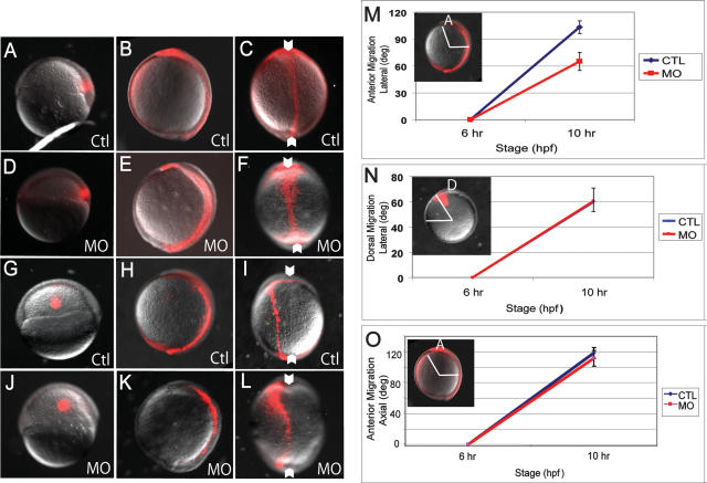
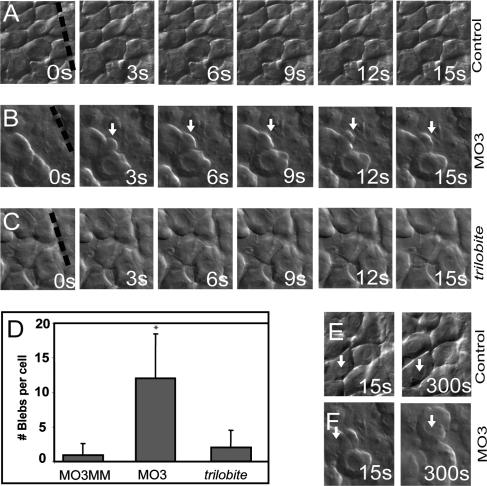
Convergent extension of the mesoderm is the major driving force of vertebrate gastrulation. During this process, mesodermal cells move toward the future dorsal side of the embryo, then radically change behavior as they initiate extension of the body axis. How cells make this transition in behavior is unknown. We have identified the scaffolding protein and tumor suppressor Gravin as a key regulator of this process in zebrafish embryos. We show that Gravin is required for the conversion of mesodermal cells from a highly migratory behavior to the medio-laterally intercalative behavior required for body axis extension. In the absence of Gravin, paraxial mesodermal cells fail to shut down the protrusive activity mediated by the Rho/ROCK/Myosin II pathway, resulting in embryos with severe extension defects. We propose that Gravin functions as an essential scaffold for regulatory proteins that suppress the migratory behavior of the mesoderm during gastrulation, and suggest that this function also explains how Gravin inhibits invasive behaviors in metastatic cells.


Figures

Figure 1.
Figure 1.
Zebrafish Gravin. A scaffolding protein expressed during early development. (A) Homology of Gravin proteins between zebrafish (Gravin), Xenopus (XGL), and human (AKAP12β). The percentage of amino acid identity is indicated between the orthologs by domain. (B) Sequence alignment of the basic regions of zebrafish and human Gravin. Charged residues (blue) and phosphorylation sites (red) implicated in membrane interactions are shown. (C–J) Expression of gravin during early development. Gravin is expressed zygotically beginning at the sphere stage (C), and remains ubiquitously expressed during the shield (D), 85% epiboly (E), and bud (F) stages. During segmentation, gravin expression becomes restricted to the presomitic mesoderm, head mesoderm, notochord, adaxial cells, vasculature, and nervous system, as seen by lateral view at the 16-somite (G) and 23-somite (H) stages, and by flat mount at the 10-somite stage (I,J).
Figure 2.
Figure 2.
Zebrafish Gravin localizes to the perinucleus and cell periphery in mammalian cells and regulates cell shape. (A–E) COS7 cells were transfected with either GFP alone (A,B) or zebrafish GFP-Gravin (C,D), fixed, and stained with DAPI and Alexa 594-phalloidin. GFP alone is shown in the left column, and merged images are in the right column. White arrows indicate sites of peripheral membrane puncta. (E) Cell flattening was assayed by calculating the cell area of GFP- or Gravin-transfected cells using AxioVision 4 software; error bars are standard deviations. (F–J) NIH 3T3 cells transfected with GFP (F,G) or GFP-Gravin (H,I) were fixed and stained with DAPI and phalloidin. F and H show only phalloidin staining, while G and I show phalloidin, GFP, and DAPI. (J) Cells were scored for visible phalloidin-stained stress fibers (see arrow in F).
Figure 3.
Figure 3.
Gravin is required for proper gastrulation of zebrafish embryos. (A,B) The phenotype of gravin-injected (B) and control morpholino-injected (A) embryos at 48 h post-fertilizaton (hpf). (C–F) The phenotype of gravin morphants was analyzed by in situ hybridization for myoD at the 12-somite stage (D,F) and compared with control-injected embryos (C,E). The arrows mark the extent of the body axis. (G, H) The phenotype of gravin morphants (H) and control embryos (G) at the bud stage stained with hgg1 (to mark the prechordal plate), shh (midline), pax2.1 (midbrain–hindbrain boundary), and dlx3 (neural plate). (I) Coinjection of 50 pg of gravin mRNA partially rescues the gastrulation defect of the splice blocking MO3 (6.5 ng). The Y-axis represents the percentage of embryos displaying a severe axis extension defect (black), a moderate phenotype (gray), or normal axis extension (white). (J–Q) Control or morphant embryos were stained with cmlc2 (J,K), insulin (L,M), foxa2 (N,O) and hhex (P,Q) to mark the heart field, pancreas, axial endoderm, and liver, respectively.
Figure 4.
Figure 4.
Gravin morphant cells converge dorsally but do not extend. (A–L) Distribution of labeled lateral or dorsal mesodermal and ectodermal cells at the shield and bud stages. The fluorescent dye was uncaged at the shield stage (left panels), and examined 4 h later at the bud stage in lateral (middle panels) and dorsal (right panels) views. (A–C) Control morpholino-injected (MO3MM, 7.5 ng) embryos, dye uncaged on the dorsal side. (D–F) Gravin morphant embryos (MO3, 7.5 ng), dye uncaged on the dorsal side. (G–I) Control morpholino-injected (MO3MM, 7.5 ng) embryos, dye uncaged on the lateral side (lateral cells were labeled 90° from dorsal). (J–L) Gravin morphant embryos (MO3, 7.5 ng), dye uncaged on the lateral side. White arrowheads indicate the notochord. (M,N) Quantification of the dorsal convergence and anterior extension of lateral cells in control and morphant embryos. (M) Anterior extension of lateral mesoderm is defined as the angle between the anterior-most labeled cell and the dorsal side (lateral view, anterior at top). (N) Dorsal migration is the angle between the site of activation at the shield stage, and the site of dye-labeled cells at the bud stage (animal pole view, dorsal at top, lateral to the left). (O) Anterior extension of axial mesoderm measured as in M. Note that both the convergence of lateral cells and the extension of dorsal cells are unaffected by loss of Gravin; however, extension of lateral cells (including presomitic mesoderm) is severely inhibited. (A) Anterior; (D) dorsal.
Figure 5.
Figure 5.
Gravin is required to inhibit protrusions during late gastrulation. Cell shape changes of mesodermal cells between the bud and two-somite stages were compared by time-lapse microscopy. Behavior of dorsal mesodermal cells in control morpholino-injected embryos (MO3MM, 7.5 ng) (A), in gravin morpholino-injected embryos (MO3, 7.5 ng) (B), and in trilobite embryos (C). White arrows indicate bleb-like protrusions, and the dashed black line marks the notochord–somite boundary. (D) Quantification of membrane blebbing behavior of paraxial mesodermal cells in the three conditions shown in A–C. The Y-axis shows the number of membrane blebs per cell during a 15-min interval. Note the large increase in protrusive activity in gravin morphants relative to control. Longer-term time lapse of cell movements of control (E) and gravin morphant (F) mesodermal cells. The arrow points to the nucleus of the same cell before and after time lapse; (s) seconds; (*) p < 0.05.
Figure 6.
Figure 6.
Ectopic protrusions in Gravin morphants are blocked by inhibition of the Rho/ROCK/Myosin pathway. (A) Embryos injected with a high dose (7.5 ng) of gravin MO3 treated with Rho kinase inhibitor-III just after the shield stage fail to produce blebs. (B) Coinjection of low doses of constitutively active rhoA (1 pg) and gravin MO3 (4 ng) results in excessive membrane blebbing of dorsal mesoderm. White arrows indicate bleb-like protrusions, and the dashed black line marks the notochord–somite boundary. (C) Quantification of the convergent extension (CE) defects caused by low-dose gravin MO3 (MO; 4 ng) and constitutively active rhoA (1 pg) or constitutively active rac1 (20 pg). (D) Quantification of the extent of membrane blebbing in embryos injected with a high dose of gravin MO3 (7.5 ng) and treated with Rho kinase inhibitor III (ROCKI) or blebbistatin (bleb) just after the shield stage. (E) Quantification of the extent of membrane blebbing in embryos injected with low doses (4 ng) of gravin MO3 and constitutively active rhoA (1 pg). Note that inhibition of ROCK or Myosin II blocks the ectopic blebbing seen in gravin morphants, and that coinjection of low doses of gravin MO3 and constitutively active rhoA mRNA results in synergistic convergent extension defects and membrane blebbing. (s) Seconds.
Figure 7.
Figure 7.
A model of Gravin’s role in regulating cell behaviors during gastrulation and metastasis. (A) Mesodermal cells undergo a series of cell behavior changes during gastrulation, progressing from nonpolar highly migratory cells, to polarized dorsal migratory cells, to intercalating cells extending few protrusions. (B) In the absence of Gravin, mesodermal cells fail to suppress their protrusive activity, leading to large-scale ectopic blebbing. These highly protrusive cells are then unable to undergo complex morphogenetic movements, and body axis extension is inhibited. (C) A model of Gravin function in tumor metastasis, where Gravin acts as a tumor suppressor by inhibiting the amoeboid-type movements in tumor cells that contribute to invasive cell behaviors by enhancing migration through the ECM.

References

    1. Bakkers J., Kramer C., Pothof J., Quaedvlieg N.E., Spaink H.P., Hammerschmidt M., Kramer C., Pothof J., Quaedvlieg N.E., Spaink H.P., Hammerschmidt M., Pothof J., Quaedvlieg N.E., Spaink H.P., Hammerschmidt M., Quaedvlieg N.E., Spaink H.P., Hammerschmidt M., Spaink H.P., Hammerschmidt M., Hammerschmidt M. Has2 is required upstream of Rac1 to govern dorsal migration of lateral cells during zebrafish gastrulation. Development. 2004;131:525–537. - PubMed
    1. Blaser H., Reichman-Fried M., Castanon I., Dumstrei K., Marlow F.L., Kawakami K., Solnica-Krezel L., Heisenberg C.P., Raz E., Reichman-Fried M., Castanon I., Dumstrei K., Marlow F.L., Kawakami K., Solnica-Krezel L., Heisenberg C.P., Raz E., Castanon I., Dumstrei K., Marlow F.L., Kawakami K., Solnica-Krezel L., Heisenberg C.P., Raz E., Dumstrei K., Marlow F.L., Kawakami K., Solnica-Krezel L., Heisenberg C.P., Raz E., Marlow F.L., Kawakami K., Solnica-Krezel L., Heisenberg C.P., Raz E., Kawakami K., Solnica-Krezel L., Heisenberg C.P., Raz E., Solnica-Krezel L., Heisenberg C.P., Raz E., Heisenberg C.P., Raz E., Raz E. Migration of zebrafish primordial germ cells: A role for Myosin contraction and cytoplasmic flow. Dev. Cell. 2006;11:613–627. - PubMed
    1. Carreira-Barbosa F., Concha M.L., Takeuchi M., Ueno N., Wilson S.W., Tada M., Concha M.L., Takeuchi M., Ueno N., Wilson S.W., Tada M., Takeuchi M., Ueno N., Wilson S.W., Tada M., Ueno N., Wilson S.W., Tada M., Wilson S.W., Tada M., Tada M. Prickle 1 regulates cell movements during gastrulation and neuronal migration in zebrafish. Development. 2003;130:4037–4046. - PubMed
    1. Charras G.T., Yarrow J.C., Horton M.A., Mahadevan L., Mitchison T.J., Yarrow J.C., Horton M.A., Mahadevan L., Mitchison T.J., Horton M.A., Mahadevan L., Mitchison T.J., Mahadevan L., Mitchison T.J., Mitchison T.J. Non-equilibration of hydrostatic pressure in blebbing cells. Nature. 2005;435:365–369. - PMC - PubMed
    1. Charras G.T., Hu C.K., Coughlin M., Mitchison T.J., Hu C.K., Coughlin M., Mitchison T.J., Coughlin M., Mitchison T.J., Mitchison T.J. Reassembly of contractile actin cortex in cell blebs. J. Cell Biol. 2006;175:477–490. - PMC - PubMed

Publication types

MeSH terms