. 2007 Aug 15;16(16):1940-50.
doi: 10.1093/hmg/ddm141. Epub 2007 Jun 16.

Genetic interaction studies link autosomal dominant and recessive polycystic kidney disease in a common pathway

Affiliations

Genetic interaction studies link autosomal dominant and recessive polycystic kidney disease in a common pathway

Miguel A Garcia-Gonzalez et al. Hum Mol Genet. .

Abstract

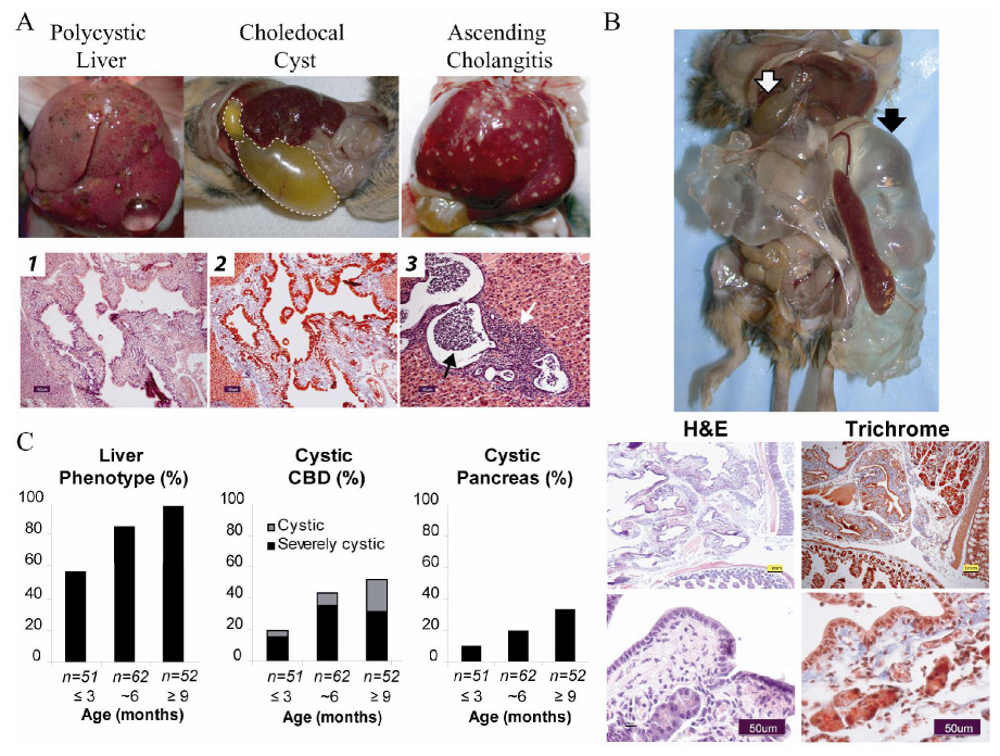
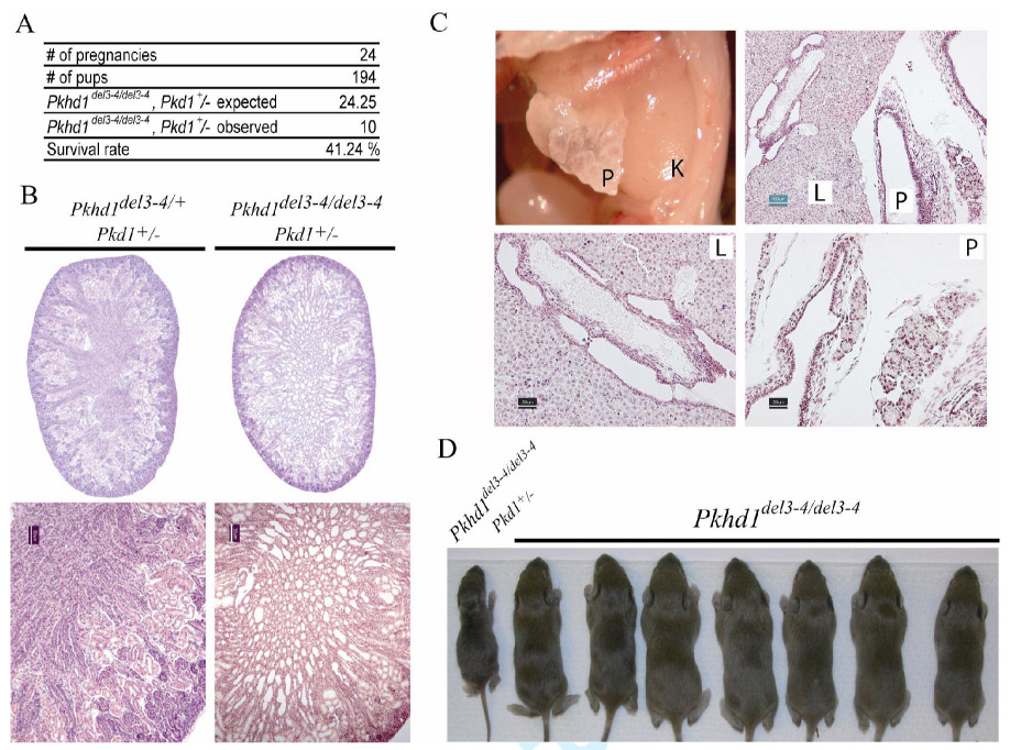
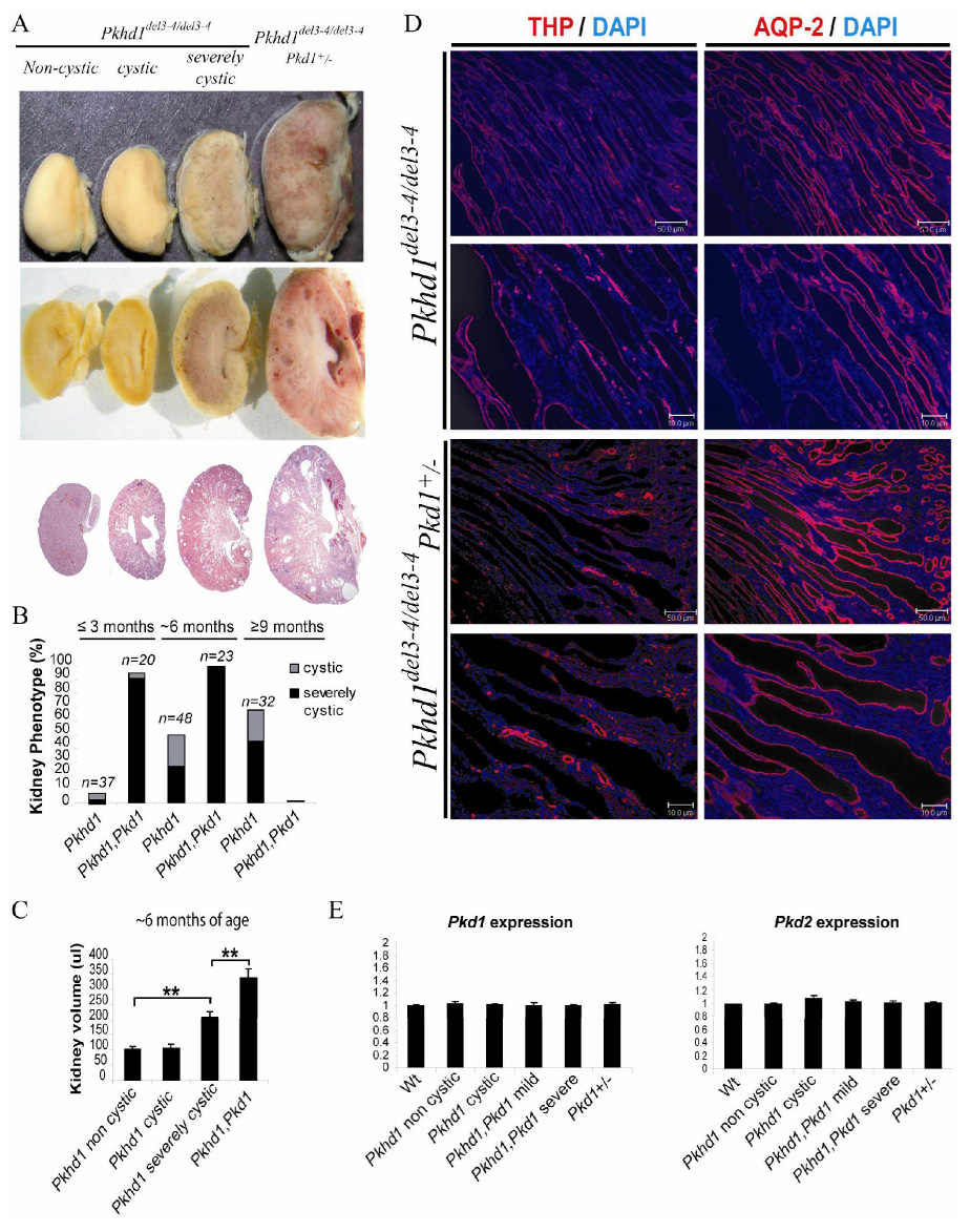
Polycystic kidney disease (PKD) describes a heterogeneous collection of disorders that differ significantly with respect to their etiology and clinical presentation. They share, however, abnormal tubular morphology as a common feature, leading to the hypothesis that their respective gene products may function cooperatively in a common pathway to maintain tubular integrity. To study the pathobiology of one major form of human PKD, we generated a mouse line with a floxed allele of Pkhd1, the orthologue of the gene mutated in human autosomal recessive PKD. Cre-mediated excision of exons 3-4 results in a probable hypomorphic allele. Pkhd1(del3-4/del3-4) developed a range of phenotypes that recapitulate key features of the human disease. Like in humans, abnormalities of the biliary tract were an invariant finding. Most mice 6 months or older also developed renal cysts. Subsets of animals presented with either perinatal respiratory failure or exhibited growth retardation that was not due to the renal disease. We then tested for genetic interaction between Pkhd1 and Pkd1, the mouse orthologue of the gene most commonly linked to human autosomal dominant PKD. Pkd1(+/-); Pkhd1(del3-4/del3-4) mice had markedly more severe disease than Pkd1(+/+); Pkhd1(del3-4/del3-4) littermates. These studies are the first to show genetic interaction between the major loci responsible for human renal cystic disease in a common PKD pathway.


Figures

Figure 1.
Figure 1.. Generation of floxed and mutant alleles of the Pkhd1 locus
(A) Schematic structures of the targeting construct (TC) and the Pkhd1 locus (Gen) before and after Cre-mediated excision of the floxed sequence. Pkhd1flox3–4 (Pkhd1tm1Ggg) and Pkhd1del3–4 (Pkhd1tm1.1Ggg) identify the floxed and post-Cre mutant alleles, respectively. The positions of primers (“a–b–c”) and probes (“5’ Ext”, “5’ Int” and “3’ Ext”) are as shown. The predicted size of each HindIII genomic fragment and PCR product is as indicated. The Pgk-Neomycin cassette, lox P, frt and HindIII sites are indicated by a blue box, red and yellow triangles, and “H” respectively. Gray ovals identify exons 2–5. (B) Characterization of the Pkhd1flox3–4 allele by Southern blot. DNA from offspring of a highly chimeric mouse was digested with HindIII, Southern blotted and probed with three markers. Each of the three probes detects the ~17kb HindIII fragment of the wild type allele (M indicates the marker, “+” a mouse that carries the targeted allele and “−“ identifies a wild type littermate). All probes also detect smaller fragments of the predicted sizes for a properly targeted locus. (C) A PCR strategy was used to identify the wild type, floxed and deleted alleles using the primers “a–b–c” in a single reaction. The ethidium bromide-stained gel shows a representative example of the PCR products amplified from each of the different genotypes. Primer pair “a–b” amplifies185bp and 254bp products from the Pkhd1flox3–4 and Pkhd1del3–4 alleles, respectively, while primer pair “a–c” amplifies a 361bp from the Pkhd1del3–4 allele and nothing from the floxed allele. (D) An ethidium bromide-stained gel showing a representative example of the RT-PCR products amplified from kidneys of individual mice with each of the different genotypes using primers from exons 2 to 6. Sample “del-3–4/del3–4(cystic)” was isolated from a cystic specimen. The data show that new splice variants are present after deletion of exons 3 and 4. Each of the new bands was excised from the gel, cloned and then sequenced. Three of the bands (red) were found to cause frame shifts either by splicing into the Pgk-Neomycin cassette or into exon 5 (fragments 650, 600 and 196). The sequence of the 87 bp fragment predicts an in-frame splicing event between exons 2–6. (E) Schematic representation of the predicted polyductin variant that results from splicing between exon 2–6. We have not been able to identify unequivocally either the full-length or mutant protein in mouse kidney using available antibodies.
Figure 2.
Figure 2.. Pkhd1del3–4/del3–4 mutants have respiratory failure and growth retardation in the absence of significant renal disease
(A) A subset of Pkhd1del3–4/del3–4 neonates died shortly after birth with dyspnea/apnea, aerophagia, and respiratory failure. Immediate necropsy confirmed aerophagia as stomach and intestines (dashed line and arrows) were filled with air. (B) A subset of Pkhd1del3–4/del3–4 mutants has severe growth retardation that is out of proportion to any other apparent organ dysfunction. In the top panel on left, gross external appearance of a wild type and Pkhd1del3–4/del3–4 mouse at P4. In right panels, gross anatomy (middle) and histopathology (far right) of the kidney (K), Liver (L) and pancreas (P, not present in middle) of the same mutant reveal little in the way of abnormal pathology. Below are representative examples of a normal and mutant P21 mouse with growth retardation as seen on gross exam (top) and by histopathologic analysis of kidney (K), liver (L) and pancreas (P). Only mild ductal plate malformations were detected (arrow). Scale bars shown correspond to 1mm (yellow) and 100µm (green).
Figure 3.
Figure 3.. Pkhd1del3–4/del3–4 mice develop abnormalities of the biliary tract and pancreas
(A) Pkhd1del3–4/del3–4 mutants develop a range of hepatobiliary abnormalities. Gross external appearance of representative examples of macroscopic cystic disease, a massive choledochal cyst (dashed line) and ascending cholangitis. Liver histopathology of a Pkhd1del3–4/del3–4 mouse showing cystic dilation of biliary ducts and dysplastic ducts (b1, Hematoxylin-eosin staining) and periportal fibrosis (b2, Masson’s trichrome). These changes are characteristic of the ductal plate malformations seen in human ARPKD. Acute ascending cholangitis (black arrow, b3) and cholecystitis also are seen (white arrow, b3). Scale bars correspond to 50 µm (black). (B) Cystic disease of the pancreas was variably present and on occasion extremely severe. Top panel shows a gross external appearance of a 6 month old Pkhd1del3–4/del3–4 mouse with severe pancreatic cystic disease (black arrow) and choledochal cyst (white arrow). Representative histopathology of pancreas stained with hematoxylin/eosin (H&E) and Masson-Trichrome showing massive dilation of the pancreatic duct, peri-ductule fibrosis (blue staining) and remnant acini. Scale bars shown correspond to 1mm (yellow) in the top and 50µm (black) in the lower panel. (C) Frequency of various phenotypes present on gross exam at different ages from a total of 165 Pkhd1del3–4/del3–4 mutant mice. Liver phenotypes include macroscopic cysts and ascending cholangitis. The common bile duct (CBD) was defined as cystic when the lumen was 3–10x fold normal diameter, and severely cystic when more than 10x.
Figure 4.
Figure 4.. Pkhd1del3–4/del3–4 mutants develop polycystic kidney disease of variable severity
(A) The kidney phenotype was classified based on its gross appearance: non-cystic, cystic (< 10 macroscopic cysts) and severely cystic (>10 cysts). (B) Histopathology confirmed the presence of cysts (lumen >3X that of a normal tubule) in the second and third groups. Two subcategories were defined in the non-cystic group based on their histopathology: kidneys with dilated tubules (lumen 1–3X that of a normal tubule) and kidneys with tubules with normal diameter. Representative examples are shown from 6 month (non-cystic and cystic) and 9 month old mice (Severely cystic). (C) Gross examination at different ages indicated that the frequency and severity of the kidney phenotype increases with the age in the Pkhd1del3–4/del3–4 mutant mice. (D) Renal volume correlated with gross phenotype and was significantly higher in the severely cystic group at 6 months of age. ** p<0.005 using the two-tail t-test.
Figure 5.
Figure 5.. Genetic interaction between the two major loci responsible for ADPKD and ARPKD
(A) The number of Pkhd1del3–4/del3–4, Pkd1+/− newborn double mutants was <50% of what was expected. (B) Newborn Pkhd1del3–4/del3–4, Pkd1+/− double mutants frequently had dilated renal tubules and cysts. 1X (top) and 10X (bottom) images of kidneys from Pkhd1del3–4/del3–4, Pkd1+/− and Pkhd1del3–4/+, Pkd1+/− newborn littermates. Scale bars correspond to 50µm (black). (C) Abnormalities including liver (L) and pancreatic (P) cysts along with fibrosis were often present on the first day of life. Scale bars shown correspond to 100µm (green) in the top and 50µm (black) in the lower panel. (D) Representative litter at 1 week of age from a cross between Pkhd1del3–4/del3–4, Pkd1+/− and Pkhd1del3–4/del3–4, Pkd1+/+ parents illustrating the gross distortion of the observed genotypes from those that were expected.
Figure 6.
Figure 6.. Genetic interaction between Pkd1 and Pkhd1 causes an accentuation of ARPKD-like disease
(A) Gross (top) and microscopic examination (bottom) of ~6 month old littermates indicates increased severity of the cystic disease in Pkhd1del3–4/del3–4, Pkd1+/− mice. Cysts radiated from papilla to cortex and were derived from distal tubule and collecting ducts in a pattern similar to that seen in human ARPKD and in severely cystic, late-stage Pkhd1del3–4/del3–4 murine kidneys (Fig 4B). (B) The incidence of the severely cystic phenotype was markedly increased in double mutant mice at all time points. All Pkhd1del3–4/del3–4, Pkd1+/− mutants died before 9 months of age. Pkhd1 and Pkhd1, Pkd1 indicate Pkhd1del3–4/del3–4 and Pkhd1del3–4/del3–4, Pkd1+/− genotypes, respectively. All comparisons were based on littermate controls. (C) Renal volume correlated with gross phenotype and was significantly higher in Pkhd1,Pkd1 double mutants than in severely cystic Pkhd1del3–4/del3–4 mice at 6 months of age. ** p<0.005 using two-tail t-test. (D) Cysts of Pkhd1del3–4/del3–4 mice universally stained positive for either aquaporin-2 (AQP-2), identifying them as derived from collecting ducts (predominant), or Tamm Horsfall protein/uromodulin (THP), identifying them as derived from the Thick Ascending Limb of Henle. Cysts of Pkhd1del3–4/del3–4, Pkd1+/− double mutants had an identical staining pattern, suggesting an accentuation of the underlying ARPKD-like phenotype. Scale bars correspond to 10µm and 50µm. (E) Ratio of expression level of Pkd1 and Pkd2 in experimental vs control (wild type) kidneys of 6 month old mice as measured by qPCR. All reactions were done in triplicate using samples from 3 mice for each genotype. Data were normalized to 18s RNA levels and then presented relative to the mean level of Pkd1 and Pkd2 in wild type specimens. All comparisons were based on littermate controls.

References

    1. Gabow PA. Autosomal Dominant Polycystic Kidney Disease. 1993;329:332–342. - PubMed
    1. Fick GM, Johnson AM, Hammond WS, Gabow PA. Causes of death in autosomal dominant polycystic kidney disease. J. Am. Soc. Nephrol. 1995;5:2048–2056. - PubMed
    1. The European Polycystic Kidney Disease Consortium. The polycystic kidney disease 1 gene encodes a 14 kb transcript and lies within a duplicated region on chromosome 16. Cell. 1994;77:881–894. - PubMed
    1. Mochizuki T, Wu G, Hayashi T, Xenophontos SL, Veldhuisen B, Saris JJ, Reynolds DM, Cai Y, Gabow PA, Pierides A, et al. PKD2, a gene for polycystic kidney disease that encodes an integral membrane protein. Science. 1996;272:1339–1342. - PubMed
    1. Sandford R, Mulroy S, Foggensteiner L. The polycystins: a novel class of membrane-associated proteins involved in renal cystic disease. Cell. Mol. Life Sci. 1999;56:567–579. - PMC - PubMed

Publication types

Substances