. 2007;35(13):4347-58.
doi: 10.1093/nar/gkm443. Epub 2007 Jun 18.

Manipulation of P-TEFb control machinery by HIV: recruitment of P-TEFb from the large form by Tat and binding of HEXIM1 to TAR

Affiliations

Manipulation of P-TEFb control machinery by HIV: recruitment of P-TEFb from the large form by Tat and binding of HEXIM1 to TAR

Stanley C Sedore et al. Nucleic Acids Res. 2007.

Abstract

Basal transcription of the HIV LTR is highly repressed and requires Tat to recruit the positive transcription elongation factor, P-TEFb, which functions to promote the transition of RNA polymerase II from abortive to productive elongation. P-TEFb is found in two forms in cells, a free, active form and a large, inactive complex that also contains 7SK RNA and HEXIM1 or HEXIM2. Here we show that HIV infection of cells led to the release of P-TEFb from the large form. Consistent with Tat being the cause of this effect, transfection of a FLAG-tagged Tat in 293T cells caused a dramatic shift of P-TEFb out of the large form to a smaller form containing Tat. In vitro, Tat competed with HEXIM1 for binding to 7SK, blocked the formation of the P-TEFb-HEXIM1-7SK complex, and caused the release P-TEFb from a pre-formed P-TEFb-HEXIM1-7SK complex. These findings indicate that Tat can acquire P-TEFb from the large form. In addition, we found that HEXIM1 binds tightly to the HIV 5' UTR containing TAR and recruits and inhibits P-TEFb activity. This suggests that in the absence of Tat, HEXIM1 may bind to TAR and repress transcription elongation of the HIV LTR.


Figures

Figure 1.
Figure 1.
HIV infection leads to activation of P-TEFb. (A) HeLa37 cells were infected with p256 HIV at an MOI of 0.1. After 4 days, the lysates of HIV infected HeLa37 cells and a parallel culture of uninfected HeLa37 cells were lysed to extract all P-TEFb and subjected to glycerol gradient sedimentation. The fractions were examined by quantitative western blotting for Cdk9 and cyclin T1 as indicated. (B) Cdk9 was quantitated for small and large forms of P-TEFb, in either uninfected or infected HeLa37 cells, from duplicate experiments and averages were calculated and plotted. Error bars represent ±1 SD of the mean.
Figure 2.
Figure 2.
HIV Tat releases P-TEFb from the large form in vivo. (A) 293T cells were transiently transfected with a HIV Tat-expressing construct containing a N-terminal FLAG tag. After 48 h, control 293T cells (β-Gal-transfected) and FLAG-Tat transfected 293T cells were lysed to extract all P-TEFb and subjected to glycerol gradient sedimentation. The gradient fractions were analyzed by quantitative western blotting for Cdk9 or FLAG-Tat, as indicated. (B) Repeat experiment, as in (A), with an additional mock transfection control (Control) and with higher transfection efficiencies.
Figure 3.
Figure 3.
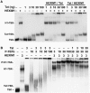
HIV Tat competes with HEXIM1 for binding to 7SK and inhibits the formation of the P-TEFb–HEXIM1–7SK complex (A) The binding of 10 ng of HEXIM1 or the indicated amounts of Tat recombinant proteins to in vitro transcribed, radiolabeled 7SK was evaluated by electrophoretic mobility shift assay (EMSA) under equilibrium conditions as described in Materials and Methods. The proteins were added to the radiolabeled 7SK individually or HEXIM1 (H1) was added 10 min before the indicated amounts of Tat (HEXIM1/Tat) was added for an additional 10 min. Reactions were also carried out with the indicated amounts of Tat added to 7SK before 10 ng of HEXIM1 (Tat/HEXIM1) was added. Complexes were resolved by gel electrophoresis on a native gel and visualized by autoradiography. (B) The ability to form a P-TEFb–HEXIM1–7SK complex or inhibit formation of the complex was evaluated by titrating the indicated amounts of P-TEFb and/or Tat onto a preformed HEXIM1–7SK complex containing 3 ng of HEXIM1 and resolving complexes as in (A).
Figure 4.
Figure 4.
HIV Tat inhibits the formation of and disrupts a preformed P-TEFb–HEXIM1–7SK complex. (A) To determine the mechanism of inhibition of complex formation, the indicated amounts of recombinant P-TEFb (P), HEXIM1 (H1) and HIV Tat (Tat) were added to radiolabeled 7SK in the presence of Zn2+, incubated for 15 min, and the complexes resolved by gel electrophoresis on a native gel. Supershifts were carried out by adding affinity-purified anti-cyclin T1 (T1) to reactions containing 32P-7SK, Tat–P-TEFb–7SK and P-TEFb–HEXIM1–7SK and incubating for an additional 10 min to verify the presence of P-TEFb. (B) Inhibition of P-TEFb–HEXIM1–7SK complex formation was evaluated with EMSA by pre-incubating the indicated amounts of P-TEFb, HEXIM1 and HIV Tat for 5 min, followed by addition of radiolabeled 7SK and incubation for an additional 15 min. To evaluate P-TEFb–HEXIM1–7SK complex disruption, the indicated amounts of P-TEFb, HEXIM1 and 7SK were pre-incubated for 10 min to allow formation of the complex, followed by addition of increasing amounts of HIV Tat and incubation for an additional 10 min. Complexes were resolved and visualized as in (A).
Figure 5.
Figure 5.
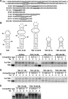
HIV Tat binds to a region of 7SK resembling TAR. (A) Analysis of the sequence of 7SK reveals three AUCUG Tat consensus-binding sites in the first 100 nt. Several structured and unstructured RNA oligos with or without this consensus sequence were designed and chemically synthesized with most coming from the native 7SK sequences indicated. 7SK (10–48 M) has one insertion and one deletion in an otherwise wild-type 7SK (10–48) sequence. (B) Predicted structures of all oligos with sufficient stability to be the predominate form at room temperature. (C) Competition EMSA analysis of Tat–7SK complex formation. Tat, 32P-labeled 7SK, and the indicated cold RNA oligos were pre-incubated and the resulting complexes were resolved by gel electrophoresis on a native gel, followed by autoradiography to visualize the 7SK shift. The dsRNA was a 25-bp double-stranded RNA unrelated to 7SK sequence described previously (53). (D) Competition EMSA comparison between 7SK (10–48) and 7SK (10–48 M). Note in this experiment the 7SK was of higher specific activity than in (C) so less Tat was needed to achieve a higher fraction of Tat–7SK complex.
Figure 6.
Figure 6.
HEXIM1 binds TAR and the HEXIM1–TAR complex recruits P-TEFb. The binding of HEXIM1, TAR and P-TEFb was evaluated under stoichiometric conditions by EMSA. The indicated components were pre-incubated and the resulting complexes were resolved by gel electrophoresis on a native gel. The gel was silver-stained to visualize protein shifts and autoradiography used to visualize the 32P-labeled RNA shifts.
Figure 7.
Figure 7.
HEXIM1–TAR inhibits the kinase activity of P-TEFb. (A) HEXIM1, TAR RNA, 7SK RNA or mixtures of HEXIM1 and TAR or 7SK found by EMSA to result in 1:1 HEXIM1–TAR or HEXIM1–7SK complexes were titrated into in vitro kinase assays containing purified P-TEFb and incorporation of 32P into the Spt5 subunit of DSIF was analyzed by SDS–PAGE followed by autoradiography. (B) Kinase activity of P-TEFb in the presence HEXIM1, TAR and 7SK as described in (A) was quantitated using an Instant Imager and plotted as a function of increasing HEXIM1.
Figure 8.
Figure 8.
Relative interaction potentials. All complexes examined in this study are indicated and the various protein–protein and protein–RNA interactions are lettered. As described in the text, two hierarchies of interaction potentials are indicated.

References

    1. Price DH. P-TEFb, a cyclin-dependent kinase controlling elongation by RNA polymerase II. Mol. Cell. Biol. 2000;20:2629–2634. - PMC - PubMed
    1. Garriga J, Grana X. Cellular control of gene expression by T-type cyclin/CDK9 complexes. Gene. 2004;337:15–23. - PubMed
    1. Peterlin BM, Price DH. Controlling the elongation phase of transcription with P-TEFb. Mol. Cell. 2006;23:297–305. - PubMed
    1. Zhu Y, Pe’ery T, Peng J, Ramanathan Y, Marshall N, Marshall T, Amendt B, Mathews MB, Price DH. Transcription elongation factor P-TEFb is required for HIV-1 tat transactivation in vitro. Genes Dev. 1997;11:2622–2632. - PMC - PubMed
    1. Shore SM, Byers SA, Maury W, Price DH. Identification of a novel isoform of Cdk9. Gene. 2003;307:175–182. - PubMed

Publication types

MeSH terms