. 2007 Jul 9;204(7):1571-82.
doi: 10.1084/jem.20070058. Epub 2007 Jun 18.

A LAD-III syndrome is associated with defective expression of the Rap-1 activator CalDAG-GEFI in lymphocytes, neutrophils, and platelets

Affiliations

A LAD-III syndrome is associated with defective expression of the Rap-1 activator CalDAG-GEFI in lymphocytes, neutrophils, and platelets

Ronit Pasvolsky et al. J Exp Med. .

Abstract

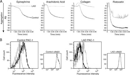
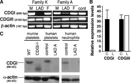
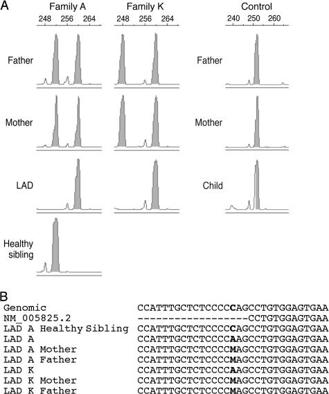
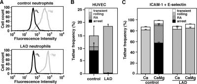
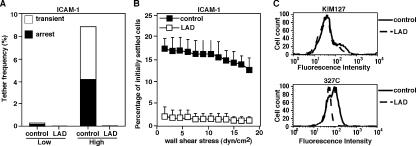
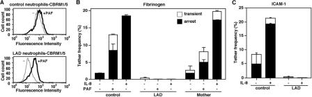
Leukocyte and platelet integrins rapidly alter their affinity and adhesiveness in response to various activation (inside-out) signals. A rare leukocyte adhesion deficiency (LAD), LAD-III, is associated with severe defects in leukocyte and platelet integrin activation. We report two new LAD cases in which lymphocytes, neutrophils, and platelets share severe defects in beta(1), beta(2), and beta(3) integrin activation. Patients were both homozygous for a splice junction mutation in their CalDAG-GEFI gene, which is a key Rap-1/2 guanine exchange factor (GEF). Both mRNA and protein levels of the GEF were diminished in LAD lymphocytes, neutrophils, and platelets. Consequently, LAD-III platelets failed to aggregate because of an impaired alpha(IIb)beta(3) activation by key agonists. beta(2) integrins on LAD-III neutrophils were unable to mediate leukocyte arrest on TNFalpha-stimulated endothelium, despite normal selectin-mediated rolling. In situ subsecond activation of neutrophil beta(2) integrin adhesiveness by surface-bound chemoattractants and of primary T lymphocyte LFA-1 by the CXCL12 chemokine was abolished. Chemokine inside-out signals also failed to stimulate lymphocyte LFA-1 extension and high affinity epitopes. Chemokine-triggered VLA-4 adhesiveness in T lymphocytes was partially defective as well. These studies identify CalDAG-GEFI as a critical regulator of inside-out integrin activation in human T lymphocytes, neutrophils, and platelets.


Figures

Figure 1.
Figure 1.
GPCR and collagen agonists fail to trigger LAD-III–derived platelet aggregation caused by their inability to trigger high affinity αIIbβ3 integrin. (A) Platelet aggregation induced by GPCR agonists, collagen, and the vWF agonist ristocetin. Figures depict treatments at time 0 of either healthy control (black line) or LAD patient K (gray line) platelets with epinephrine (first panel), arachidonic acid (second panel), collagen (third panel), or ristocetin (fourth panel), as indicated in the Materials and methods. (B) FACS staining of resting (gray line) and agonist-stimulated (black line) platelets derived from a healthy (age-matched) donor and LAD patient K with the αIIbβ3 ligand mimetic mAb PAC-1. Vertical lines depict the mean intensity of background staining with an isotype-matched control mAb. Small graphs depict the total surface αIIbβ3 levels on these stimulated platelets. Similar results were obtained with LAD patient A.
Figure 2.
Figure 2.
Loss of CalDAG-GEFI mRNA and protein in blood samples of two LAD patients. (A) CalDAG-GEFI amplified by RT-PCR from peripheral blood DNA derived from patients A and K and their parents using the primers listed in Fig. S1. Levels of CalDAG-GEFIII and actin transcripts are shown as controls. The data shown are representative of five independent experiments. (B) Relative CalDAG-GEFI and CalDAG-GEFIII mRNA expression levels were determined by Q-RT-PCR in LAD patient A (L) and an age-matched control (C). GusB was used for normalization for each gene, and normalization to HRPT1 gave similar results. The data are representative of three independent experiments. (C) Western blot analysis of platelet and neutrophil lysates derived from an age-matched control donor and LAD patient. CalDAG-GEFI (CDGI) levels were probed using the mAb 18B11. Left columns depict CalDAG-GEFI levels in wt and CalDAG-GEFI knockout murine platelets probed with the same mAb. Actin levels are shown as control.
Figure 3.
Figure 3.
Both LAD patients are homozygous for a c>a mutation in a splice junction of the CalDAG-GEFI gene. (A) A microsatellite marker containing 11.75 TATC perfect repeats located on chromosome 11q13.1, which is located 159,253 bp away from the CalDAG-GEFI gene, was amplified in single plex reaction by touchdown PCR using genomic DNA prepared from the patients (A and K), their parents, and one healthy sibling from family A, as well as from an unrelated control family. The separated fluorescently labeled PCR products were analyzed using GeneScan analysis software. The allele length distribution in each detected individual is shown. (B) Multiple alignment of genomic DNA surrounding the putative disease-causing mutation. The reference genome (National Center for Biotechnology Information 36; chr11:64253078-64253108), the mRNA (NM_005825), and genomic sequence from the patients, their parents, and one healthy sibling were aligned surrounding the mutation (in bold). Note that M is the IUPAC-IUB ambiguity code for A or C. The full trace alignments are depicted, in color, in Fig. S3.
Figure 4.
Figure 4.
Firm β2-mediated adhesion, but not capture or rolling, are defective in LAD neutrophils. (A) FACS staining of LFA-1 and Mac-1 integrins on control and LAD patient A neutrophils using the mAbs TS2.4 (anti-αL integrin subunit) and CBRM1/2 (anti-αM integrin subunit), respectively. Background antibody stainings had fluorescence intensity values of <5. (B) Neutrophil accumulation and development of firm arrest on TNFα-activated HUVECs under physiological shear flow. Healthy age-matched donor (control) or patient A (LAD) neutrophils were perfused for 1 min at 0.75 dyn/cm2 over the HUVEC monolayers, and accumulated leukocytes were subjected to a shear stress of 5 dyn/cm2 for a 10-s period. The number of adherent leukocytes that either continued to roll on the monolayer or came to full arrest immediately or after a short period of rolling (RA, rolling then arrest) was determined during the 10-s period. The values shown correspond to fractions of the original leukocyte flux in immediate contact with the endothelial layer. (C) Frequency and type of tethers generated by control and LAD (patient A) neutrophils interacting with 0.2 μg/ml E-selectin-Fc coimmobilized with 5 μg/ml ICAM-1-Fc on a protein A–coated surface. Neutrophils were perfused at 1.5 dyn/cm2 over the substrate either in the presence of Ca2+ alone (Ca) or in regular binding medium containing both Ca2+ and Mg2+ (CaMg), and the frequencies of each of the indicated tethers were determined. Results in B and C are each representative of three independent experiments. Data represent the mean ± the range of two fields of view. Similar results were obtained in LAD patient K.
Figure 5.
Figure 5.
Reduced levels of spontaneous β2 integrin adhesiveness and β2 activation states in LAD neutrophils. (A) β2 integrin–mediated capture and arrest on ICAM-1 are defective in LAD (patient A) neutrophils. The categories of the different type of adhesive tethers (transient or arrest) measured at a shear stress of 0.5 dyn/cm2 are expressed as the percentage of the total neutrophils in direct contact with the indicated ICAM-1–coated substrates. Low and high density ICAM-1–coated fields (380 and 1,900 sites/μm2, respectively) were prepared by overlaying 1 and 5 μg/ml of ICAM-1-Fc, respectively, on protein A precoated at 1 and 20 μg/ml. (B) Spontaneous β2-mediated neutrophil adhesion to the high density ICAM-1 depicted in A developed upon 1-min static contact. Level and strength of adhesion were determined by the relative resistance of either healthy (age-matched) or LAD neutrophils to detachment by the indicated incremented shear stresses. Results in A and B are representative of four independent experiments. (C) Basal expression of the β2 extension epitope KIM127 (top) and the β2 I-like domain activation epitope 327C (bottom) on either control or LAD (patient A) neutrophils analyzed by FACS staining. Background antibody stainings had fluorescence intensity values of <5. Results are given as the mean ± the range of determinations in two fields of view, and the experiment shown is representative of three independent determinations. Similar results were obtained in LAD patient K.
Figure 6.
Figure 6.
Chemoattractants fail to trigger Mac-1 adhesiveness in LAD neutrophils at subsecond contacts. (A) Normal PAF-triggered induction of the activation Mac-1 neoepitope CBRM1/5 on LAD (patient A) neutrophils. Control or LAD neutrophils were either left intact (−) or stimulated with 100 nM PAF (+). Background antibody stainings had fluorescence intensity values of <5. (B) Capture and arrest of neutrophils on low density fibrinogen (coated at 0.5 μg/ml) coated alone or in the presence of immobilized 100 nM PAF or 2 μg/ml IL-8. Control or LAD neutrophils were perfused over the substrates at a shear stress of 0.5 dyn/cm2. Analysis of neutrophils derived from patient's mother and analyzed on identical substrates is included. Results are given as the mean ± the range. The data shown are representative of three experiments. (C) Capture and arrest of neutrophils on ICAM-1 coated at 5 μg/ml alone or in the presence of 2 μg/ml IL-8. Analysis was conducted as in B. The experiment shown is representative of three. In B and C, the number of leukocytes transiently or stably interacting with the substrate was determined in two fields of view and expressed in frequency units as in Figs. 4 and 5. The experiment shown is representative of three. Similar results were obtained in LAD patient K.
Figure 7.
Figure 7.
Defective chemokine triggering of LFA-1– and VLA-4–mediated PBL arrest on ICAM-1 and VCAM-1. (A) Western blot analysis of T lymphocyte lysates derived from a C57BL mouse, a healthy donor (control), and LAD patient A (LAD). CalDAG-GEFI (CDGI) levels were probed with an anti–CalDAG-GEFI mAb (top). Actin levels are shown as controls (bottom). (B) FACS staining of LFA-1, VLA-4, and CXCR4 on control and LAD PBLs using the αL subunit–specific mAb TS2.4, the α4 subunit-specific mAb HP1.2, and 12G5, respectively. (C) LFA-1 extension epitope detected by the reporter mAb KIM127 (top row) and high affinity LFA-1 epitope detected by the mAb 327C (bottom row) in intact (−) and CXCL12-treated (+) control and LAD PBLs, assessed by FACS staining. Background antibody stainings in B and C had fluorescence intensity values of <5. (D) Tethering and immediate arrest of control (left) and LAD (right) PBLs on ICAM-1-Fc (coated at 2 μg/ml) triggered by immobilized CXCL12 (2 μg/ml). Control or LAD PBLs were perfused over the substrates at a shear stress of 0.5 dyn/cm2, and both the frequency and strength of all tethers were determined in two fields. Results are given as the mean ± the range. All adhesive tethers were blocked in the presence of the LFA-1 blocking mAb, TS1.18, or EDTA. The experiment shown is representative of three. (E) Tethering and arrest of control and LAD PBLs measured at a shear stress of 0.5 dyn/cm2 on sVCAM-1 (coated at 2 μg/ml) triggered by immobilized CXCL12 (2 μg/ml). The lifetimes of the transient tethers are depicted above the bars. All tethers were blocked in the presence of the VLA-4 blocking mAb HP1.2, or the low mol wt ligand, Bio1211 (not depicted). The different tether categories were determined as in D.

References

    1. Springer, T.A. 1994. Traffic signals for lymphocyte recirculation and leukocyte emigration: the multistep paradigm. Cell. 76:301–314. - PubMed
    1. Carman, C.V., and T.A. Springer. 2003. Integrin avidity regulation: are changes in affinity and conformation underemphasized? Curr. Opin. Cell Biol. 15:547–556. - PubMed
    1. Campbell, J.J., and E.C. Butcher. 2000. Chemokines in tissue-specific and microenvironment-specific lymphocyte homing. Curr. Opin. Immunol. 12:336–341. - PubMed
    1. Ley, K. 2003. Arrest chemokines. Microcirculation. 10:289–295. - PubMed
    1. Ginsberg, M.H., A. Partridge, and S.J. Shattil. 2005. Integrin regulation. Curr. Opin. Cell Biol. 17:509–516. - PubMed

Publication types