. 2007 Jun 18;177(6):1005-15.
doi: 10.1083/jcb.200702062.

Spindly, a novel protein essential for silencing the spindle assembly checkpoint, recruits dynein to the kinetochore

Affiliations

Spindly, a novel protein essential for silencing the spindle assembly checkpoint, recruits dynein to the kinetochore

Eric R Griffis et al. J Cell Biol. .

Abstract

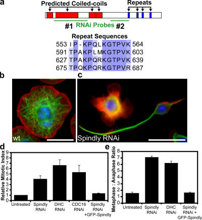
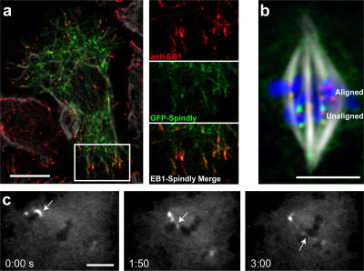
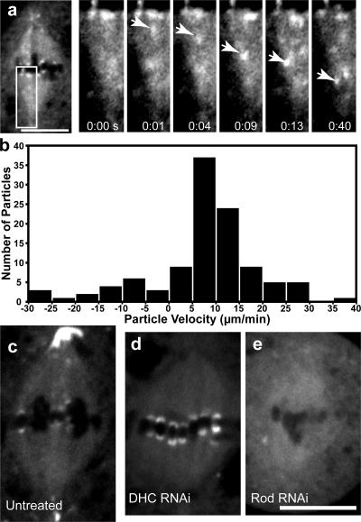
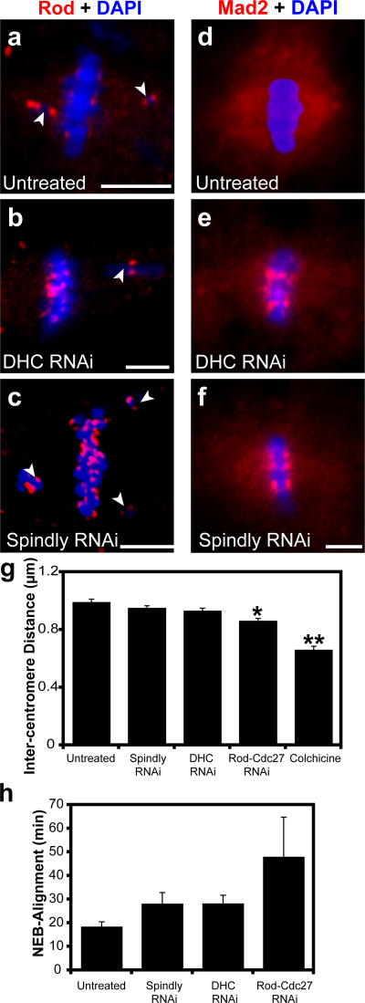
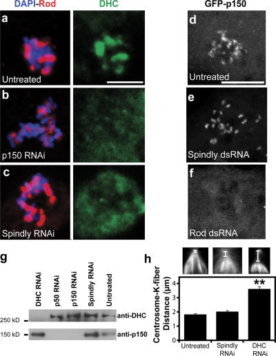
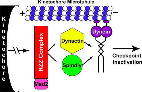
The eukaryotic spindle assembly checkpoint (SAC) monitors microtubule attachment to kinetochores and prevents anaphase onset until all kinetochores are aligned on the metaphase plate. In higher eukaryotes, cytoplasmic dynein is involved in silencing the SAC by removing the checkpoint proteins Mad2 and the Rod-Zw10-Zwilch complex (RZZ) from aligned kinetochores (Howell, B.J., B.F. McEwen, J.C. Canman, D.B. Hoffman, E.M. Farrar, C.L. Rieder, and E.D. Salmon. 2001. J. Cell Biol. 155:1159-1172; Wojcik, E., R. Basto, M. Serr, F. Scaerou, R. Karess, and T. Hays. 2001. Nat. Cell Biol. 3:1001-1007). Using a high throughput RNA interference screen in Drosophila melanogaster S2 cells, we have identified a new protein (Spindly) that accumulates on unattached kinetochores and is required for silencing the SAC. After the depletion of Spindly, dynein cannot target to kinetochores, and, as a result, cells arrest in metaphase with high levels of kinetochore-bound Mad2 and RZZ. We also identified a human homologue of Spindly that serves a similar function. However, dynein's nonkinetochore functions are unaffected by Spindly depletion. Our findings indicate that Spindly is a novel regulator of mitotic dynein, functioning specifically to target dynein to kinetochores.


Figures

Figure 1.
Figure 1.
RNAi of Spindly alters cell morphology and causes mitotic arrest in Drosophila S2 cells. (a) Domains of the Spindly protein showing predicted coiled-coil sequences in red and repeated motifs in blue; sequence alignment of residues in the repeat motifs is shown below. The locations of two nonoverlapping dsRNAs used to deplete Spindly are shown in green. A third dsRNA to the 3′ UTR was also used (not depicted). (b and c) Wild-type (wt) S2 cells show a uniformly spread morphology (b), whereas Spindly RNAi-treated cells (c) show marked defects in the actin lamellae as well as increased numbers of cells with long microtubule-rich projections. Actin, red; microtubules, green; DNA, blue. (d) The mitotic index of S2 cells is increased after the depletion of Spindly, dynein heavy chain (DHC), or a subunit of the APC (Cdc16; mean ± SEM [error bars]; n = 3 experiments, with 1,000–3,000 cells counted per experiment). Values are expressed as a ratio of RNAi-treated to untreated cells (untreated cells have a mitotic index of 1–3%). (e) The ratio of metaphase to anaphase cells (scored manually after staining with anti-tubulin and antiphosphohistone antibodies; see Materials and methods) reveals a selective increase in metaphase cells after Spindly and DHC RNAi (mean ± SEM; n = 2 experiments, with >200 mitotic spindles scored per experiment). (d and e) The expression of GFP-tagged Spindly can rescue the mitotic phenotype after endogenous Spindly is depleted using a dsRNA that targets the Spindly 3′ UTR. Bars, 10 μm.
Figure 2.
Figure 2.
Spindly binds to microtubule plus ends in interphase and to kinetochores in mitosis. (a) GFP-Spindly–expressing cells were fixed with methanol-formaldehyde and stained with anti-EB1 and anti-tubulin antibodies. The insets (magnified image of boxed area) show GFP-Spindly and anti-EB1 antibodies colocalized on the tips of microtubules. (b) Stably expressed GFP-Spindly (green) localizes to kinetochores (marked by the CENP-A homologue Cid; red) but substantially enriches on kinetochores that have not yet aligned on the metaphase plate (compare top kinetochores [aligned kinetochores] with bottom kinetochores [unaligned kinetochores]). (c) A time-lapse sequence of a GFP-Spindly–expressing cell starting in prometaphase. The arrows show an unaligned chromosome that is captured by microtubules from the opposite pole and then dragged to the metaphase plate. As this chromosome reaches the metaphase plate, the kinetochore levels of GFP-Spindly decrease until they reach the levels of the aligned chromosomes (the minutes and seconds elapsed are shown at the bottom). The video (Video 2) is available at http://www.jcb.org/cgi/content/full/jcb.200702062/DC1. Bars (a and b), 10 μm; (c) 5 μm.
Figure 3.
Figure 3.
GFP-Spindly is moved from kinetochores toward the spindle pole in a dynein-dependent manner. (a) By live cell microscopy, a particle of GFP-Spindly (arrows in the insets [magnified images of the boxed area]) can be seen moving from the kinetochore to the centrosome. The seconds elapsed are shown at the bottom (see Video 4, available at http://www.jcb.org/cgi/content/full/jcb.200702062/DC1). (b) Kymograph analysis was performed on the GFP-Spindly particles, and a histogram of the rates of 110 GFP-Spindly particles during episodes of continuous motion was produced (data were obtained from four separate spindles). The mean speed was 11.9 ± 6.9 μm/min (±SD). (c–e) GFP-Spindly in live cells was imaged by spinning disc confocal microscopy in untreated (c), dynein (DHC) RNAi-treated (d), or Rod RNAi-treated (e) cells. Dynein depletion caused Spindly to accumulate at high levels on aligned kinetochores, whereas Rod depletion blocked the recruitment of Spindly to the kinetochore. Bars, 5 μm.
Figure 4.
Figure 4.
Spindly is required for removing Rod and Mad2 from kinetochores. (a) Immunofluorescence of Rod (red in this overlay) enriches on chromosomes (blue) that are not aligned on the metaphase plate (arrowheads indicate misaligned chromosomes). (b and c) In DHC and Spindly-depleted cells, the levels of Rod are similar on aligned and unaligned kinetochores. (d) Similarly, Mad2 (red) is barely detectable on aligned kinetochores but is present throughout the spindle. (e and f) DHC (e) and Spindly (f) depletion causes the accumulation of Mad2 on aligned chromosomes (blue) and a decrease in Mad2 staining on the spindle. (g) Intercentromere tension, which was measured as the distance between Cid-stained centromeres, was measured in untreated cells and cells treated with the indicated dsRNAs or 6 μg/ml colchicine (4-h treatment; n ≥ 25 for each condition; error bars represent SEM; *, P < 5 × 10⁻⁵, **, P < 5 × 10⁻¹⁰). (h) As a second measure of kinetochore function, the time required for untreated and dsRNA-treated cells to form a metaphase spindle after nuclear envelope breakdown (NEB) was measured from time-lapse videos (n ≥ 6 cells for each condition). Bars, 5 μm.
Figure 5.
Figure 5.
Spindly depletion blocks the recruitment of DHC but not dynactin to kinetochores. (a) In mitotic cells treated with colchicine to depolymerize the mitotic spindle, Rod (red) and DHC (green) colocalize on chromosomes (blue). (b and c) In cells depleted of p150Glued (b) or Spindly (c), Rod remains bound to kinetochores, but the DHC is displaced. (d–f) S2 cells stably expressing GFP-p150Glued (a dynactin subunit) were treated with 6 μg/ml colchicine for 4 h, and the localization of the protein was assayed after RNAi treatment. In untreated (d) and Spindly-depleted (e) cells, GFP-p150Glued still bound to the kinetochore, whereas the depletion of Rod (f) prevented the protein from associating with the kinetochore. Images are maximum intensity z projections of 2-μm-thick stacks of images taken of live cells. (g) Immunoblots of lysates from RNAi-treated S2 cells show that Spindly RNAi did not affect dynein (DHC) or dynactin (p150Glued) protein levels. (h) The distance between the minus ends of kinetochore (K) fibers and the centrosome (see insets) was measured for untreated (left inset), Spindly RNAi (middle inset), and DHC RNAi (right inset) cells (n ≥ 69 for each condition; error bars represent SEM; **, P < 5 × 10⁻¹⁰), revealing a defect with dynein but not Spindly depletion. Bars, 5 μm.
Figure 6.
Figure 6.
Identification of a human Spindly homologue that is also required for targeting dynein to the kinetochore. (a) The mitotic index was determined (n = 3 wells per condition and >1,000 cells per well counted; error bars represent SEM) 24 or 48 h after siRNAs targeting the indicated proteins were transfected into HeLa cells. (b–d) The ratio of metaphase to anaphase cells for these treatments is shown (n = 2 experiments; at least 75 cells per condition). NP_060255 was localized using crude antisera in HeLa cells treated with colchicine to enrich for the protein on kinetochores, and we found that NP_060255 colocalizes with the centromere marker CENP-A (c). Colchicine treatment helped to accumulate NP_060255 on kinetochores; without this treatment, background spindle staining with the NP_060255 antibody made it difficult to unambiguously visualize kinetochore localization, even on prometaphase chromosome. To confirm the specificity of kinetochore localization in colchicine-treated cells, we depleted NP_060255 with siRNA oligonucleotides and found that the colocalization with CENP-A was eliminated (d). (e and f) The dynein intermediate chain (DIC) was localized in control and NP_060255 siRNA–transfected cells that had been treated with 6 μg/ml colchicine for 4 h to depolymerize all microtubules. The insets (magnified images of boxed areas) show that NP_060255 depletion eliminated the colocalization between CENP-A and DIC, demonstrating that NP_060255 is required for bringing dynein to the kinetochore. Bars, 5 μm.
Figure 7.
Figure 7.
A model of Spindly activity. During mitosis, the RZZ complex binds to the outer kinetochore region and recruits Mad2, Spindly, and the dynactin complex. Spindly and dynactin then cooperatively work to recruit dynein, which then transports the whole complex toward the spindle pole and silences SAC signaling on the kinetochore. See Discussion for details.

References

    1. Acquaviva, C., and J. Pines. 2006. The anaphase-promoting complex/cyclosome: APC/C. J. Cell Sci. 119:2401–2404. - PubMed
    1. Alexander, S.P., and C.L. Rieder. 1991. Chromosome motion during attachment to the vertebrate spindle: initial saltatory-like behavior of chromosomes and quantitative analysis of force production by nascent kinetochore fibers. J. Cell Biol. 113:805–815. - PMC - PubMed
    1. Bailey, T.L., and C. Elkan. 1994. Fitting a mixture model by expectation maximization to discover motifs in biopolymers. Proc. Int. Conf. Intell. Syst. Mol. Biol. 2:28–36. - PubMed
    1. Bailey, T.L., and M. Gribskov. 1998. Combining evidence using p-values: application to sequence homology searches. Bioinformatics. 14:48–54. - PubMed
    1. Baker, D.J., J. Chen, and J.M. van Deursen. 2005. The mitotic checkpoint in cancer and aging: what have mice taught us? Curr. Opin. Cell Biol. 17:583–589. - PubMed

Publication types