. 2007 Jun 18;177(6):1105-17.
doi: 10.1083/jcb.200611031.

TGF-beta signaling is essential for joint morphogenesis

Affiliations

TGF-beta signaling is essential for joint morphogenesis

Anna Spagnoli et al. J Cell Biol. .

Abstract

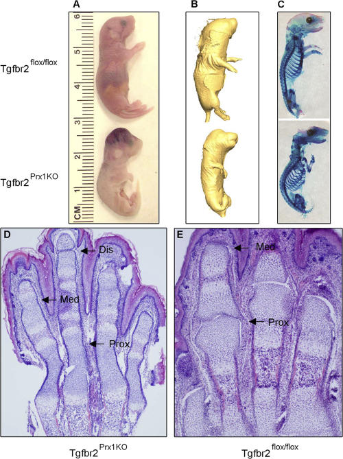
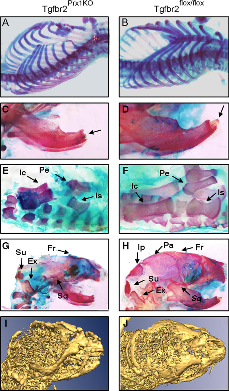
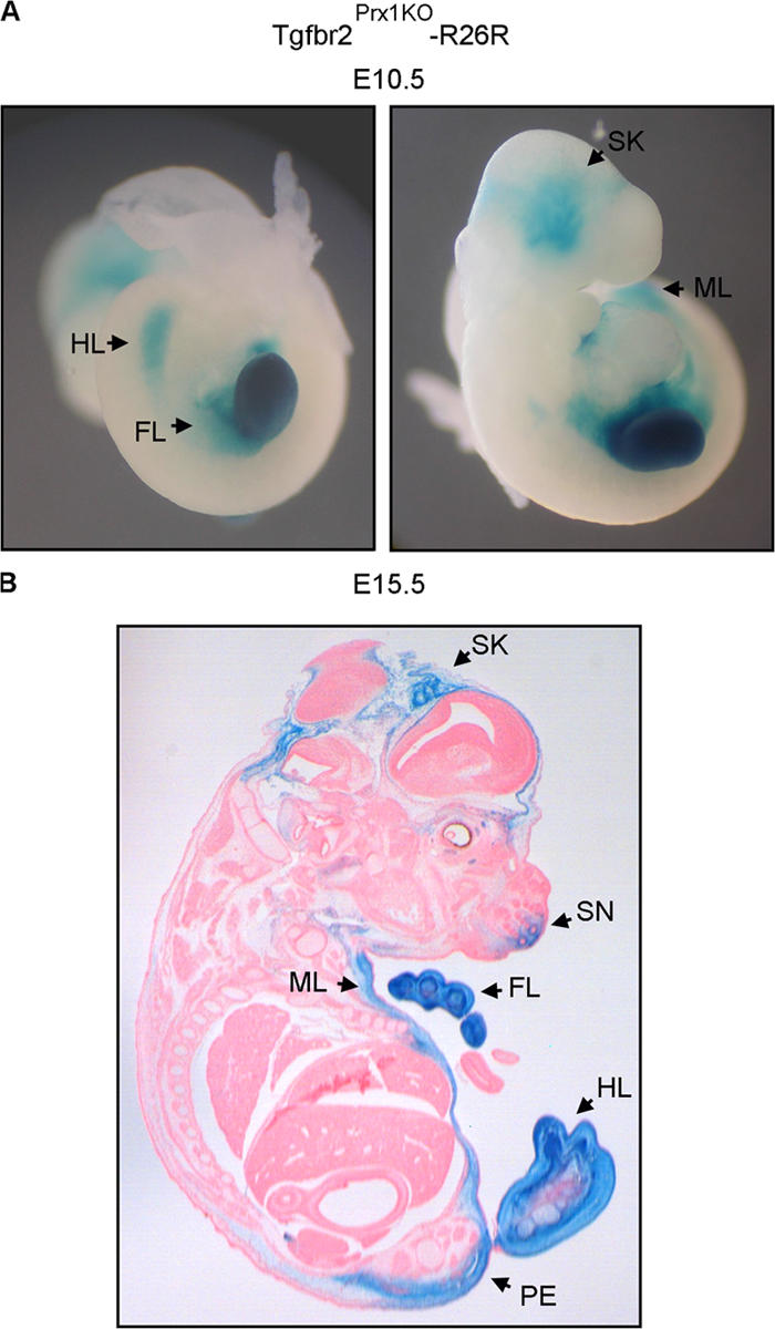
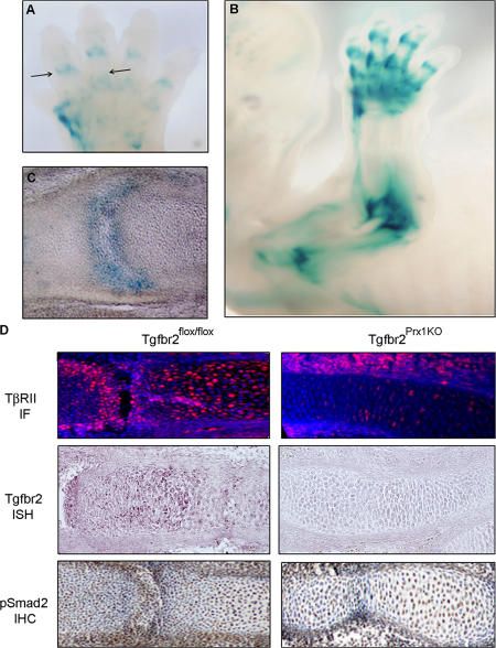
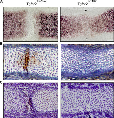
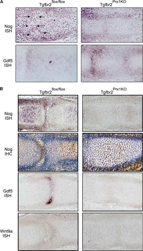
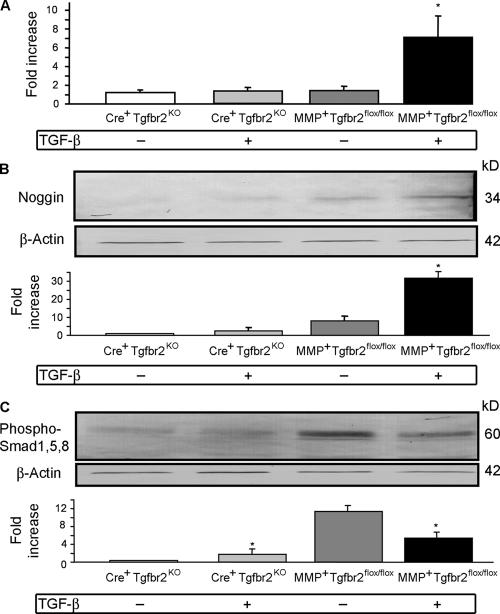
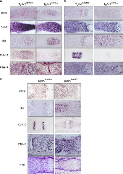
Despite its clinical significance, joint morphogenesis is still an obscure process. In this study, we determine the role of transforming growth factor beta (TGF-beta) signaling in mice lacking the TGF-beta type II receptor gene (Tgfbr2) in their limbs (Tgfbr2(PRX-1KO)). In Tgfbr2(PRX-1KO) mice, the loss of TGF-beta responsiveness resulted in the absence of interphalangeal joints. The Tgfbr2(Prx1KO) joint phenotype is similar to that in patients with symphalangism (SYM1-OMIM185800). By generating a Tgfbr2-green fluorescent protein-beta-GEO-bacterial artificial chromosome beta-galactosidase reporter transgenic mouse and by in situ hybridization and immunofluorescence, we determined that Tgfbr2 is highly and specifically expressed in developing joints. We demonstrated that in Tgfbr2(PRX-1KO) mice, the failure of joint interzone development resulted from an aberrant persistence of differentiated chondrocytes and failure of Jagged-1 expression. We found that TGF-beta receptor II signaling regulates Noggin, Wnt9a, and growth and differentiation factor-5 joint morphogenic gene expressions. In Tgfbr2(PRX-1KO) growth plates adjacent to interphalangeal joints, Indian hedgehog expression is increased, whereas Collagen 10 expression decreased. We propose a model for joint development in which TGF-beta signaling represents a means of entry to initiate the process.


Figures

Figure 1.
Figure 1.
TGF-β signaling regulates limb development and is essential for interphalangeal joint formation. (A–C) External morphology (A), micro-CT analysis (B), and Alizarin red/Alcian blue staining of newborn Tgfbr2Prx1KO mutant (C, bottom) and Tgfbr2flox/flox control (C, top) demonstrating that the mutant is smaller and has severe stylopod, zeugopod, and autopod defects. (D and E) Morphometric analysis of sectioned autopod forelimbs of newborn Tgfbr2Prx1KO mutant (D) and Tgfbr2flox/flox control (E) by hematoxylin and eosin, indicating that the mutant lacked the proximal (Prox) and medial (Med) interphalangeal joints; sporadically, a distal interphalangeal joint (Dis) is observed (as in the third digit).
Figure 2.
Figure 2.
TGF-β signaling regulates the morphological features of limb stylopods, zeugopods, and autopods. (A–D) Alizarin red/Alcian blue limb skeletons were prepared from newborn Tgfbr2Prx1KO mutants and Tgfbr2flox/flox control mice. Mutants (left) showed smaller forelimbs (A) and hindlimbs (B) with bended zeugopods and autopods. Tgfbr2Prx1KO humerus and femur stylopods (C and D) were shorter, had a middle concavity, and showed dysplastic poorly mineralized metaphyses. Humerus (C) lacked the deltoid tuberosity.
Figure 3.
Figure 3.
Lack of TGF-β signaling determines axial, pelvic bones, and calvaria defects. (A–H) Alizarin red/Alcian blue ribcage (A and B), mandibular (C and D), pelvis (E and F), and calvaria (G and H) skeletons were prepared from newborn Tgfbr2Prx1KO mutants and Tgfbr2flox/flox control mice. In Tgfbr2Prx1KO mice, the ribcage is open, lacking sternum development (A); the incisors were hypoplastic (C), and the ischial (Is), pelvic (Pe), and iliac (Ic) bones (marked by arrows) were smaller and chondrodysplastic (D). Tgfbr2Prx1KO mice lacked the parietal (Pa) and interparietal (Ip) bones, whereas the supraoccipital (Su), exoccipital (Ex), frontal (Fr) and squamosal (Sq) bones were present but hypoplastic (G). (I and J) The lack of the parietal and interparietal bones was confirmed by micro-CT analyses of living newborn Tgfbr2Prx1KO mice, indicating that it was not caused by the accidental removal of the vault during the Alizarin red/Alcian blue staining procedure (I).
Figure 4.
Figure 4.
Expression pattern of Prx-1–mediated Cre recombination in whole mount and sectioned Tgfbr2Prx1KO-R26R embryos. (A and B) X-galactosidase staining of a whole mount E10.5 Tgfbr2Prx1KO-R26R embryo (A) and section of X-galactosidase–stained E15.5 Tgfbr2Prx1KO-R26R embryo (B). Histological sections were lightly counterstained with Nuclear Fast red. In the E10.5 mutant embryo, the X-galactosidase blue staining, indicating a Cre-catalyzed recombination event, is visible through the forelimb (FL) and hindlimb (HL) buds, skull (SK), and anterior midline region (ML). In the E15.5 mutant embryo, Cre activity is additionally present in the pelvis (PE) and snout (SN) region (regions are marked by arrows). The expression pattern of PRX-1–mediated Cre recombination closely matches the regions where the Tgfbr2Prx1KO mutant presents substantial skeletal abnormalities.
Figure 5.
Figure 5.
In the developing interphalangeal joint interzone, the TβRII is highly and specifically expressed, and the R-Smad signaling is activated. (A–C) E12.5 (A) and E16.5 (B and C) Tgfbr2-GFP-β−GEO-BAC forelimbs were subjected to X-galactosidase staining. Intense staining is present in the proximal and medial interphalangeal joints (A and B) as well as in the elbow and shoulder joints (B). (C) Cryosection of an E16.5 forelimb medial interphalangeal joint confirmed intense staining. (D) Paraffin sections of an E16.5 medial interphalangeal joint from the Tgfbr2flox/flox control and Tgfbr2Prx1KO mutant were subjected to TβRII immunofluorescence (IF; top), Tgfbr2 in situ hybridization (ISH; middle), and immunohistochemistry for phosphorylated Smad-2 (IHC; bottom). Immunofluorescence for TβRII was visualized by Cy3 fluorescence (red), and nuclei were counterstained with Hoechst 33258 (blue). In Tgfbr2flox/flox, Tgfbr2 was expressed in the joint cells and in the phalangeal prehypertrophic chondrocytes. The Tgfbr2Prx1KO mutants lacked Tgfbr2 expression in the joints and chondrocytes. Nuclear immunostaining for phosphorylated Smad-2 was visualized in brown, and sections were counterstained using hematoxylin. Nuclei of joint cells from Tgfbr2flox/flox mice showed an intense phosphorylated Smad-2 staining that was abrogated in Tgfbr2Prx1KO mutant joints and largely in the phalangeal chondrocytes, providing evidence that TβRII signaling is activated in normal joints but abrogated in mutants. Studies were performed in at least two sections for each antibody and six sections for the Tgfbr2 probe; sections were obtained from at least two mutant and control embryos.
Figure 6.
Figure 6.
TGF-β signaling is needed for chondrocyte segmentation and interzone cell apoptosis. (A) Paraffin sections of an E16.5 hindlimb medial interphalangeal joint from the Tgfbr2Prx1KO mutant and Tgfbr2flox/flox control were subjected to in situ hybridization for Collagen 2. In Tgfbr2Prx1KO mutants, at the site where the presumptive joints should have formed, a persistence of Collagen 2 expression was found, as indicated by asterisks. (B) Paraffin sections of an E13.5 medial interphalangeal joint from the Tgfbr2Prx1KO mutant and Tgfbr2flox/flox control were subjected to immunohistochemistry analyses for Jagged-1 expression. The sections were lightly counterstained with hematoxylin. In control embryos, an intense Jagged-1 expression was detected in the interzone cells, whereas mutants lacked any detectable Jagged-1 in the presumptive joints. The increased cell density noted at the presumptive joint sites of the mutant is a sporadic finding most likely caused by a technical problem during the sectioning process. (C) Paraffin sections of an E13.5 medial interphalangeal joint from the Tgfbr2Prx1KO mutant and Tgfbr2flox/flox control were subjected to TUNEL assay, and fragmented DNA of apoptotic cells were visualized as brown nuclei. The sections were lightly counterstained with hematoxylin. In control embryos, an intense staining of apoptotic nuclei was visualized in the interzone, Tgfbr2Prx1KO mutants lacked the interzone, and no sign of apoptosis was detected in the presumptive joint. Studies were performed in at least two sections for Jagged-1 antibody and TUNEL assay and at least six sections for the Collagen 2 probe; sections were obtained from at least two mutant and control embryos.
Figure 7.
Figure 7.
TGF-β signaling is essential for Noggin, Gdf-5, and Wnt9a expression in the interphalangeal joints. (A and B) Paraffin sections of an E13.5 (A) and E16.5 (B) hindlimb medial interphalangeal joint from the Tgfbr2Prx1KO mutant and Tgfbr2flox/flox control were subjected to in situ hybridization (ISH) for Noggin (Nog; A and B, top), immunohistochemistry (IHC) for Noggin (B, second row), in situ hybridization for Gdf-5 (B, third row), and Wnt9a (B, fourth row). Tgfbr2Prx1KO mutants showed a down-regulation of Noggin at E13.5 and E14.5; Gdf-5 was up-regulated at E13.5, whereas it was down-regulated at E16.5; Wnt9a was down-regulated at E16.5. Arrows indicate the control developing interzone joint. Studies were performed in at least two sections for Noggin antibody and at least six sections for the Noggin, Gdf-5, and Wnt9a probes; sections were obtained from at least two mutant and control embryos.
Figure 8.
Figure 8.
TGF-β signaling up-regulates Noggin and down-regulates BMP activity. (A–C) Micromass cultures of mesenchymal limb bud cells were obtained from E11.5 Tgfbr2flox/flox mice. To knockout Tgfbr2, cultures were retrovirally infected either with the HR-MMPCreGFP vector (Cre recombinase) to generate Cre+Tgfbr2KO micromasses or with MMP-GFP empty vector to generate MMP+Tgfbr2flox/flox micromasses. mRNA and cell lysates were obtained from cultures treated with or without 20 ng/ml TGF-β for 36 h and were subjected to quantitative RT-PCR (A) or WIB (B) for Noggin expression or WIB (C) for phosphorylated Smad-1, -5, and -8. Noggin expression was normalized to glyceraldehyde-3-phosphate dehydrogenase expression and expressed as fold of increases compared with the lowest value found in the untreated control, which was given an arbitrary value of 1. TGF-β induced Noggin mRNA and protein expressions, and the effect was abrogated in Cre+Tgfbr2KO micromasses. In accordance with the increase of Noggin expression, we found that in MMP+Tgfbr2flox/flox cultures, TGF-β induced a down-regulation of BMP activity as documented by a decrease of Smad-1, -5, and -8 phosphorylations; consistently, TGF-β action was abolished in Cre+Tgfbr2KO micromasses. Membranes were probed with anti–β-actin antibody as an internal control for the protein amount loaded. For WIB densitometric analysis, band intensity was quantified, and background intensity was subtracted and normalized by the respective β-actin band intensity. Plots are represented, and mean ± SD (error bars) as the fold increase over control is given. *, P < 0.05 versus untreated MMP+Tgfr2flox/flox by one-way analysis of variance; n = 3.
Figure 9.
Figure 9.
TGF-β signaling in cartilage development: lack of joint development is associated with selective effects on the adjacent chondrogenesis. (A–C) Paraffin sections of E13.5 (A), E16.5 (B), and P0 (C) growth plates adjacent to the (presumptive) interphalangeal joint of the Tgfbr2Prx1KO mutant and Tgfbr2flox/flox control were subjected to in situ hybridization for Sox-9 (A and B, top), Collagen 2 (A and B, second rows; and C, top), Ihh (A and B, third rows; and C, second row), Collagen 10 (A and B, fourth rows; and C, third row), PTH-rP (A and B, bottom; and C, fourth row), or hematoxylin and eosin staining (C, bottom). Tgfbr2Prx1KO mutants showed an up-regulation of Ihh and a down-regulation of Collagen 10 expressions at all stages. Collagen 2 and Sox-9 expressions were similar in Tgfbr2Prx1KO and control, but Collagen 2–expressing cells at P0 display a less organized columnar distribution than controls. PTH-rP expression in E13.5 Tgfbr2Prx1KO mutants is similar to the control, whereas at E16.5 and P0, it is increased in the perichondrium and especially in the prehypertrophic/upper proliferative cells. Studies were performed in at least six sections for each probe that were obtained from at least two mutant and control embryos.
Figure 10.
Figure 10.
TGF-β is a master signaling center within the joint interzone. Proposed model for the role of TβRII signaling in interphalangeal joint development. TβRII is specifically expressed by the joint-developing cells, and lack of TGF-β signaling results in the failure of interzone formation by lack of the survival of interzone-forming cells, persistence of differentiated chondrocytes in the joint region, and derangement of joint morphogenic gene expressions.

Similar articles

Cited by

References

    1. Ando, K., S. Kanazawa, T. Tetsuka, S. Ohta, X. Jiang, T. Tada, M. Kobayashi, N. Matsui, and T. Okamoto. 2003. Induction of Notch signaling by tumor necrosis factor in rheumatoid synovial fibroblasts. Oncogene. 22:7796–7803. - PubMed
    1. Antonopoulou, I., L.A. Mavrogiannis, A.O. Wilkie, and G.M. Morriss-Kay. 2004. Alx4 and Msx2 play phenotypically similar and additive roles in skull vault differentiation. J. Anat. 204:487–499. - PMC - PubMed
    1. Archer, C.W., G.P. Dowthwaite, and P. Francis-West. 2003. Development of synovial joints. Birth Defects Res. C. Embryo Today. 69:144–155. - PubMed
    1. Baffi, M.O., E. Slattery, P. Sohn, H.L. Moses, A. Chytil, and R. Serra. 2004. Conditional deletion of the TGF-beta type II receptor in Col2a expressing cells results in defects in the axial skeleton without alterations in chondrocyte differentiation or embryonic development of long bones. Dev. Biol. 276:124–142. - PubMed
    1. Bhowmick, N.A., A. Chytil, D. Plieth, A.E. Gorska, N. Dumont, S. Shappell, M.K. Washington, E.G. Neilson, and H.L. Moses. 2004. TGF-beta signaling in fibroblasts modulates the oncogenic potential of adjacent epithelia. Science. 303:848–851. - PubMed

Publication types

MeSH terms

Substances