. 2007 Sep;27(17):6038-52.
doi: 10.1128/MCB.00522-07. Epub 2007 Jun 18.

Double knockouts reveal that protein tyrosine phosphatase 1B is a physiological target of calpain-1 in platelets

Affiliations

Double knockouts reveal that protein tyrosine phosphatase 1B is a physiological target of calpain-1 in platelets

Shafi M Kuchay et al. Mol Cell Biol. 2007 Sep.

Abstract

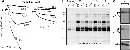
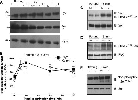
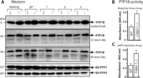
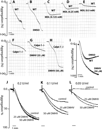
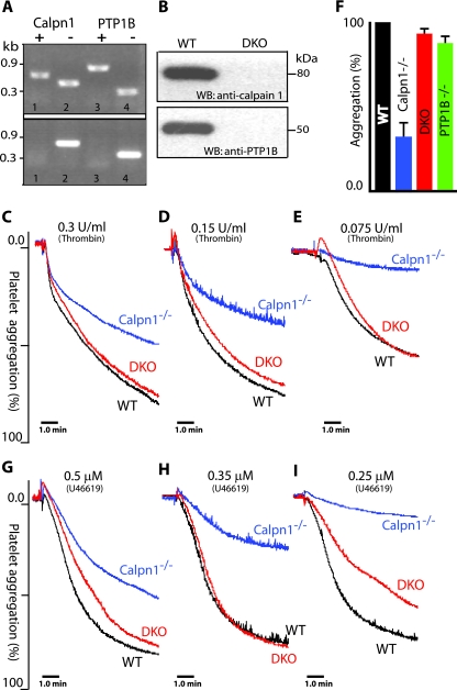
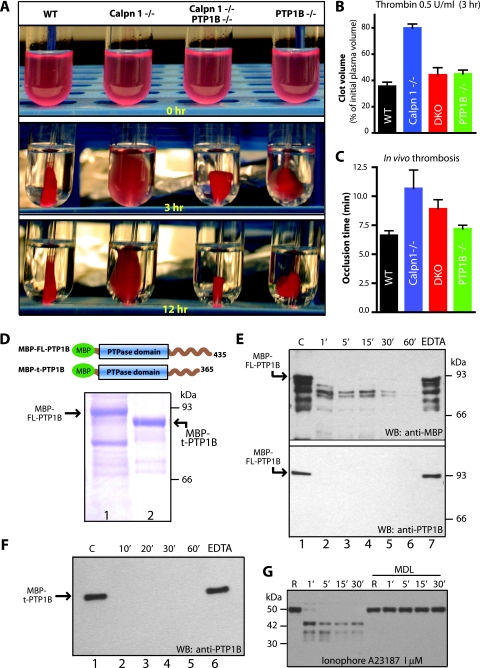
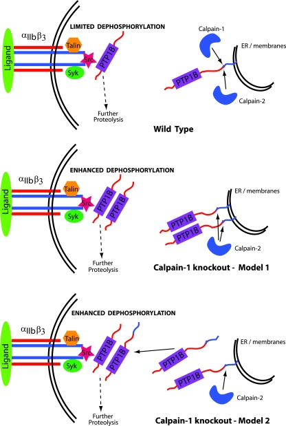
Calpains are ubiquitous calcium-regulated cysteine proteases that have been implicated in cytoskeletal organization, cell proliferation, apoptosis, cell motility, and hemostasis. Gene targeting was used to evaluate the physiological function of mouse calpain-1 and establish that its inactivation results in reduced platelet aggregation and clot retraction potentially by causing dephosphorylation of platelet proteins. Here, we report that calpain-1 null (Capn1-/-) platelets accumulate protein tyrosine phosphatase 1B (PTP1B), which correlates with enhanced tyrosine phosphatase activity and dephosphorylation of multiple substrates. Treatment of Capn1-/- platelets with bis(N,N-dimethylhydroxamido)hydroxooxovanadate, an inhibitor of tyrosine phosphatases, corrected the aggregation defect and recovered impaired clot retraction. More importantly, platelet aggregation, clot retraction, and tyrosine dephosphorylation defects were rescued in the double knockout mice lacking both calpain-1 and PTP1B. Further evaluation of mutant mice by the ferric chloride-induced arterial injury model suggests that the Capn1-/- mice are relatively resistant to thrombosis in vivo. Together, our results demonstrate that PTP1B is a physiological target of calpain-1 and suggest that a similar mechanism may regulate calpain-1-mediated tyrosine dephosphorylation in other cells.


Figures

FIG. 1.
FIG. 1.
Platelet aggregation and tyrosine phosphorylation of cytoskeletal proteins in calpain-1 null platelets. (A) Gel-filtered (3 × 10⁸/ml) platelets were analyzed for platelet aggregation assay in response to different doses of thrombin. (B) Gel-filtered platelets (5 × 10⁸/ml) from WT (+/+) and calpain-1 null (−/−) mice were activated by 0.15 U/ml of thrombin, and tyrosine phosphorylation of the cytoskeletal proteins was analyzed by Western blotting using the 4G10 antibody (WB: 4G10). The cytoskeleton extract was normalized after activation with thrombin for up to 5.0 min (5′) using β-actin as a control antibody. 30″, 30 s. (C) Tyrosine phosphorylation of the β3 subunit of αIIbβ3 integrin was evaluated after 5 min of thrombin activation (0.15 U/ml) of cells from WT (+/+) and calpain-1 null (−/−) mice using antiphosphotyrosine specific antibodies as described in the text. The antibody against β-actin served as a control.
FIG. 2.
FIG. 2.
Evaluation of protein tyrosine kinase activity in calpain-1 null platelets. (A) Gel-filtered (3 × 10⁸/ml) platelets from WT (+/+) and calpain-1 null (−/−) mice were analyzed for the presence of different tyrosine kinases (Syk, Fyn, and c-Yes) by Western blotting with specific antibodies in the resting platelets and platelets activated by thrombin (0.15 U/ml) for defined time periods (30 s [30″ and 1 min [1′]). The Fyn antibody epitope is conserved in the Lyn tyrosine kinase and therefore can detect both tyrosine kinases. (B) Total protein tyrosine kinase activity was measured in the resting and thrombin-activated platelets. (C) Src tyrosine kinase was immunoprecipitated (IP) using a mouse monoclonal antibody, and the immunoprecipitated Src was probed with an antibody specific for phosphotyrosine-416 antibody (PhosY416 Src). The bottom gel shows the total amount of Src in the platelet lysate used to immunoprecipitate Src. IB, immunoblotting. (D) In the resting and thrombin-activated platelet lysates, FAK activation was analyzed using an antibody against phosphotyrosine 577 in FAK. The bottom gel shows the total amount of FAK in the platelet lysate. (E) Western blot analysis of Src in the resting and thrombin-activated platelet lysates. An antibody specific for the dephosphorylation site in Src at position 527 was used. The bottom gel shows the total amount of Src in platelet lysate.
FIG. 3.
FIG. 3.
Comparison of phosphatase activity in the resting and thrombin-activated platelets. (A and B) Platelet lysates from gel-filtered platelets were tested for total phosphatase activity. Inhibition of tyrosine phosphatases in calpain-1 null lysates was performed by incubation with 1.0 mM sodium vanadate in the phosphatase assay buffer. Total phosphatase activity in the double knockout (DKO) platelet lysate was included to demonstrate the rescue of enhanced phosphatase activity in calpain-1 null platelets. (C) The serine-threonine phosphatase activity of calcineurin was measured calorimetrically as described in the text. (D) Total protein tyrosine phosphatase (PTP) activity was measured in thrombin-activated platelets. (E and F) Immunodepletion of PTP1B was carried out with polyclonal antibodies against PTP1B using protein G-conjugated agarose beads. Resting and thrombin-activated platelet lysates were analyzed for total phosphatase by the pNPP hydrolysis assay in the WT, calpain-1 null, and calpain-1 null lysate that was immunodepleted as previously described.
FIG. 4.
FIG. 4.
Characterization of PTP1B in mouse platelets. (A) Gel-filtered platelets from WT (+/+) and calpain-1 null (−/−) mice were activated with 0.15 U/ml of thrombin, and the amount of PTP1B was examined using three different antibodies (Ab) targeted to defined epitopes at the C and N termini (C-term and N-term) of PTP1B. Western blot analysis of SH-PTP1 and SH-PTP2 indicated no changes in their protein level and served as an internal control. 30″, 30 s; 1′, 1 min. (B and C) Gel-filtered platelets were activated with 0.15 U/ml of thrombin, and PTP1B was immunoprecipitated using a polyclonal antibody directed against the N terminus of PTP1B. The data show PTP1B enzyme activity in the mouse platelets activated for 3.0 min by thrombin.
FIG. 5.
FIG. 5.
Effects of pharmacological inhibitors of calpain-1 and PTP1B on platelet aggregation. Gel-filtered platelets were analyzed for platelet aggregation in response to synthetic inhibitors of calpains and PTP1B. The platelet aggregation was induced by 0.15 U/ml of thrombin. (A to E) WT platelets (A), WT platelets incubated with vehicle DMSO (B), and WT platelets incubated with the indicated concentrations of MDL (C to E). (F) Platelet aggregation response of calpain-1 null platelets. It is noteworthy that at 25 μM DMHV, although the final extent of platelet aggregation was not altered in calpain-1 null mice (G), the overall shape of the aggregation tracing without DMHV appears to be distinct (F). (H) The reduced platelet aggregation in calpain-1 null mice was rescued by incubation with 50 μM DMHV, a pharmacological inhibitor of tyrosine phosphatases, including PTP1B. (I) In the WT mouse platelets, 50 μM DMHV enhanced platelet aggregation at 0.075 U/ml thrombin activation. (J to L) Washed human platelets (2 × 10⁸/ml) were incubated with 25 μM and 50 μM DMHV for 30 min at room temperature for aggregation response to thrombin activation.
FIG. 6.
FIG. 6.
Generation of double knockout mice lacking calpain-1 and PTP1B and correction of the platelet aggregation defect. (A) Genomic DNA was analyzed for the corresponding genotype by PCR. Mice heterozygous (top gel) for both calpain-1 (lanes 1 and 2) and PTP1B (lanes 3 and 4), and double knockout mice (bottom gel) for calpain-1 (lane 2) and PTP1B (lane 4) are shown. (B) Western blot (WB) analysis of erythrocyte membrane lysates with anti-calpain-1 and anti-PTP1B antibodies. The erythrocytes were from WT and double knockout (DKO) mice. (C to I) Correction of platelet aggregation defect in the double knockout (DKO) mice. Gel-filtered platelets were analyzed for platelet aggregation response with thrombin (C to E) and with U46619, a stable thromboxane A2 analogue (G to I). (F) Quantification of platelet aggregation at various concentrations of thrombin in the WT, calpain-1 null (Calpn1−/−), double knockout (DKO), and PTP1B null (PTP1B−/−) mice (P < 0.05). For statistical analysis, the extent of platelet aggregation in the WT mouse platelets at each dose was taken as 100%. The platelet aggregation response at comparable doses in the mutant platelets was normalized to their respective dose in the WT platelets. Each bar graph shows the mean platelet aggregation plus SEM for a mouse genotype. Data were analyzed using one-way analysis of variance by GraphPad Prism software.
FIG. 7.
FIG. 7.
Correction of clot retraction defect in the double knockout mice. Platelet-rich plasma samples were pooled, and platelet counts were normalized. (A) Representative photographs of clot retraction induced by thrombin. (B) Quantification of fibrin clot retraction 3.0 h after thrombin and CaCl2 treatment. Clot volumes are expressed as a percentage of initial platelet-rich plasma volume. (C) Measurement of in vivo thrombosis by ferric chloride injury assay. Calpain-1 null mice are less susceptible to thrombus development. The mean plus SEM (error bar) for each group are shown. Data were analyzed using one-way analysis of variance by GraphPad prism software (P = 0.01). (D) Coomassie blue staining of recombinant PTP1B. Bacterially expressed full-length PTP1B (FL-PTP1B) (lane 1) and truncated PTP1B (t-PTP1B) (lane 2) were purified by affinity chromatography. The boundaries of each fusion protein with MBP at the N terminus are shown in the schematic diagram. (E) In vitro proteolysis of full-length recombinant PTP1B (FL-PTP1B) by commercial calpain-1 for indicated time intervals (from 1 min [1′] to 60 min [60′]) (see Materials and Methods for details). Lane 1 contains a negative control where calpain-1 was not added. Lanes 2 to 6 show various incubation times of PTP1B with calpain-1. Lane 7 shows incubation of PTP1B with calpain-1 in the presence of 2.0 mM EDTA for 60 min to inhibit calpain-1. The top blot was probed with a rabbit polyclonal antibody raised against MBP. The bottom blot was probed with a commercially available polyclonal antibody against PTP1B. WB, Western blotting. (F) In vitro proteolysis of truncated PTP1B by calpain-1. Lane 1 contains a negative control with no calpain-1, and lanes 2 to 5 show incubation times (from 10 min [10′] to 60 min [60′]) with calpain-1. Lane 6 shows incubation with calpain-1 in the presence of 2.0 mM EDTA for 60 min. Western blotting (WB) was carried out using an anti-PTP1B antibody. (G) Gel-filtered platelets (3 × 10⁸/ml) from WT mice were incubated with either DMSO or calpain inhibitor MDL 28170 (350 μM) in DMSO for 30 min at room temperature. PTP1B proteolysis was initiated by incubation of platelets with calcium ionophore A23187, and PTP1B degradation was monitored by Western blotting using a polyclonal antibody directed against mouse PTP1B. This antibody, kindly provided by B. Neel, recognizes intact and cleaved forms of PTP1B under these conditions R, resting.
FIG. 8.
FIG. 8.
Proposed model for the regulation of PTP1B by calpains. See text for details. ER, endoplasmic reticulum.

Similar articles

Cited by

References

    1. Arias-Salgado, E. G., F. Haj, C. Dubois, B. Moran, A. Kasirer-Friede, B. C. Furie, B. Furie, B. G. Neel, and S. J. Shattil. 2005. PTP-1B is an essential positive regulator of platelet integrin signaling. J. Cell Biol. 170:837-845. - PMC - PubMed
    1. Arthur, J. S. C., J. S. Elce, C. Hegadorn, K. Williams, and P. A. Greer. 2000. Disruption of the murine calpain small subunit gene, Capn4: calpain is essential for embryonic development but not for cell growth and division. Mol. Cell. Biol. 20:4474-4481. - PMC - PubMed
    1. Azam, M., S. S. Andrabi, K. E. Sahr, L. Kamath, A. Kuliopulos, and A. H. Chishti. 2001. Disruption of the mouse μ-calpain gene reveals an essential role in platelet function. Mol. Cell. Biol. 21:2213-2220. - PMC - PubMed
    1. Bence, K. K., M. Delibegovic, B. Xue, C. Z. Gorgun, G. S. Hotamisligil, B. G. Neel, and B. B. Kahn. 2006. Neuronal PTP1B regulates body weight, adiposity and leptin action. Nat. Med. 12:917-924. - PubMed
    1. Born, G. V. 2005. Light on platelets. J. Physiol. 568:713-714. - PMC - PubMed

Publication types

MeSH terms

Substances

LinkOut - more resources