. 2007 Sep;27(17):6229-42.
doi: 10.1128/MCB.02246-06. Epub 2007 Jun 18.

BNIP3 is an RB/E2F target gene required for hypoxia-induced autophagy

Affiliations

BNIP3 is an RB/E2F target gene required for hypoxia-induced autophagy

Kristin Tracy et al. Mol Cell Biol. 2007 Sep.

Abstract

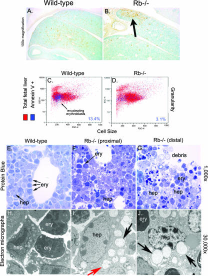
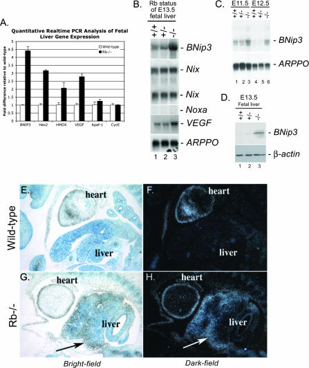
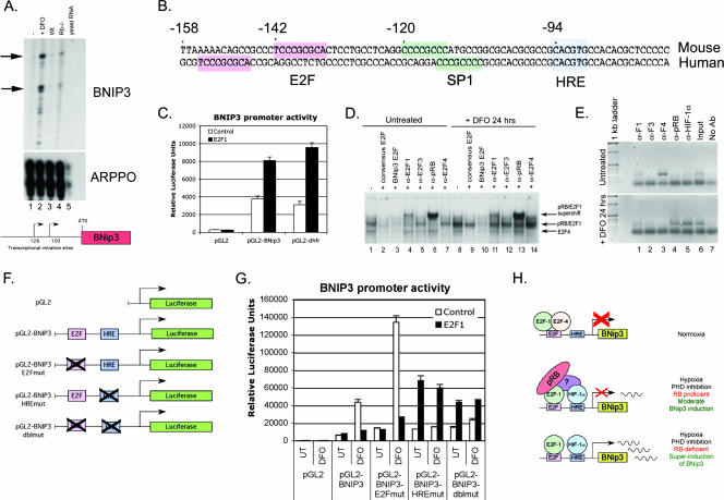
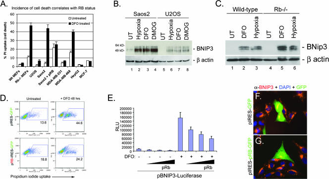
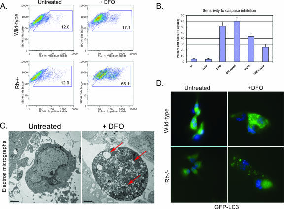
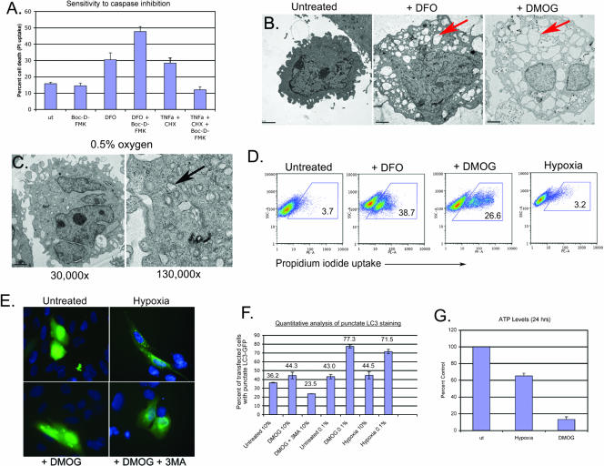
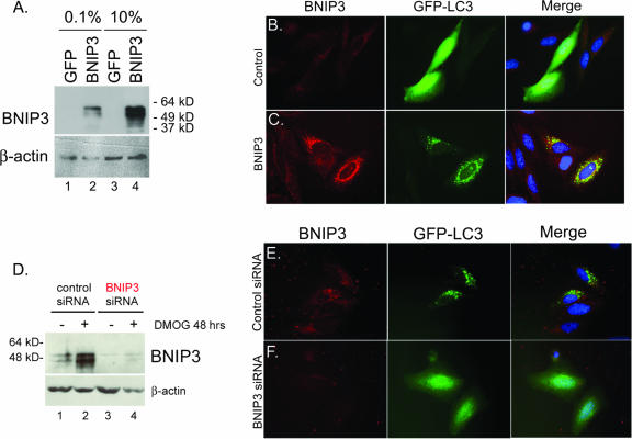
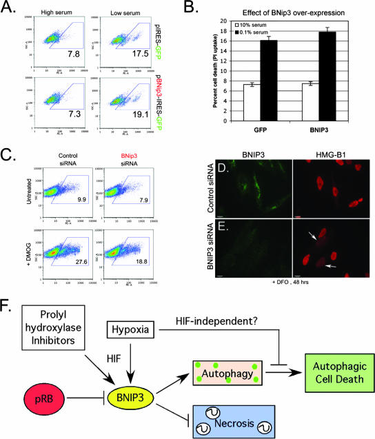
Hypoxia and nutrient deprivation are environmental stresses governing the survival and adaptation of tumor cells in vivo. We have identified a novel role for the Rb tumor suppressor in protecting against nonapoptotic cell death in the developing mouse fetal liver, in primary mouse embryonic fibroblasts, and in tumor cell lines. Loss of pRb resulted in derepression of BNip3, a hypoxia-inducible member of the Bcl-2 superfamily of cell death regulators. We identified BNIP3 as a direct target of pRB/E2F-mediated transcriptional repression and showed that pRB attenuates the induction of BNIP3 by hypoxia-inducible factor to prevent autophagic cell death. BNIP3 was essential for hypoxia-induced autophagy, and its ability to promote autophagosome formation was enhanced under conditions of nutrient deprivation. Knockdown of BNIP3 reduced cell death, and remaining deaths were necrotic in nature. These studies identify BNIP3 as a key regulator of hypoxia-induced autophagy and suggest a novel role for the RB tumor suppressor in preventing nonapoptotic cell death by limiting the extent of BNIP3 induction in cells.


Figures

FIG. 1.
FIG. 1.
Nonapoptotic cell death in the midgestational FLs of Rb null mice. (A and B) TUNEL assays to detect cell death in wild-type and Rb null E13.5 FLs. (C and D) Flow cytometric analysis of annexin V-fluorescein isothiocyanate-stained FL cells. (E to G) Light microscopic analysis of protein-blue-stained FL sections showing increased vacuolation in Rb null FL (F and G, arrows) compared to the level of vacuolation in wild-type FL (E). Dark-staining erythroblasts (F, ery) were not overtly affected. Cellular debris is evident in distal regions of Rb null FL (G). (H to J) Electron microscopic analysis of wild-type (H) and Rb null FLs (I and J). Extensive vacuolation in Rb null hepatocytes (hep) is associated with increased mitochondrial swelling (I and J, black arrows). Double-membrane-bound vesicles also are seen in Rb null hepatocytes (I and J, red arrow).
FIG. 2.
FIG. 2.
BNip3 expression is up-regulated in the Rb null FL. (A) Quantitative real-time PCR for known HIF target genes (encoding BNIP3, hexokinase 2, heme oxygenase, and VEGF) and two known E2F target genes (encoding Apaf-1 and cyclin E2 [CycE]) carried out on cDNA derived from E13.5 wild-type and Rb null FLs. (B) Total RNA from FLs from wild-type (lane 1), Rb heterozygous (lane 2), or Rb null (lane 3) E13.5 mice was examined by Northern blotting for expression of BNip3, Nix, Noxa, VEGF, and ARPPO (a control for loading). (C) Northern blot analysis of BNip3 mRNA expression in wild-type (lanes 1 and 4), Rb heterozygous (lanes 2 and 5), and Rb null (lanes 3 and 6) FLs at E11.5 (lanes 1 to 3) or E12.5 (lanes 4 to 6). (D) BNip3 protein expression in wild-type (lane 1), Rb heterozygous (lane 2), and Rb null (lane 3) FLs determined by Western blotting. (E to H) BNip3 mRNA was detected in wild-type (E and F) or Rb null (G and H) E13.5 mouse embryo sections by in situ hybridization. BNip3 mRNA was visualized at 100× magnification by bright-field (E and G) and by dark-field (F and H) microscopy on toluidine blue-counterstained sections.
FIG. 3.
FIG. 3.
The BNIP3 promoter contains a functional E2F binding site that interacts functionally with the HRE. (A) Transcriptional initiation sites in the mouse BNIP3 promoter were determined by RNase protection assays using RNA from untreated (lane 1) or DFO-treated (lane 2) MEFs and wild-type (lane 3) FL (Wt), Rb null (lane 4) FL, or control yeast (lane 5) RNA. A diagrammatic representation of the two major initiation sites in the mouse BNIP3 promoter is shown. (B) Sequence alignment of the proximal regions of the mouse and human BNIP3 promoters highlighting conserved regulatory elements that include an HRE (blue) at −94 bp relative to the start site of transcription and an E2F site (pink) at −142 bp (mouse) and −155 bp (human). (C) The proximal 500 bp of the mouse BNIP3 promoter was cloned upstream of the luciferase reporter gene and tested for its activity in U2OS cells in response to overexpression of E2F-1, and its activity then was compared to that of the control reporter construct pGL2 lacking a promoter and that of the characterized E2F-responsive mouse dhfr promoter. (D) The binding activity of the BNIP3 promoter E2F site was tested by EMSA using nuclear lysates from untreated (lanes 1 to 7) or DFO-treated (lanes 8 to 14) U2OS cells in the presence or absence of excess cold oligonucleotide competitor encoding either the consensus E2F recognition sequence (lanes 2 and 9) or the BNip3 E2F site (lanes 3 and 10). The effect of supershifting antibodies to E2F-1 (α-E2F1) (lanes 4 and 11), E2F-3 (α-E2F3) (lanes 5 and 12), pRB (α-pRB) (lanes 6 and 13), or E2F-4 (α-E2F4) (lanes 7 and 14) on DNA-protein complex migration also was examined. (E) The identity of E2Fs bound to the endogenous BNIP3 promoter in untreated (top panel) or DFO-treated (bottom panel) U2OS cells was examined by ChIPs using antibodies to E2F-1 (α-F1), E2F-3 (α-F3), E2F-4 (α-F4), pRB (α-pRB), or HIF-1α (α-HIF-1α) and compared to results with no-antibody negative control samples (No Ab) or to those with input positive control samples (Input). (F) Diagrammatic representation of mutations introduced into the mouse BNIP3 promoter to test the functional interaction between E2F and HIF in regulating BNip3 transcription. (G) Reporter (luciferase) gene assay for BNIP3 promoter activity in U2OS cells following DFO treatment, E2F-1 overexpression, or mutation of either the E2F site, the HRE, or both sites in the context of the proximal 500 bp of the mouse BNIP3 promoter. UT, untreated. (H) Schematic representation of the proposed interaction between pRB, E2F family members, and HIF at the BNIP3 promoter.
FIG. 4.
FIG. 4.
RB status and BNIP3 expression correlate with cell death. (A) The incidence of cell death of primary MEFs and different human tumor cell lines was assessed after 48 h of treatment with DFO by measuring uptake of PI, which was taken up only by dying cells. Wt, wild type. (B) Western blot analysis of BNIP3 induction in Saos2 and U2OS cells by hypoxia, DMOG treatment, or DFO treatment. (C) Western blot analysis of BNip3 induction in wild-type and Rb null MEFs by DFO treatment or hypoxia. UT, untreated. (D) Saos2 cells were transiently transfected with pIRES-GFP or pRB-IRES-GFP and treated with DFO for 48 h. The incidence of cell death was determined by flow cytometric analysis of PI uptake. (E) The effect of pRB overexpression for BNIP3 promoter activity was determined for Saos2 cells by a luciferase assay for reporter gene expression. Results presented are the averages of at least three independent experiments. RLU, relative luciferase units. (F and G) The effect of pRB expression on the levels of BNIP3 protein was determined by immunofluorescent staining for BNIP3 (red) in cells that expressed GFP by virtue of transfection with either pIRES-GFP (F) or pRB-IRES-GFP (G). DAPI, 4′,6′-diamidino-2-phenylindole.
FIG. 5.
FIG. 5.
Nonapoptotic cell death of Rb null MEFs. (A) Wild-type and Rb null MEFs were cultured in the presence of 260 μM DFO for 48 h, and the viability of MEFs was compared to that of untreated controls as determined by flow cytometric analysis of PI uptake. (B) DFO-induced cell death of Rb null MEFs is resistant to the pancaspase inhibitor z-vad-fmk, in contrast to death induced by TNF-α and cycloheximide. (C) Electron microscopy of untreated and DFO-treated Rb null MEFs reveals small double-membrane-bound vesicles in hypoxic cells (red arrow), indicative of autophagy. (D) GFP-LC3 staining of untreated and DFO-treated (48 h) wild-type and Rb null MEFs reveals increased punctate GFP-LC3 staining in Rb null MEFs, consistent with increased autophagy.
FIG. 6.
FIG. 6.
Nonapoptotic cell death induced by PHD inhibition. (A) DMOG-induced cell death of Saos2 cells is resistant to the pancaspase inhibitor Boc-D-FMK, in contrast to death induced by TNF-α and cycloheximide. (B) Electron microscopy of Saos2 revealed extensive vacuolation in DFO- and DMOG-treated cells (red arrow) compared to the level of vacuolation seen with untreated Saos2 cells. (C) Electron microscopy of Saos2 cells revealed small double-membrane-bound vesicles in hypoxic cells (black arrow), indicative of autophagy. The vacuolation observed with DMOG (B) is not apparent. (D) Viability of Saos2 cells exposed to 260 μM DFO, 1 mM DMOG, or 0.5% oxygen (hypoxia) for 48 h was determined by flow cytometric analysis of PI uptake. (E) GFP-LC3 staining of untreated Saos2 cells or Saos2 cells exposed to 0.5% oxygen, 1 mM DMOG, or 1 mM DMOG plus 3-methyladenine (3MA). (F) The incidence of autophagy in either hypoxic or DMOG-treated Saso2 cell cultures was quantified microscopically by counting the number of cells that exhibited punctate GFP-LC3 staining and expressing that as a percentage of the total number of cells that expressed GFP-LC3 (punctate and diffuse). (G) ATP levels in Saos2 cells were determined after treatment with DMOG or exposure to 0.5% oxygen for 24 h.
FIG. 7.
FIG. 7.
BNIP3 is required for hypoxia-induced autophagy. (A) Western blot analysis of BNIP3 in lysates from Saos2 cells that had been transfected with the control plasmid (pIRES-GFP) or with the BNIP3 expression plasmid (pBNIP3-IRES-GFP) under conditions of low-concentration (0.1%) or high-concentration (10%) serum. (B and C) The effect of BNIP3 overexpression (C, red) for the incidence of punctate GFP-LC3 staining (green) was assessed in Saos2 cells (grown in low-concentration serum) and compared to that observed in control cultures transfected with empty vector (B). (D) Western blot analysis of BNIP3 in lysates from Saos2 cells that had been transfected with either control siRNA or BNIP3 siRNA from untreated cells or cells treated with DMOG for 48 h. (E and F) Knockdown of BNIP3 (F, red) in hypoxic, serum-starved Saos2 cells blocked autophagy, as determined by loss of punctate GFP-LC3 staining (green), whereas no effect was observed with control siRNA.
FIG. 8.
FIG. 8.
BNIP3 is necessary, but not sufficient, for cell death induced by DFO or DMOG. (A and B) The effect of BNIP3 overexpression on the incidence of cell death induced by 48 h of DFO treatment, in the presence or absence of serum, was determined by flow cytometric analysis of PI uptake. (C) Flow cytometric analysis of the effect of BNIP3 knockdown on cell death induced by PHD inhibition, as determined by PI uptake. (D and E) Immunofluorescent staining for HMG-B1 release from DFO-treated cells that had been transfected with either control siRNA (D) or BNIP3 siRNA (E) reveals increased release of the nuclear protein HMG-B1 from cells in which BNIP3 is knocked down (E, arrows). (F) Model summarizing the role of RB and BNIP3 in responses to hypoxia and the chemical inhibitors of PHDs (DFO and DMOG).

References

    1. Bindra, R. S., S. L. Gibson, A. Meng, U. Westermark, M. Jasin, A. J. Pierce, R. G. Bristow, M. K. Classon, and P. M. Glazer. 2005. Hypoxia-induced down-regulation of BRCA1 expression by E2Fs. Cancer Res. 65:11597-11604. - PubMed
    1. Bindra, R. S., and P. M. Glazer. 2007. Repression of RAD51 gene expression by E2F4/p130 complexes in hypoxia. Oncogene 26:2048-2057. - PubMed
    1. Bruick, R. K. 2000. Expression of the gene encoding the proapoptotic Nip3 protein is induced by hypoxia. Proc. Natl. Acad. Sci. USA 97:9082-9087. - PMC - PubMed
    1. Budde, A., N. Schneiderhan-Marra, G. Petersen, and B. Brune. 2005. Retinoblastoma susceptibility product pRB activates hypoxia-inducible factor (HIF-1). Oncogene 24:1802-1808. - PubMed
    1. Chau, B. N., and J. Y. J. Wang. 2003. Coordinated regulation of life and death by RB. Nat. Rev. Cancer 3:130-138. - PubMed

Publication types

MeSH terms

LinkOut - more resources