. 2007 Jun 26;104(26):10894-9.
doi: 10.1073/pnas.0704044104. Epub 2007 Jun 18.

Canonical Wnt signaling is a positive regulator of mammalian cardiac progenitors

Affiliations

Canonical Wnt signaling is a positive regulator of mammalian cardiac progenitors

Chulan Kwon et al. Proc Natl Acad Sci U S A. .

Abstract

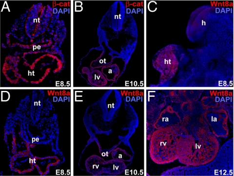
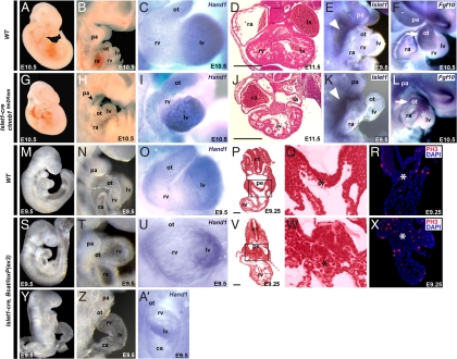
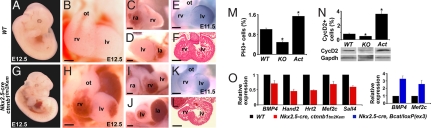
Guiding multipotent cells into distinct lineages and controlling their expansion remain fundamental challenges in developmental and stem cell biology. Members of the Wnt pathway control many pivotal embryonic events, often promoting self-renewal or expansion of progenitor cells. In contrast, canonical Wnt ligands are thought to negatively regulate cardiomyogenesis in several species. However, the cell-autonomous role of canonical Wnt signaling within precardiac mesoderm, through its obligatory transcriptional mediator, beta-catenin, is unknown. Using tissue-specific in vivo genetic manipulation, we found that beta-catenin is required for development of cardiac progenitors and is a positive regulator of proliferative expansion of such progenitor cells. At discrete windows of development in embryonic stem cells, activation of canonical Wnt signaling promoted expansion of cardiac progenitors after initial commitment and was required for cardiac differentiation. Together, these data provide in vivo and in vitro evidence that canonical Wnt signaling promotes the expansion of cardiac progenitors and differentiation of cardiomyocytes.


Conflict of interest statement

The authors declare no conflict of interest.

Figures

Fig. 1.
Fig. 1.
Wnt/β-catenin signaling is active in the developing heart. (A and B) Immunohistochemistry of mouse embryos for β-catenin protein (β-cat, red) at E8.5 (A) and E10.5 (B). (C) Lateral view of whole-mount immunostained E8.5 mouse embryo for Wnt8a (red) protein expression. (D–F) Immunohistochemistry with antibodies against Wnt8a (red) at E8.5 (D), E10.5 (E), and E12.5 (F). DAPI (blue) was used to counterstain the nuclei. All cryosections were transverse. a, atria; h, head; ht, heart tube; la, left atrium; lv, left ventricle; nt, neural tube; ot, outflow tract; pe, pharyngeal region of foregut endoderm; ra, right atrium; and rv, right ventricle.
Fig. 2.
Fig. 2.
β-Catenin is required for expansion of SHF cardiac progenitors. (A–F and M–R) WT embryos. (G–L) Islet1-cre, ctnnb1tm2Kem homozygous embryos. (S–A′) Islet1-cre, β-catenin/loxP(ex3) heterozygous embryos. (A and G) Lateral views of E10.5 embryos. (B and H) Lateral view focused on cardiac and pharyngeal arch (pa) regions showing hypoplasia of pharyngeal arches (pa) (arrowhead) and RV (rv). (C and I) Frontal view of E10.5 hearts hybridized with Hand1 riboprobe marking left ventricle (lv) and outflow tract (ot). (D and J) Transverse sections through the heart. (Scale bars, 250 μm.) (E, F, K, and L) Lateral views of embryos hybridized with SHF markers, Islet1 (R and K, E9.5) and Fgf10 (F and L, E10.5) riboprobes. Arrowheads indicate SHF; arrows indicate Fgf10 in ot. (M, S, and Y) Lateral views of E9.5 embryos. (N, T, and Z) Lateral view focused on cardiac and pa regions showing significantly enlarged rv region. (O, U, and A′) Frontal view of E9.0 hearts hybridized with Hand1 riboprobe showing expanded rv. (P, Q, V, and W) Transverse sections of E9.25 embryos (P and V) with enlargement of boxed areas (Q and W) showing hyperplasia of SHF cardiac progenitors (asterisk) ventral to the pharyngeal region of foregut endoderm (pe). (Scale bars, 50 μm.) (R and X) PH3 immunostaining (red) of transverse sections through the ot region. DAPI was used to counterstain the nuclei. ca, common atrium; la, left atrium; nt, neural tube; ra, right atrium.
Fig. 3.
Fig. 3.
β-Catenin regulates proliferation of ventricular cardiomyocytes. (A–F) WT embryos. (G–L) Nkx2.5-cre, ctnnb1tm2Kem homozygous embryos. (A and G) Lateral embryonic view of E12.5 embryo. (B–D and H–J) Closeup frontal, right lateral, and left lateral views of the heart, respectively. (E and K) In situ hybridization with Hand1 riboprobe. (F and L) Transverse section of E11.5 heart. (M) Percentage of PH3-positive cells in WT, Nkx2.5-cre, ctnnb1tm2Kem homozygous (KO) and Nkx2.5-cre, β-catenin/loxP(ex3) heterozygous (Act) embryos at E11.5. (N) Percentage of Cyclin D2 (CycD2)-positive cells and Western blot of ventricles using CyclinD2 antibody in indicated mutants. GAPDH antibody was used as a control. (O) Quantitative real-time RT-PCR of indicated genes from ventricles of WT and mutant embryos at E12.5 (Left) or E13.5 (Right). ∗, P < 0.01. Error bars indicate standard deviations. la, left atrium; lv, left ventricle; ot, outflow tract; ra, right atrium; rv, right ventricle. (Scale bars, 250 μm.)
Fig. 4.
Fig. 4.
Canonical Wnt signaling regulates cardiac expansion and differentiation in ESCs. (A) Brachyury expression profiles, determined by quantitative real-time PCR (qPCR), of undifferentiated ESCs on day 0 and EBs harvested on days 3, 6, and 9 after early Wnt3a or Dkk-1 treatment compared with control. (B) Beating EBs grown in suspension were counted from days 8 to 12. Percentage of beating EBs after early (days 4–6) or late (days 7–9) treatment with Wnt3a or Dkk-1. Asterisks indicate significant difference in beating percentage of treatment groups versus untreated EBs (P < 0.01). (C) Percentage of Nkx2.5-gfp+ cells at days 6, 9, and 12 in untreated EBs or those treated with Wnt3a or Dkk1 from days 4–6. (Bars represent percent of Nkx2.5-gfp+ cells from 4 × 10⁴ cells analyzed). (D) Immunohistochemistry of day12 EBs with anti-Tropomyosin. (E) Cardiac gene expression after early (days 4–6, Upper) or late (days 7–9, Lower) treatment with Wnt3a or Dkk-1. EBs were harvested on days 3, 6, and 9 (early treatment) or 6, 9, and 12 (late treatment). Fold change in expression of all indicated genes (y axis) in EBs with respect to undifferentiated ESCs was assessed by qPCR. NS, not significant. ∗, P < 0.01.

References

    1. Olson EN. Science. 2006;313:1922–1927. - PMC - PubMed
    1. Srivastava D. Cell. 2006;126:1037–1048. - PubMed
    1. Cai CL, Liang X, Shi Y, Chu PH, Pfaff SL, Chen J, Evans S. Dev Cell. 2003;5:877–889. - PMC - PubMed
    1. Buckingham M, Meilhac S, Zaffran S. Nat Rev Genet. 2005;6:826–835. - PubMed
    1. Kelly RG, Brown NA, Buckingham ME. Dev Cell. 2001;1:435–440. - PubMed

Publication types

LinkOut - more resources