. 2007 Aug;6(4):577-91.
doi: 10.1111/j.1474-9726.2007.00308.x. Epub 2007 Jun 18.

Dynamic assembly of chromatin complexes during cellular senescence: implications for the growth arrest of human melanocytic nevi

Affiliations

Dynamic assembly of chromatin complexes during cellular senescence: implications for the growth arrest of human melanocytic nevi

Debdutta Bandyopadhyay et al. Aging Cell. 2007 Aug.

Abstract

The retinoblastoma (RB)/p16(INK4a) pathway regulates senescence of human melanocytes in culture and oncogene-induced senescence of melanocytic nevi in vivo. This senescence response is likely due to chromatin modifications because RB complexes from senescent melanocytes contain increased levels of histone deacetylase (HDAC) activity and tethered HDAC1. Here we show that HDAC1 is prominently detected in p16(INK4a)-positive, senescent intradermal melanocytic nevi but not in proliferating, recurrent nevus cells that localize to the epidermal/dermal junction. To assess the role of HDAC1 in the senescence of melanocytes and nevi, we used tetracycline-based inducible expression systems in cultured melanocytic cells. We found that HDAC1 drives a sequential and cooperative activity of chromatin remodeling effectors, including transient recruitment of Brahma (Brm1) into RB/HDAC1 mega-complexes, formation of heterochromatin protein 1 beta (HP1 beta)/SUV39H1 foci, methylation of H3-K9, stable association of RB with chromatin and significant global heterochromatinization. These chromatin changes coincide with expression of typical markers of senescence, including the senescent-associated beta-galactosidase marker. Notably, formation of RB/HP1 beta foci and early tethering of RB to chromatin depends on intact Brm1 ATPase activity. As cells reached senescence, ejection of Brm1 from chromatin coincided with its dissociation from HP1 beta/RB and relocalization to protein complexes of lower molecular weight. These results provide new insights into the role of the RB pathway in regulating cellular senescence and implicate HDAC1 as a likely mediator of early chromatin remodeling events.


Figures

Fig. 1
Fig. 1
Overexpression of HDAC1 results in expression of senescent makers in vitro and correlates with senescence of intradermal melanocytic nevi in vivo. (A) HDAC1 is prominently detected in residual, senescent nevi but not in proliferating recurrent nevi. Melan-A highlights recurrent melanocytes in the epidermis and residual dermal nests of melanocytes (left and right column, first row). Note the positive staining of normal intra-epidermal melanocytes with Melan-A along the dermal–epidermal (right column, first row, red arrow). Recurrent melanocytic nevus show confluent proliferation of atypical melanocytes in the epidermis along the dermal–epidermal junction (E) and underlying scar tissue in the dermis (D, left column). Corresponding residual dermal nests of nevomelanocytes are located adjacent to the scar tissue and separated from the area of recurrence (D, right column). Recurrent nevi and scattered basal keratinocytes exhibit cyclin A immunoreactivity whereas the residual nevi are virtually cyclin A negative. There is absence of HDAC1 staining in the recurrent nevus cells along the dermal–epidermal junction (a white line was drawn to highlight a nevus nest) compared to the nuclear expression of HDAC1 in epidermal keratinocytes, which serve as a positive internal control (left column, third row). A higher magnification of recurrent melanocytes confirms that the nuclei of the recurrent nevi are essentially HDAC1 negative. Brown arrows highlight melanin pigment (left column, fourth row). In contrast, residual dermal nests of nevomelanocytes display positive nuclear immunoreactivity for HDAC1 in foci-like structures (right column, third and fourth row and insert). p16INK4a staining demonstrated focal weak immunoreactivity in the recurrent nevi (left column, last row and insert) compared to strong nuclear and cytoplasmic expression staining in the residual nevus cells (right column, last row and insert). Virtually identical results were obtained in two additional sets of recurrent/residual nevi and in three independent intradermal melanocytic nevi. (B) Western blot showing inducible HDAC1 levels by the Tet-On system in UCD-Mel-N melanoma cell line. Numbers above the figure indicate Dox concentrations. (C) Growth curves of UCD-Empty vector control and UCD-HDAC1 cells before and after treatment with Dox. (D) Induction of the SA-β-gal marker in irreversibly quiescent cells 3 week after HDAC1 induction. Software enhancement of color was used to show the cell morphology of the SA-β-gal-negative cells in the inserts of the right and left upper figures. (E) Senescence induced by HDAC1 reproduces features of normal melanocyte senescence. Protein extracts from cells were electrophoresed, transferred to nitrocellulose and probed with the antibodies shown in the figure. PD, population doublings; S, senescent melanocytes (normal melanocytes usually senesce after 12–14 PD).
Fig. 2
Fig. 2
Overexpression of HDAC1 triggers a time-dependent increase in HP1β foci and association of RB and HP1β with chromatin. (A) HDAC1-induced cells show formation of HP1β foci in a dose-dependent manner. The cells were processed for immunocytochemistry 3 days after Dox treatments. (B) HDAC1 induces HP1β foci in a time-dependent manner. The experiments shown herein and in subsequent figures used 2 µg mL−1 Dox to induce HDAC1. (C) Increased affinity of HP1β with senescent chromatin. Left panel: chromatin from senescent melanocytes shows increased resistance to micrococcal nuclease (MNase) digestion (see Experimental procedures). Right panel: an in vitro assay (Experimental procedures) using MNase-digested chromatin with a size range of 146 bp–2 kbp demonstrated that HP1β has increased affinity for senescent chromatin. (D) A cell-based assay (Experimental procedures) confirmed and extended results from the in vitro assay by showing that HDAC1 increases the association of both HP1β and RB with chromatin. M, DNA marker. T.E., total protein extract from uninduced UCD-HDAC1 cells. Right panel: Ponceau red staining indicating protein loading.
Fig. 4
Fig. 4
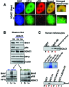
Induction of HDAC1 results in transient association of Brm1 with RB/HP1 complexes. (A) HDAC1 induces the association of HP1β with RB in nuclear foci. The boxes indicate areas zoomed to visualize the foci present in HDAC1-induced but not in control cells. The UCD-HDAC1 cells were treated as described in the legend of Fig. 3. (B) Western blot showing HDAC1 induction and concomitant changes in Brm1, RB, HDAC1, and HP1β protein levels. Immunoprecipitations with HP1β and HDAC1 antibodies suggest that large amounts of Brm1 are transiently associated with RB/HP1β complexes. (C) GST pull-down assays using extracts from proliferating (P) and senescent (S) melanocytes (see text for details). Upper panel: Increased association of HDAC1 with RB in senescent melanocytes. Middle panel: coomassie blue staining of total extracts (TE) and purified GST and GST-RB proteins. Bottom panel: GST-RB pulls down Brm1 from proliferating melanocytes whereas minimal levels are pulled down from senescent cultures. GST-NPC-RB and GST-LXCXE-RB were used as negative controls.
Fig. 3
Fig. 3
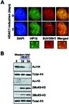
HDAC1 induces features of heterochromatinization. (A) Colocalization of HP1β and SUV39H1 in nuclear foci. UCD-HDAC1 cells were left untreated or treated with 2 µg mL−1 doxycline for 2 days, fixed and processed for immunofluorescence using HP1β and SUV39H1 as described in Experimental procedures. The boxes indicate areas zoomed (bottom figures) to show colocalization of HP1β with SUV39H1. (B) HDAC1 induces deacetylation of H4 and H3, and di- and trimethylation of H3-K9. 2MeK9-H3, dimethylated K9 on histone H3; 3MeK9-H3, trimethylated K9 on histone H3.
Fig. 5
Fig. 5
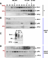
Formation of transient mega-complexes containing Brm1/RB/HP1β precedes growth inhibition. HPLC fractionation, Western blotting and coimmunoprecipitations of control cells (0 day) and cells induced to express HDAC1 for 2 days or 3 weeks (see Experimental procedures). IP, immunoprecipitation.
Fig. 6
Fig. 6
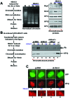
Brm1 ATPase activity is required for the association of RB and HP1β with chromatin and formation of HP1β foci. (A) Transient association of Brm1 but not Brg1 with chromatin 2 days after HDAC1 induction. (B) Transient expression of dnBrm1 prevented the association of RB and HP1β with chromatin. Transfection of wild-type Brg1, a highly homologous Brm1 protein, did not compensate dnBrm1 dominant negative activity. (C) dnBrm1 prevents formation of HP1β foci. UCD-HDAC1 cells were transiently transfected with pcDNA or dnBrm1. Cotransfection with a GFP-expressing plasmid was used to identify cells expressing an empty vector (pcDNA) or dnBrm1. After 8 h, cells were left untreated or treated with Dox for 2 days (to induce HDAC1). Coverslips were processed for immunofluorescence as described in Experimental procedures. The rectangular marquees indicate areas zoomed at the bottom to show that dnBrm1 curtails foci induced by HDAC1 (compare Fig. 6C second panel with Fig. 6C fourth panel).
Fig. 7
Fig. 7
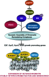
Changes in the stoichiometry of RB/HDAC1 complexes regulate the onset of senescence. We propose a model by which up-regulation of p16INK4a converts RB into its active, nonphosphorylated form which in turns recruits HDAC1, initiating a chain of dynamic chromatin events eventually leading to expansion of heterochromatin and cellular senescence (see text). A dotted arrow indicates that insertion of histone variants, changes in the nucleosome structure and yet to be defined molecular events including post-translational modifications of chromatin regulators contribute to the formation of stable heterochromatization and irreversibility of the senescent state.

Comment in

Similar articles

Cited by

References

    1. Aagaard L, Laible G, Selenko P, Schmid M, Dorn R, Schotta G, Kuhfittig S, Wolf A, Lebersorger A, Singh PB, Reuter G, Jenuwein T. Functional mammalian homologues of the Drosophila PEV-modifier Su(var)3-9 encode centromere-associated proteins which complex with the heterochromatin component M31. EMBO J. 1999;18:1923–1938. - PMC - PubMed
    1. Angelov D, Molla A, Perche PY, Hans F, Cote J, Khochbin S, Bouvet P, Dimitrov S. The histone variant macroH2A interferes with transcription factor binding and SWI/SNF nucleosome remodeling. Mol. Cell. 2003;11:1033–1041. - PubMed
    1. Angus SP, Mayhew CN, Solomon DA, Braden WA, Markey MP, Okuno Y, Cardoso MC, Gilbert DM, Knudsen ES. RB reversibly inhibits DNA replication via two temporally distinct mechanisms. Mol. Cell. Biol. 2004;24:5404–5420. - PMC - PubMed
    1. Bandyopadhyay D, Medrano EE. The emerging role of epigenetics in cellular and organismal aging. Exp. Gerontol. 2003;38:1299–1307. - PubMed
    1. Bandyopadhyay D, Mishra A, Medrano EE. Overexpression of histone deacetylase 1 confers resistance to sodium butyrate-mediated apoptosis in melanoma cells through a p53-mediated pathway. Cancer Res. 2004;64:7706–7710. - PubMed

Publication types

MeSH terms