. 2007 Jul 1;21(13):1687-700.
doi: 10.1101/gad.1552207. Epub 2007 Jun 19.

A functional genomics strategy reveals clockwork orange as a transcriptional regulator in the Drosophila circadian clock

Affiliations

A functional genomics strategy reveals clockwork orange as a transcriptional regulator in the Drosophila circadian clock

Akira Matsumoto et al. Genes Dev. .

Abstract

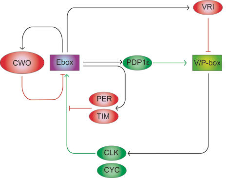
The Drosophila circadian clock consists of integrated autoregulatory feedback loops, making the clock difficult to elucidate without comprehensively identifying the network components in vivo. Previous studies have adopted genome-wide screening for clock-controlled genes using high-density oligonucleotide arrays that identified hundreds of clock-controlled genes. In an attempt to identify the core clock genes among these candidates, we applied genome-wide functional screening using an RNA interference (RNAi) system in vivo. Here we report the identification of novel clock gene candidates including clockwork orange (cwo), a transcriptional repressor belonging to the basic helix-loop-helix ORANGE family. cwo is rhythmically expressed and directly regulated by CLK-CYC through canonical E-box sequences. A genome-wide search for its target genes using the Drosophila genome tiling array revealed that cwo forms its own negative feedback loop and directly suppresses the expression of other clock genes through the E-box sequence. Furthermore, this negative transcriptional feedback loop contributes to sustaining a high-amplitude circadian oscillation in vivo. Based on these results, we propose that the competition between cyclic CLK-CYC activity and the adjustable threshold imposed by CWO keeps E-box-mediated transcription within the controllable range of its activity, thereby rendering a Drosophila circadian clock capable of generating high-amplitude oscillation.


Figures

Figure 1.
Figure 1.
A functional genomics strategy revealed cwo as a clock component. (A) Genome-wide tissue-specific knockdown analysis of clock-controlled genes in Drosophila. UAS-IR transgenic lines to express dsRNA for the target gene under the control of UAS were established (Pili-Floury et al. 2004). Usually two independent insertion lines are established for one target gene. Each of the UAS-IR lines was mated to tim(UAS)-gal4 to drive the expression of dsRNA specifically within clock cells. The locomotor activity of RNAi transgenic flies for 133 candidates among 200 clock-controlled genes was recorded in DD. UAS-TATA sequence or UAS sequence (yellow rectangle), SV40 late polyA site (white rectangle), ∼500-bp fragment of a target gene (red arrow), a tim promoter region (green rectangle), and gal4 gene (pink arrow) are represented. (B) Typical locomotor activity in wild-type, knockdown flies of known clock genes (left) and five new candidates (right). The names of the knocked-down genes are described at each actogram.
Figure 2.
Figure 2.
Temporal and spatial expression pattern of CWO protein. (A) cwo encodes a bHLH-ORANGE family protein. The CWO protein, 685 amino acids in length, is multiply aligned with mouse DEC1, DEC2, and HES5 proteins. Conserved amino acids in bHLH (green) and ORANGE (orange) domains are marked by blue and gaps are represented as “−”. (B) Temporal expression profiles of cwo (red square), per (black circle), and tim (blue triangle) mRNA in wild-type flies under LD and DD. Relative mRNA levels of the indicated genes were measured using a Q-PCR assay. GAPDH2 was used as an internal control. Data were normalized so that the average copy number (n = 2) over 12 time points is 1.0. (C) Spatial expression pattern of CWO in adult brains. (Panel 1) CWO immunostaining (green) in a 12-μm optical Z-stack through the right hemisphere of a wild-type brain. Three clusters of CWO-positive cells are detected in the dorsal brain. A magnified view of the boxed region is shown in panel 1m. Arrows denote DN1s, DN2s, and DN3s. Arrowheads denote additional CWO immunostaining. (Panel 2) PER immunostaining (red) in the same region shown in panel 1. Three clusters of PER-positive cells are detected in the dorsal brain. A magnified view of the boxed region is shown in panel 2m. Arrows denote the same cells described in panel 1m. (Panel 3) CWO and PER coimmunostaining in the same region shown in panels 1 and 2. Colocalization of CWO and PER immunofluorescence in this superimposed dual laser image is shown in yellow. A magnified view of the boxed region is shown in panel 3m. Arrows denote the same cells described in panels 1m and 2m. Arrowheads denote CWO staining in cells not expressing PER. (Panel 4) CWO immunostaining in a 22-μm optical Z-stack through the right hemisphere of a wild-type brain. Three clusters of CWO-positive cells are detected in the lateral brain. A magnified view of the boxed region is shown in panel 4m. Arrows denote clusters of LNds, lLNvs, and sLNvs. Arrowheads denote additional CWO immunostaining. (Panel 5) PER immunostaining in the same region shown in panel 4. Three clusters of PER-positive cells are detected in the lateral brain. A magnified view of the boxed region is shown in panel 5m. Arrows denote the same cells described in panel 4m. (Panel 6) CWO and PER coimmunostaining in the same region shown in panels 4 and 5. Colocalization of CWO and PER immunofluorescence is shown in yellow. A magnified view of the boxed region is shown in panel 6m. Arrows denote the same cells described in panels 4m and 5m. Arrowheads denote CWO staining in cells not expressing PER. (Panel 7) CWO immunostaining in an 8-μm optical Z-stack through the right hemisphere of a f05073 mutant brain. No specific CWO immunostaining is detected. A magnified view of the boxed dorsal brain region is shown in panel 7m. (Panel 8) PER immunostaining in the same region shown in panel 7. PER-positive DNs are detected in the dorsal brain. A magnified view of the boxed region is shown in panel 8m. (Panel 9) CWO and PER coimmunostaining in the same region shown in panels 7 and 8. Only PER immunofluorescence in DNs is seen in this superimposed dual laser image. A magnified view of the boxed region is shown in panel 9m. (Panel 10) CWO immunostaining in a 24-μm optical Z-stack through the right hemisphere of a f05073 mutant brain. No specific CWO immunostaining is detected. A magnified view of the boxed region is shown in panel 10m. (Panel 11) PER immunostaining in the same region shown in panel 10. Three clusters of PER-positive cells are detected in the lateral brain. A magnified view of the boxed region is shown in panel 11m. Arrows denote clusters of LNds, lLNvs, and sLNvs. (Panel 12) CWO and PER coimmunostaining in the same region shown in panels 10 and 11. Only PER immunofluorescence is detected. A magnified view of the boxed region is shown in panel 6m. Arrows denote the same cells described in panel 11m.
Figure 3.
Figure 3.
cwo is directly controlled by circadian clock. (A) The averaged expression levels of cwo, per, and tim mRNA in wild-type and arrhythmic mutant flies (per01 and ClkJrk). Temporal expression profiles in mutant flies are shown in Supplementary Figure 4. (B) Evolutionarily conserved E-box sequences in cwo promoter region. Relative positions of E-boxes to the transcription start site of cwo are indicated. Multiple alignments of seven Drosophila species and its conservation score are represented (University of California at Santa Cruz Genome Browser Database [Hinrichs et al. 2006] version dm2). (C) The cwo promoter is activated by CLK. (Top panel) The three tandem repeats of an 18-bp fragment containing wild-type and mutant E-boxes were fused to a TATA sequence driving a destabilized luciferase (dLuc) reporter gene. Well-conserved base pairs (bold) and mutated base pairs (red) are also indicated. Relative luciferase activities of cwo promoter, wild-type, and mutant E-box reporters in the presence of 0 and 100 ng of Clk are shown in the bottom panel. Signals were normalized with Rluc activity. Error bars represent the SEM (n = 3).
Figure 4.
Figure 4.
CWO protein directly targets known clock genes. (A) Chromosomal view of potential CWO-binding sites (black vertical bar on each chromosome) identified by ChIP assay on a Drosophila genome tiling array. The locations of vri, Pdp1, and cwo are indicated as a green vertical bar. (B) Close view of potential cwo-binding sites on vri, Pdp1, and cwo genes. (Top panel) The fold changes between signal (Flag) and background (V5) were plotted for each probe with the potential binding sites (blue box). (Bottom panel) Gene structures are also indicated. We identified two independent binding sites in the Pdp1 promoter (Pdp1 #1 and #2). (C) Independent verification of ChIP experiments. The relative abundance of immunoprecipitated chromosome regions was measured using a Q-PCR assay. Input product (genomic DNA without ChIP) was used as an internal control. Act57B is used as a negative control. Error bars represent the SEM (n = 3). cwo, Pdp1, and vri promoters are significantly bound by CWO protein. (*) p < 0.05 in t-test. (D) Canonical E-box (CACGTG) recognized by CWO protein. The DNA sequence overrepresented in potential CWO-binding sites was identified by Weeder and drawn by EnoLOGOS.
Figure 5.
Figure 5.
CWO protein regulates transcription of known clock genes. (A) CWO protein suppresses transcription of known clock genes. Relative luciferase activities of per-luc, tim-luc, vri-luc, and Pdp1-luc in the presence of 0 and 100 ng of Clk and 0, 100, 400, and 800 ng of cwo are represented. Signals were normalized with Rluc activity. Error bars represent the SEM (n = 3). (B) Expression of per, tim, vri, and Pdp1 mRNA at the trough level (ZT3) in wild-type (black) and cwo RNAi (red) flies under LD. Relative mRNA levels of each genes were measured with Q-PCR assay. GAPDH2 was used as an internal control. Data were normalized so that the average copy number in cwo RNAi flies is 1.0. Error bars represent the SEM (n = 3). (C) Temporal expression profiles of per, tim, vri, and Pdp1 mRNA in wild-type (black circle) and cwo RNAi transgenic (red rectangle) flies under LD. The relative mRNA levels of each gene were measured with a Q-PCR assay. GAPDH2 was used as an internal control. Error bars represent the SEM (n = 2). We note that sampling time points (ZT1, ZT5, ZT9, ZT13, ZT17, and ZT21) of temporal expression profile are different from ZT3 in B. The expected trough phase calculated from these temporal expression data is almost ZT3, suggesting that the two data are consistent with each other. Since ZT1 and ZT5 are not exactly the trough phase, expression levels of clock genes at these timings are equal or slightly higher in wild-type flies than those of cwo RNAi flies. (D) CWO protein suppresses its own transcription. Relative luciferase activities of cwo-luc, and E-box2-luc in the presence of 0 and 100 ng of Clk and 0, 100, 400, and 800 ng of cwo are represented. Signals were normalized with Rluc activity. Error bars represent the SEM (n = 3). (E) Temporal expression profiles of cwo mRNA in wild-type (black circle) and cwo RNAi transgenic (red rectangle) flies under DD. Relative mRNA levels of each gene were measured with a Q-PCR assay. GAPDH2 was used as an internal control. Error bars represent the SEM (n = 2).
Figure 6.
Figure 6.
Transcriptional circuit underlying Drosophila circadian clock. Ellipsoids represent clock proteins, and rectangles represent time-of-day-specific DNA elements. Activators and repressors are represented in green and red, respectively. CWO protein directly binds to E-boxes and functions as a repressor.

Similar articles

Cited by

References

    1. Allada R., White N.E., So W.V., Hall J.C., Rosbash M., White N.E., So W.V., Hall J.C., Rosbash M., So W.V., Hall J.C., Rosbash M., Hall J.C., Rosbash M., Rosbash M. A mutant Drosophila homolog of mammalian Clock disrupts circadian rhythms and transcription of period and timeless. Cell. 1998;93:791–804. - PubMed
    1. Blau J., Young M.W., Young M.W. Cycling vrille expression is required for a functional Drosophila clock. Cell. 1999;99:661–671. - PubMed
    1. Cawley S., Bekiranov S., Ng H.H., Kapranov P., Sekinger E.A., Kampa D., Piccolboni A., Sementchenko V., Cheng J., Williams A.J., Bekiranov S., Ng H.H., Kapranov P., Sekinger E.A., Kampa D., Piccolboni A., Sementchenko V., Cheng J., Williams A.J., Ng H.H., Kapranov P., Sekinger E.A., Kampa D., Piccolboni A., Sementchenko V., Cheng J., Williams A.J., Kapranov P., Sekinger E.A., Kampa D., Piccolboni A., Sementchenko V., Cheng J., Williams A.J., Sekinger E.A., Kampa D., Piccolboni A., Sementchenko V., Cheng J., Williams A.J., Kampa D., Piccolboni A., Sementchenko V., Cheng J., Williams A.J., Piccolboni A., Sementchenko V., Cheng J., Williams A.J., Sementchenko V., Cheng J., Williams A.J., Cheng J., Williams A.J., Williams A.J., et al. Unbiased mapping of transcription factor binding sites along human chromosomes 21 and 22 points to widespread regulation of noncoding RNAs. Cell. 2004;116:499–509. - PubMed
    1. Ceriani M.F., Hogenesch J.B., Yanovsky M., Panda S., Straume M., Kay S.A., Hogenesch J.B., Yanovsky M., Panda S., Straume M., Kay S.A., Yanovsky M., Panda S., Straume M., Kay S.A., Panda S., Straume M., Kay S.A., Straume M., Kay S.A., Kay S.A. Genome-wide expression analysis in Drosophila reveals genes controlling circadian behavior. J. Neurosci. 2002;22:9305–9319. - PMC - PubMed
    1. Claridge-Chang A., Wijnen H., Naef F., Boothroyd C., Rajewsky N., Young M.W., Wijnen H., Naef F., Boothroyd C., Rajewsky N., Young M.W., Naef F., Boothroyd C., Rajewsky N., Young M.W., Boothroyd C., Rajewsky N., Young M.W., Rajewsky N., Young M.W., Young M.W. Circadian regulation of gene expression systems in the Drosophila head. Neuron. 2001;32:657–671. - PubMed

Publication types

MeSH terms