. 2007 Jun 26;104(26):11014-9.
doi: 10.1073/pnas.0704002104. Epub 2007 Jun 19.

Roles of DNA topoisomerase II isozymes in chemotherapy and secondary malignancies

Affiliations

Roles of DNA topoisomerase II isozymes in chemotherapy and secondary malignancies

Anna M Azarova et al. Proc Natl Acad Sci U S A. .

Abstract

Drugs that target DNA topoisomerase II (Top2), including etoposide (VP-16), doxorubicin, and mitoxantrone, are among the most effective anticancer drugs in clinical use. However, Top2-based chemotherapy has been associated with higher incidences of secondary malignancies, notably the development of acute myeloid leukemia in VP-16-treated patients. This association is suggestive of a link between carcinogenesis and Top2-mediated DNA damage. We show here that VP-16-induced carcinogenesis involves mainly the beta rather than the alpha isozyme of Top2. In a mouse skin carcinogenesis model, the incidence of VP-16-induced melanomas in the skin of 7,12-dimethylbenz[a]anthracene-treated mice is found to be significantly higher in TOP2beta(+) than in skin-specific top2beta-knockout mice. Furthermore, VP-16-induced DNA sequence rearrangements and double-strand breaks (DSBs) are found to be Top2beta-dependent and preventable by cotreatment with a proteasome inhibitor, suggesting the importance of proteasomal degradation of the Top2beta-DNA cleavage complexes in VP-16-induced DNA sequence rearrangements. VP-16 cytotoxicity in transformed cells expressing both Top2 isozymes is, however, found to be primarily Top2alpha-dependent. These results point to the importance of developing Top2alpha-specific anticancer drugs for effective chemotherapy without the development of treatment-related secondary malignancies.


Conflict of interest statement

The authors declare no conflict of interest.

Figures
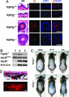
Fig. 1.
Fig. 1.
VP-16 induces melanomas in the skin of DMBA-treated mice. (A) Absence of Top2β in the epidermis (Upper) and hair follicles (Lower) of skin-specific top2β-knockout mice (samples denoted TOP2β). Cryosections of the skin of TOP2β+ and TOP2β mice (8–10 μm thick) were stained with H&E (labeled HE, first column), anti-Top2β antibody (labeled 2β, second column), or DAPI (third column). The merged images of 2β- and DAPI-stained sections are shown in the fourth column (labeled 2β/DAPI). (Scale bars: 10 μm.) (B) PCR-based genotyping of TOP2β+ and TOP2β mice. Genomic DNA samples from tail snippets were genotyped by PCR using primer sets specific for various alleles. Examples are shown here for results with samples from top2β+/flox2 (lane 1), K14-Cre top2β+/flox2 (lane 2), and K14-Cre top2βflox2/flox2 (lane 3) mice. PCR fragments characteristic of the TOP2β+, top2βflox2, top2βΔ2 (top2β), and K14-Cre alleles are depicted; skin cells of K14-Cre top2βflox2/flox2 are phenotypically TOP2β, and those from top2β+/flox2 and K14-Cre top2β+/flox2 mice are TOP2β+ (see the absence of the TOP2β+ fragment in lane 3 and the presence of the same fragment in lanes 1 and 2). (C) VP-16-induced melanomas in the skin of TOP2β+ and skin-specific top2β-knockout mice (TOP2β). Representative photos of DMBA-initiated mice treated with DMSO (vehicle control), VP-16, or phorbol 12-tetradecanoate 13-acetate (TPA) are shown. The blue arrow points to a typical melanoma. (D) Histological and immunohistochemical analyses of melanomas in the mouse skin. Consecutive sections of skin melanomas were stained with either H&E or melanoma-specific antibodies. Representative pictures of H&E staining (Upper) and melanoma antibody staining (Lower) are shown. The red arrow points to a melanoma mass, the blue arrow points to the epidermis, and the green arrow points to a hair follicle. (Scale bars: 100 μm.)
Fig. 2.
Fig. 2.
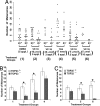
VP-16 induces fewer skin melanomas in the absence of Top2β. (A) The number of melanomas in the skin of each mouse is plotted for various treatment groups. The symbols “2β+” and “2β” denote, respectively, TOP2β+ and skin-specific top2β-knockout (TOP2β) mice. The six groups and their treatment descriptions (see numbers in parenthesis) are indicated at the bottom of the graph. (B and C) The average number of melanomas per mouse for each of the treatment groups denoted by numerals 1–6. Comparing with the DMSO-treated animals (group 1), the differences between the TOP2β+ and TOP2β pairs in the average number of melanomas per mouse are statistically significant for groups 2, 3, 5, and 6 (∗, P < 0.05).
Fig. 3.
Fig. 3.
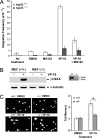
VP-16 induces Top2β-dependent plasmid integration and DSBs. (A) Effect of VP-16 on plasmid integration. SV40-transformed top2β+/− and top2β−/− MEFs were transfected with linearized pUCSV-BSD plasmid DNA in the presence (0.5 μM) or absence of VP-16, and/or the proteasome inhibitor MG132 (2 μM), as indicated. Integration frequency was measured as described in Materials and Methods. (B) VP-16 induces Top2β-dependent formation of phosphorylated histone H2AX (γ-H2AX), a DNA damage signal for DSBs. Primary TOP2β+/+ and top2β−/− MEFs, denoted by +/+ and −/−, respectively, were treated with VP-16 (250 μM, 2 h), and cell lysates were immunoblotted after gel electrophoresis by using anti-γ-H2AX as well as anti-α-tubulin antibody (the latter for loading assessment). The expression levels of Top2α and Top2β in primary TOP2β+/+ and top2β−/− MEFs were similarly assessed (Inset). (C) VP-16 induces DNA DSBs as measured by the neutral comet assay. Primary TOP2β+/+ and top2β−/− MEFs (denoted, respectively, by +/+ and −/− in the figure) were treated with VP-16 (250 μM, 1.5 h). The neutral comet assay was then performed as described in ref. (Left), and the average tail moments were quantified and plotted (Right) (error bars indicate SEM; ∗, P < 0.001 in comparing the +/+ and −/− data).
Fig. 4.
Fig. 4.
VP-16 poisons both Top2 isozymes equally, but Top2β in the trapped Top2β-DNA complexes is preferentially degraded to reveal the hidden DSBs. (A) VP-16 poisons Top2 isozymes equally in vitro. DNA cleavage assays were performed as described in ref. ; VP-16 concentrations were 0, 0, 2.0, 20, and 200 μM in the five samples from left to right. (B) VP-16 effectively traps both Top2α and Top2β cleavage complexes in vivo. Transformed MEFs expressing both Top2 isozymes were treated with VP-16 (0, 10, 50, and 250 μM) for 15 min, and the amounts of Top2 (2α and 2β) cleavage complexes were measured by the band-depletion assay as described in ref. (Upper). The results are quantified and the percentage free Top2 is plotted for each treatment (Lower). VP-16-induced Top2-DNA cleavage complexes are reversed by a further incubation in VP-16-free medium for 50 min (lane 5). (C) VP-16 induces preferential down-regulation of Top2β. Transformed MEFs expressing both Top2 isozymes were treated with VP-16 (50 μM, 2 h) in the presence or absence of the proteasome inhibitor MG132 (2 μM). The DNA cleavage complexes in the treated cells were reversed by an additional incubation in the absence of VP-16 and MG132 (37°C, 30 min), and then alkaline lysis and S7 nuclease treatment (43) were applied. The amounts of Top2 isozymes were measured by Western blotting.
Fig. 5.
Fig. 5.
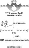
A model for VP-16-induced carcinogenesis. In this model, VP-16 stabilizes the Top2β isozyme covalently trapped on chromosomal DNA, and the trapped isozyme is then preferentially degraded relative to the trapped 2α isozyme by a proteasome pathway; the proteasomal degradation exposes topoisomerase-concealed DSBs for repair by nonhomologous end-joining (NHEJ), which in turn results in DNA sequence rearrangements and carcinogenesis.

References

    1. Felix CA. Biochim Biophys Acta. 1998;1400:233–255. - PubMed
    1. Felix CA. Med Pediatr Oncol. 2001;36:525–535. - PubMed
    1. Pedersen-Bjergaard J, Andersen MK, Christiansen DH, Nerlov C. Blood. 2002;99:1909–1912. - PubMed
    1. Cimino G, Moir DT, Canaani O, Williams K, Crist WM, Katzav S, Cannizzaro L, Lange B, Nowell PC, Croce CM, et al. Cancer Res. 1991;51:6712–6714. - PubMed
    1. Djabali M, Selleri L, Parry P, Bower M, Young BD, Evans GA. Nat Genet. 1992;2:113–118. - PubMed

Publication types

MeSH terms