

Placebo effects on human mu-opioid activity during pain
- PMID: 17578917
- PMCID: PMC1894566
- DOI: 10.1073/pnas.0702413104
Placebo effects on human mu-opioid activity during pain
Abstract
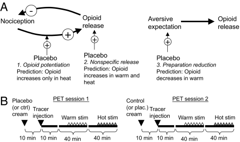
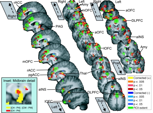
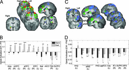
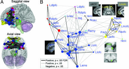
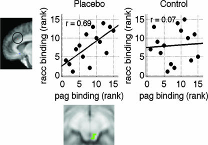
Placebo-induced expectancies have been shown to decrease pain in a manner reversible by opioid antagonists, but little is known about the central brain mechanisms of opioid release during placebo treatment. This study examined placebo effects in pain by using positron-emission tomography with [(11)C]carfentanil, which measures regional mu-opioid receptor availability in vivo. Noxious thermal stimulation was applied at the same temperature for placebo and control conditions. Placebo treatment affected endogenous opioid activity in a number of predicted mu-opioid receptor-rich regions that play central roles in pain and affect, including periaqueductal gray and nearby dorsal raphe and nucleus cuneiformis, amygdala, orbitofrontal cortex, insula, rostral anterior cingulate, and lateral prefrontal cortex. These regions appeared to be subdivided into two sets, one showing placebo-induced opioid activation specific to noxious heat and the other showing placebo-induced opioid reduction during warm stimulation in anticipation of pain. These findings suggest that a mechanism of placebo analgesia is the potentiation of endogenous opioid responses to noxious stimuli. Opioid activity in many of these regions was correlated with placebo effects in reported pain. Connectivity analyses on individual differences in endogenous opioid system activity revealed that placebo treatment increased functional connectivity between the periaqueductal gray and rostral anterior cingulate, as hypothesized a priori, and also increased connectivity among a number of limbic and prefrontal regions, suggesting increased functional integration of opioid responses. Overall, the results suggest that endogenous opioid release in core affective brain regions is an integral part of the mechanism whereby expectancies regulate affective and nociceptive circuits.
Conflict of interest statement
The authors declare no conflict of interest.
Figures
References
-
- De Pascalis V, Chiaradia C, Carotenuto E. Pain. 2002;96:393–402. - PubMed
-
- Clark WC. J Abnorm Psychol. 1969;74:363–371. - PubMed
-
- Price DD, Milling LS, Kirsch I, Duff A, Montgomery GH, Nicholls SS. Pain. 1999;83:147–156. - PubMed
-
- Vase L, Robinson ME, Verne GN, Price DD. Pain. 2003;105:17–25. - PubMed
-
- Voudouris NJ, Peck CL, Coleman G. Pain. 1989;38:109–116. - PubMed
Publication types
MeSH terms
Substances
Grants and funding
LinkOut - more resources
Full Text Sources
Other Literature Sources
Medical
Research Materials
