

IL-4-STAT6 signal transduction-dependent induction of the clinical phase of Sjögren's syndrome-like disease of the nonobese diabetic mouse
- PMID: 17579059
- PMCID: PMC2856075
- DOI: 10.4049/jimmunol.179.1.382
IL-4-STAT6 signal transduction-dependent induction of the clinical phase of Sjögren's syndrome-like disease of the nonobese diabetic mouse
Abstract
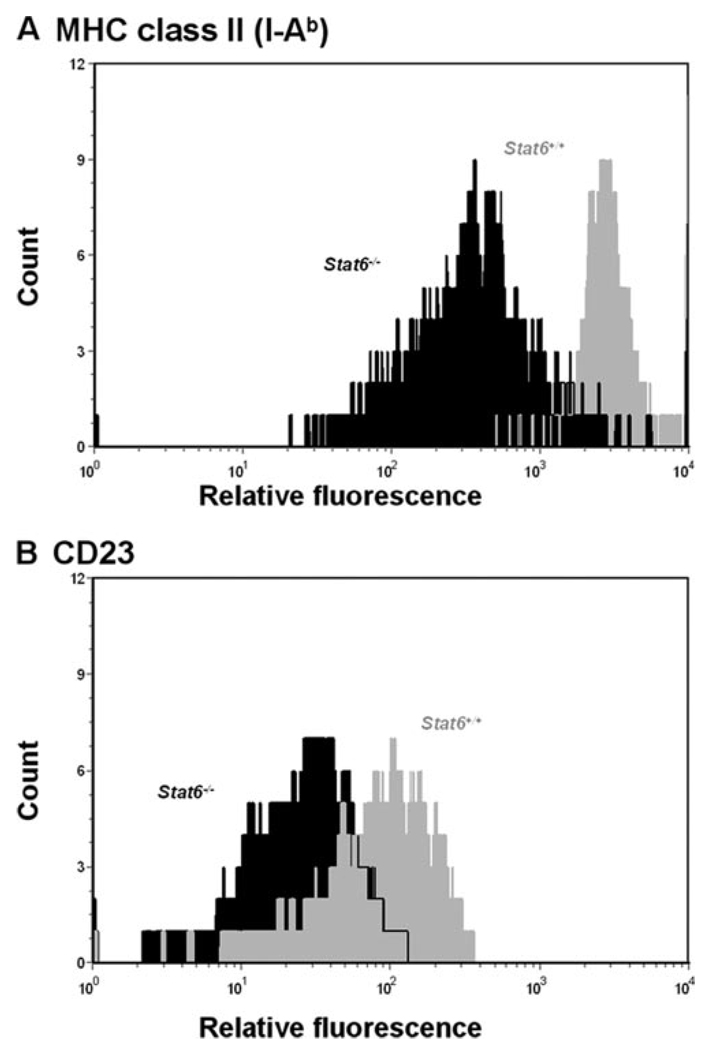
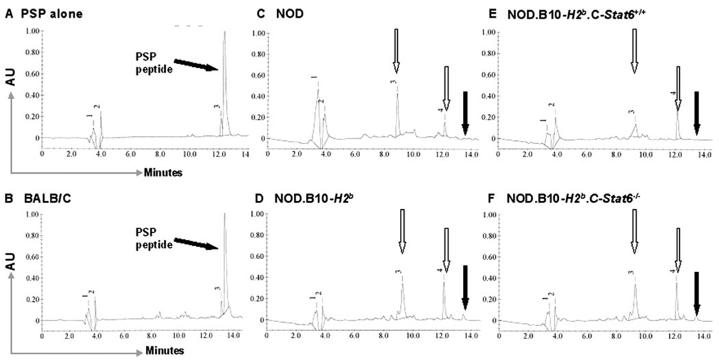
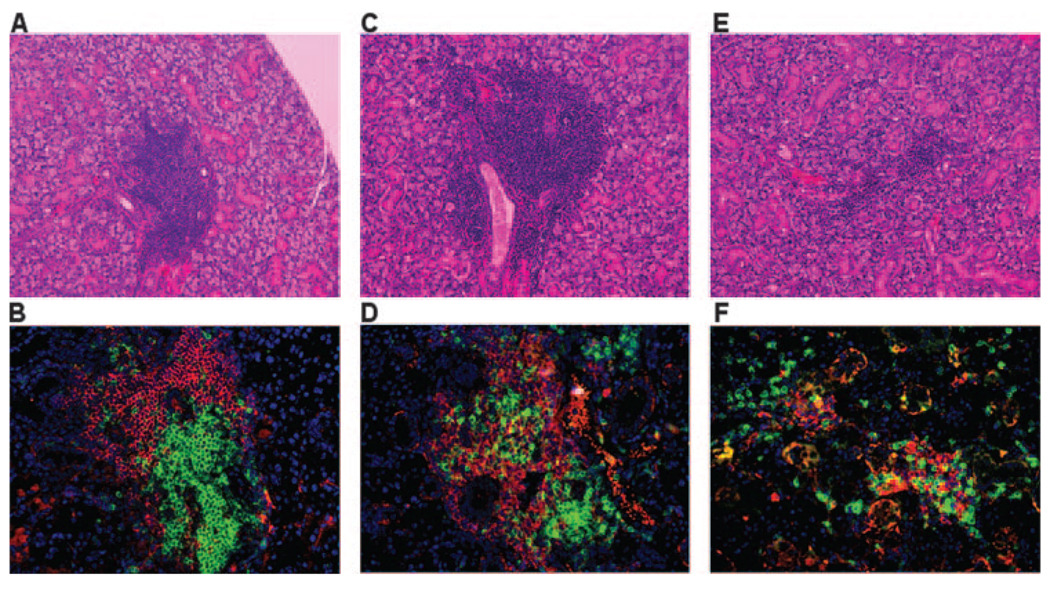
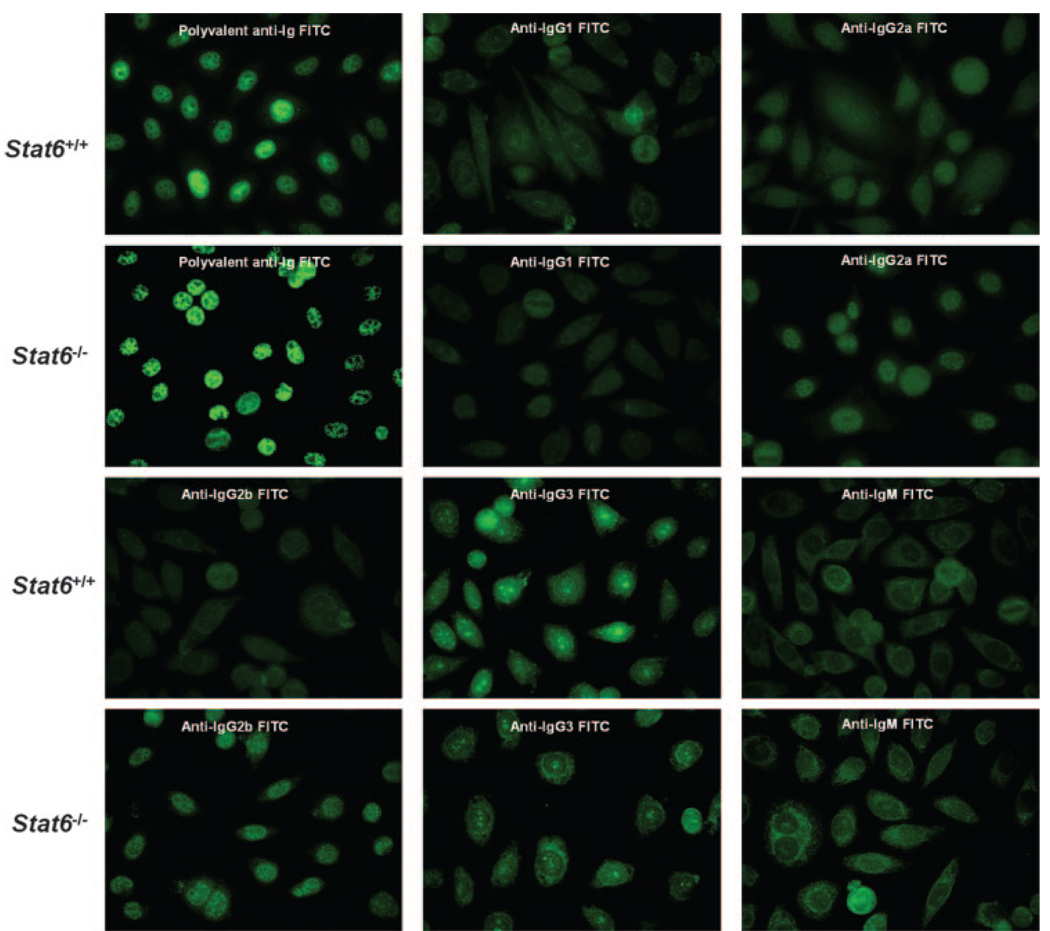
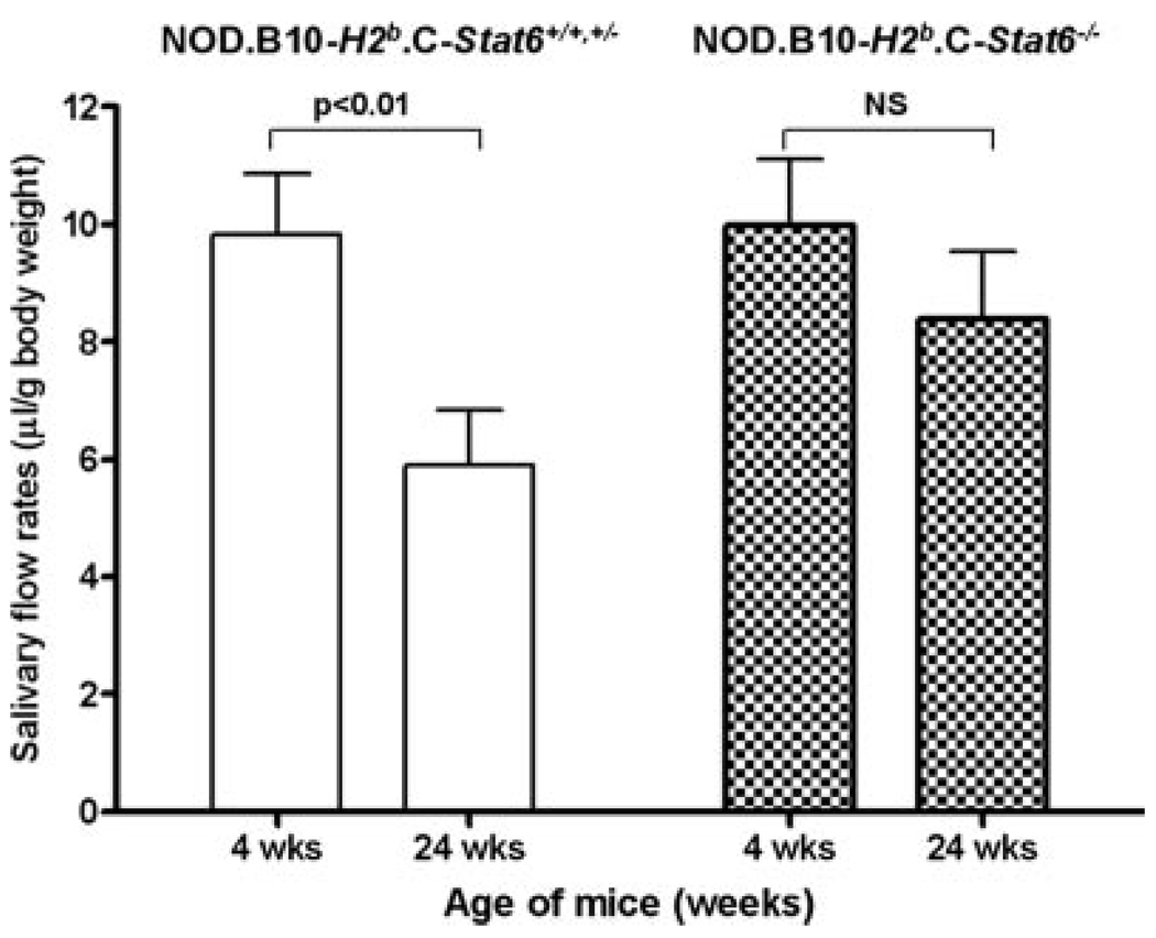
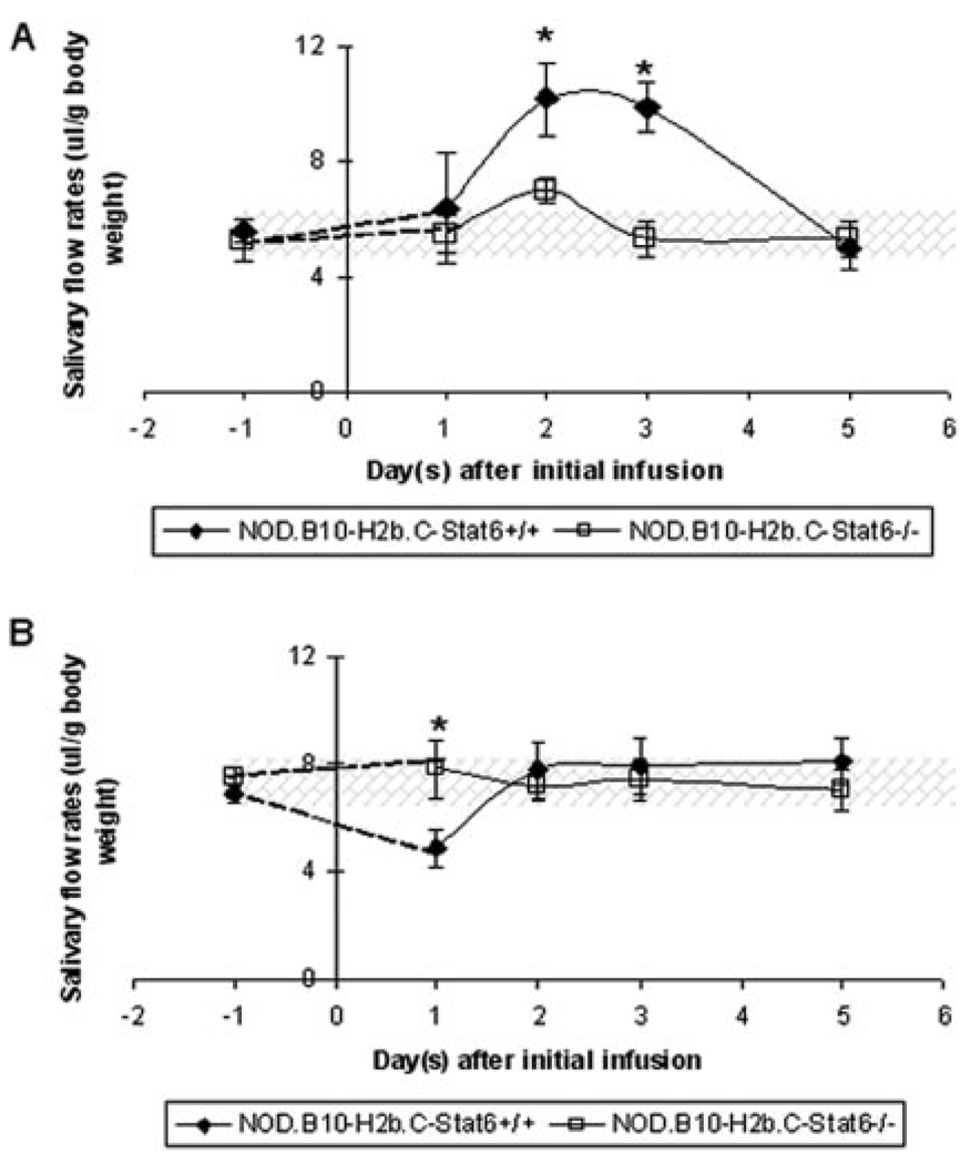
NOD.B10-H2(b) and NOD/LtJ mice manifest, respectively, many features of primary and secondary Sjögren's syndrome (SjS), an autoimmune disease affecting primarily the salivary and lacrimal glands leading to xerostomia (dry mouth) and xerophthalmia (dry eyes). B lymphocytes play a central role in the onset of SjS with clinical manifestations dependent on the appearance of autoantibodies reactive to multiple components of acinar cells. Previous studies with NOD.IL4(-/-) and NOD.B10-H2(b).IL4(-/-) mice suggest that the Th2 cytokine, IL-4, plays a vital role in the development and onset of SjS-like disease in the NOD mouse model. To investigate the molecular mechanisms by which IL-4 controls SjS development, a Stat6 gene knockout mouse, NOD.B10-H2(b).C-Stat6(-/-), was constructed and its disease profile was defined and compared with that of NOD.B10-H2(b).C-Stat6(+/+) mice. As the NOD.B10-H2(b).C-Stat6(-/-) mice aged from 4 to 24 wk, they exhibited leukocyte infiltration of the exocrine glands, produced anti-nuclear autoantibodies, and showed loss and gain of saliva-associated proteolytic enzymes, similar to NOD.B10-H2(b).C-Stat6(+/+) mice. In contrast, NOD.B10-H2(b).C-Stat6(-/-) mice failed to develop glandular dysfunction, maintaining normal saliva flow rates. NOD.B10-H2(b).C-Stat6(-/-) mice were found to lack IgG1 isotype-specific anti-muscarinic acetylcholine type-3 receptor autoantibodies. Furthermore, the IgG fractions from NOD.B10-H2(b).C-Stat6(-/-) sera were unable to induce glandular dysfunction when injected into naive recipient C57BL/6 mice. NOD.B10-H2(b).C-Stat6(-/-) mice, like NOD.B10-H2(b).IL4(-/-) mice, are unable to synthesize IgG1 Abs, an observation that correlates with an inability to develop end-stage clinical SjS-like disease. These data imply a requirement for the IL-4/STAT6-pathway for onset of the clinical phase of SjS-like disease in the NOD mouse model.
Conflict of interest statement
The authors have no financial conflict of interest.
Figures
Similar articles
-
Sjögren's syndrome in the NOD mouse model is an interleukin-4 time-dependent, antibody isotype-specific autoimmune disease.J Autoimmun. 2006 Mar;26(2):90-103. doi: 10.1016/j.jaut.2005.11.004. Epub 2006 Jan 18. J Autoimmun. 2006. PMID: 16413168
-
Role of complement and B lymphocytes in Sjögren's syndrome-like autoimmune exocrinopathy of NOD.B10-H2b mice.Mol Immunol. 2006 Mar;43(9):1332-9. doi: 10.1016/j.molimm.2005.09.003. Epub 2005 Oct 10. Mol Immunol. 2006. PMID: 16221495
-
Time course of cytokine upregulation in the lacrimal gland and presence of autoantibodies in a predisposed mouse model of Sjögren's Syndrome: the influence of sex hormones and genetic background.Exp Eye Res. 2014 Nov;128:15-22. doi: 10.1016/j.exer.2014.09.001. Epub 2014 Sep 11. Exp Eye Res. 2014. PMID: 25218176 Free PMC article.
-
Sjögren's syndrome (SjS)-like disease of mice: the importance of B lymphocytes and autoantibodies.Front Biosci. 2007 Jan 1;12:1767-89. doi: 10.2741/2187. Front Biosci. 2007. PMID: 17127420 Review.
-
Progress in understanding autoimmune exocrinopathy using the non-obese diabetic mouse: an update.Crit Rev Oral Biol Med. 2002;13(1):5-16. doi: 10.1177/154411130201300103. Crit Rev Oral Biol Med. 2002. PMID: 12097234 Review.
Cited by
-
Biomarker profiles in serum and saliva of experimental Sjögren's syndrome: associations with specific autoimmune manifestations.Arthritis Res Ther. 2008;10(1):R22. doi: 10.1186/ar2375. Epub 2008 Feb 20. Arthritis Res Ther. 2008. PMID: 18289371 Free PMC article.
-
Decreased expression of CD200 and CD200 receptor in Alzheimer's disease: a potential mechanism leading to chronic inflammation.Exp Neurol. 2009 Jan;215(1):5-19. doi: 10.1016/j.expneurol.2008.09.003. Epub 2008 Sep 24. Exp Neurol. 2009. PMID: 18938162 Free PMC article.
-
Contributions of Major Cell Populations to Sjögren's Syndrome.J Clin Med. 2020 Sep 22;9(9):3057. doi: 10.3390/jcm9093057. J Clin Med. 2020. PMID: 32971904 Free PMC article. Review.
-
Anti-IL-7 receptor-α treatment ameliorates newly established Sjögren's-like exocrinopathy in non-obese diabetic mice.Biochim Biophys Acta Mol Basis Dis. 2018 Jul;1864(7):2438-2447. doi: 10.1016/j.bbadis.2018.04.010. Epub 2018 Apr 19. Biochim Biophys Acta Mol Basis Dis. 2018. PMID: 29680668 Free PMC article.
-
Myd88 is required for disease development in a primary Sjögren's syndrome mouse model.J Leukoc Biol. 2017 Dec;102(6):1411-1420. doi: 10.1189/jlb.3A0717-311R. Epub 2017 Sep 26. J Leukoc Biol. 2017. PMID: 28951424 Free PMC article.
References
-
- Fox RI, Kang HI. Pathogenesis of Sjögren’s syndrome. Rheum. Dis. Clin. North Am. 1992;18:517–538. - PubMed
-
- Fox RI, Michelson P. Approaches to the treatment of Sjögren’s syndrome. J. Rheumatol. 2000;61 Suppl:15–21. - PubMed
-
- Jonsson R, Haga HJ, Gordon TP. Current concepts on diagnosis, autoantibodies and therapy in Sjögren’s syndrome. Scand. J. Rheumatol. 2000;29:341–348. - PubMed
-
- Vitali C, Bombardieri S, Jonsson R, Moutsopoulos HM, Alexander EL, Carsons SE, Daniels TE, Fox PC, Fox RI, Kassan SS, et al. Classification criteria for Sjögren’s syndrome: a revised version of the European criteria proposed by the American-European Consensus Group. Ann. Rheum. Dis. 2002;61:554–558. - PMC - PubMed
-
- Robinson CP, Brayer J, Yamachika S, Esch TR, Peck AB, Stewart CA, Peen E, Jonsson R, Humphreys-Beher MG. Transfer of human serum IgG to non-obese diabetic Igµ null mice reveals a role for autoantibodies in the loss of secretory function of exocrine tissues in Sjögren’s syndrome. Proc. Natl. Acad. Sci. USA. 1998;95:7538–7543. - PMC - PubMed
Publication types
MeSH terms
Substances
Grants and funding
LinkOut - more resources
Full Text Sources
Medical
Molecular Biology Databases
Research Materials
Miscellaneous
