Comparative Study
. 2007 Jul 1;179(1):455-62.
doi: 10.4049/jimmunol.179.1.455.

Localization and characterization of the mannose-binding lectin (MBL)-associated-serine protease-2 binding site in rat ficolin-A: equivalent binding sites within the collagenous domains of MBLs and ficolins

Affiliations
Comparative Study

Localization and characterization of the mannose-binding lectin (MBL)-associated-serine protease-2 binding site in rat ficolin-A: equivalent binding sites within the collagenous domains of MBLs and ficolins

Umakhanth Venkatraman Girija et al. J Immunol. .

Abstract

Ficolins and mannose-binding lectins (MBLs) are the first components of the lectin branch of the complement system. They comprise N-terminal collagen-like domains and C-terminal pathogen-recognition domains (fibrinogen-like domains in ficolins and C-type carbohydrate-recognition domains in MBLs), which target surface-exposed N-acetyl groups or mannose-like sugars on microbial cell walls. Binding leads to activation of MBL-associated serine protease-2 (MASP-2) to initiate complement activation and pathogen neutralization. Recent studies have shown that MASP-2 binds to a short segment of the collagen-like domain of MBL. However, the interaction between ficolins and MASP-2 is relatively poorly understood. In this study, we show that the MASP-2 binding site on rat ficolin-A is also located within the collagen-like domain and encompasses a conserved motif that is present in both MBLs and ficolins. Characterization of this motif using site-directed mutagenesis reveals that a lysine residue in the X position of the Gly-X-Y collagen repeat, Lys(56) in ficolin-A, which is present in all ficolins and MBLs known to activate complement, is essential for MASP-2 binding. Adjacent residues also make important contributions to binding as well as to MASP activation probably by stabilizing the local collagen helix. Equivalent binding sites and comparable activation kinetics of MASP-2 suggest that complement activation by ficolins and MBLs proceeds by analogous mechanisms.


Figures

Figure 1
Figure 1. Aligned sequences of the collagenous domains of MBLs and ficolins
A, Domain organization of MBLs (top) and ficolins (bottom). The carbohydrate-recognition domain of MBL is labeled as CRD. All mammalian MBLs, but only certain ficolins have an interruption or kink within the Gly-X-Y repeat of the collagen-like domain. B, Collagen helical wheel of the putative MASP-binding motif. C, Aligned sequences of the collagen-like domains of MBLs and ficolins. Numbering of triplets is based on the sequence of rat MBL-A. The putative MASP-binding motif is shaded. Lysine residues within the Y position of the Gly-X-Y repeat are underlined. All such residues are at least partially hydroxylated and glycosylated in rat MBL-A and MBL-C (22, 23). Most of the proline residues in the Y positions are represented as hydroxyproline (O) based on the sequences of rat MBL-A and MBL-C, in which all such residues are at least partially derivatized, except for the proline residue immediately preceding the kink, which is unmodified in each case.
Figure 2
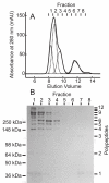
Figure 2. Gel filtration and SDS-polyacrylamide gel electrophoresis of recombinant ficolin-A
A, Gel filtration elution profile of purified rat ficolin-A. Data were fitted to Gaussian curves, using the software Origin (Microcal). The best fit was achieved with four curves, which are shown by dotted lines. Addition of extra parameters did not improve the fits significantly. B, SDS-polyacrylamide gel (4-12% linear gradient gel) under non-reducing conditions. Proteins were stained with Coomassie blue. Numbers above the gel correspond to elution fractions from the gel filtration column (in A above).
Figure 3
Figure 3. Velocity sedimentation ultracentrifugation of ficolin-A
A, Distribution of sedimenting species c(s) analysed using the software package SEDFIT. Minor differences in the proportions of ficolin oligomers from those detected by gel filtration (in Fig. 2) are probably due to differences in the extinction coefficients of oligomers at 280 nm (gel filtration) and 230 nm (sedimentation). B, Example of data from five absorbance scans. Solid lines show the global fit to the data.
Figure 4
Figure 4. Binding of MASP-2 to immobilised ficolin-A analysed by surface plasmon resonance
Comparable amounts of wild type (A) and K56P, M57O ficolin-A (B) (4489 and 4331 response units, respectively) were immobilized on separate channels of the same sensor chip, and MASP-2A was injected at 0.04, 0.08, 0.16 and 0.33 μM. In A, dotted lines show the fit to a two-complex, parallel reaction-binding model and dashed lines show the global fit to a single component binding model. Residuals to the fits are shown in the panels below. The symbol to the left of the residual plots corresponds to 20 response units. The Chi-squared values were 2.54 and 22.1, respectively. Because ficolin-A consists of four different oligomers, more than two different ficolin·MASP complexes are probably formed on the sensor chip. Nevertheless, reasonable fits were achieved using the relatively simple parallel reaction-binding model, implying either that certain ficolin oligomers bind to MASP-2 only weakly or that different oligomeric forms bind to the MASP with comparable kinetics. It has been shown previously that trimers and tetramers of MBL subunits bind to MASP-2 with similar affinities, dimers bind ∼2-fold more weakly whereas single subunits have very low affinities (24). By analogy, we suggest that the observed interactions probably reflect MASP-2 binding to immobilized ficolin dimers, trimers and tetramers.
Figure 5
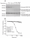
Figure 5. Kinetics of ficolin·MASP-2 activation analysed by SDS-polyacrylamide gel electrophoresis
A, SDS-polyacrylamide gel (4-12% linear gradient gel) of ficolin·MASP-2K complexes, incubated with GlcNAc-Sepharose. Proteins were separated under reducing conditions and were stained with Coomassie blue. The N-terminal fragment of MASP-2K runs as a double band due to differential glycosylation. Activation was measured by quantifying cleavage of the MASP polypeptide. B, Comparison of MASP-2K activation in wild type and ficolin-A K56P, M57O complexes. There was no detectable difference in activation of zymogen MASP-2K in the presence or absence of GlcNAc-Sepharose.
Figure 6
Figure 6. Biophysical properties of ficolin-A mutants
A, Gel filtration of wild type and mutant ficolins. B, Covalent structure of ficolin-A mutants by SDS-polyacrylamide gel electrophoresis, under non-reducing conditions.
Figure 7
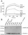
Figure 7. Binding of wild-type and mutant ficolins to MASP-2
A, Surface plasmon resonance of wild-type and mutant ficolins binding to immobilized MASP-2A. Ficolins (at 0.05 mg/ml) were injected sequentially on to the same sensor chip. B, Ficolin·MASP-2A complexes were pelleted on GlcNAc-Sepharose and separated by SDS-polyacrylamide gel electrophoresis (4-12% linear gradient gel), under reducing conditions. Relative amounts of MASP-2 bound to the ficolin (compared to wild-type complexes) were: K56P, M57O - 0.00; O54A - 0.69; K56A - 0.00; M57E - 0.11; M57S - 1.09; P59A - 0.38, from two separate experiments. MASP marker is the starting amount of MASP-2A used in each experiment. MASP - no ficolin shows the amount of MASP-2A associated with GlcNAc-Sepharose in the absence of ficolin, demonstrating that MASP-2 only binds to GlcNAc-Sepharose through its interaction with ficolin-A.
Figure 8
Figure 8. MASP-2 activation by wild-type and mutant ficolins
Proteins were incubated with GlcNAc-Sepharose and were separated by gel electrophoresis.

References

    1. Turner MW. Mannose-binding lectin: the pluripotent molecule of the innate immune system. Immunology Today. 1996;17:532–540. - PubMed
    1. Carroll MC. The complement system in regulation of adaptive immunity. Nat. Immunol. 2004;5:981–986. - PubMed
    1. Garred P, Madsen HO, Balslev U, Hofmann B, Pedersen C, Gerstoft J, Svejgaard A. Susceptibility to HIV infection and progression of AIDS in relation to varient alleles of mannose-binding lectin. Lancet. 1997;349:236–240. - PubMed
    1. Neth O, Hann I, Turner MW, Klein NJ. Deficiency of mannose-binding lectin and burden of infection in children with malignancy: a prospective study. The Lancet. 2001;358:614–618. - PubMed
    1. Kilpatrick DC. Mannan-binding lectin: clinical significance and applications. Biochim. Biophys. Acta. 2002;1572:401–413. - PubMed

Publication types

MeSH terms