. 2007 Jun 20;2(6):e538.
doi: 10.1371/journal.pone.0000538.

Control of pre-mRNA splicing by the general splicing factors PUF60 and U2AF(65)

Affiliations

Control of pre-mRNA splicing by the general splicing factors PUF60 and U2AF(65)

Michelle L Hastings et al. PLoS One. .

Abstract

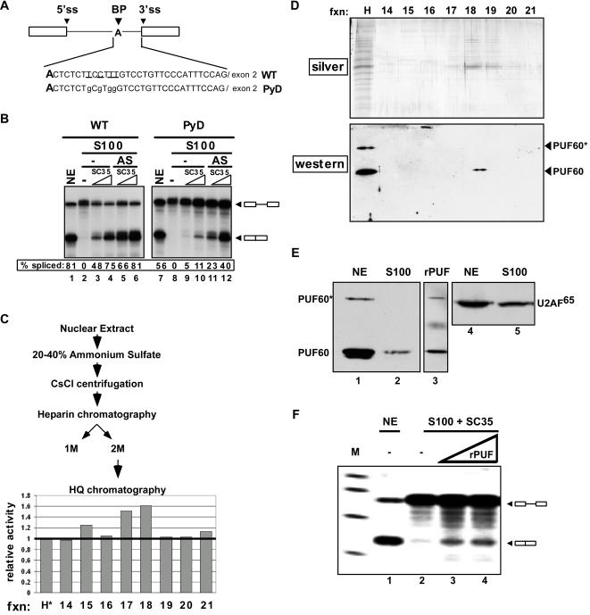
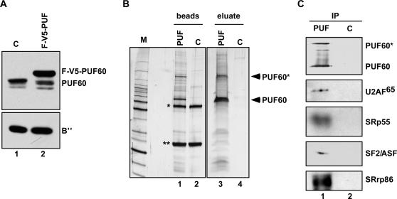
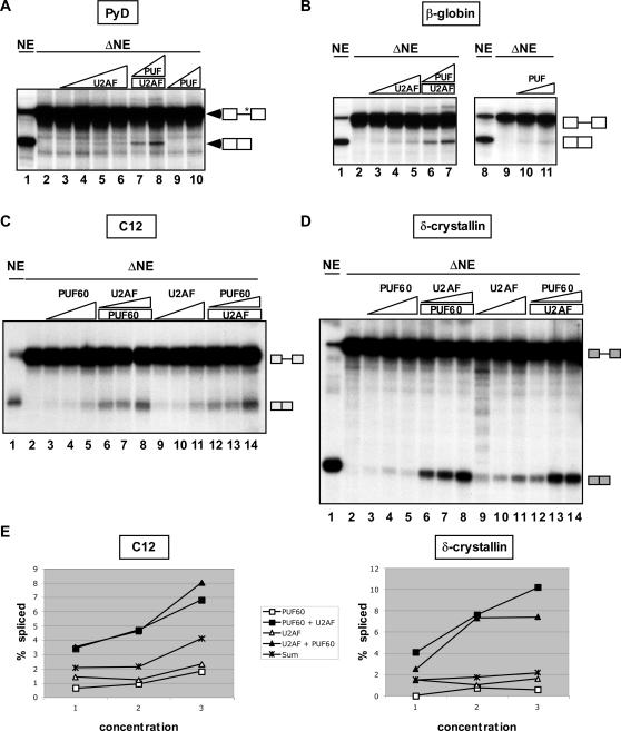
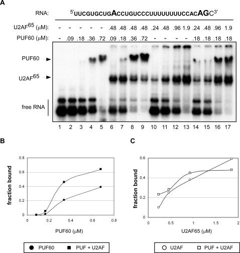
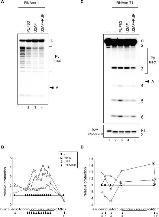
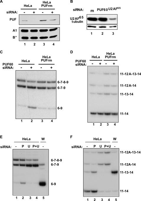
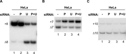
Pre-mRNA splicing is a crucial step in gene expression, and accurate recognition of splice sites is an essential part of this process. Splice sites with weak matches to the consensus sequences are common, though it is not clear how such sites are efficiently utilized. Using an in vitro splicing-complementation approach, we identified PUF60 as a factor that promotes splicing of an intron with a weak 3' splice-site. PUF60 has homology to U2AF(65), a general splicing factor that facilitates 3' splice-site recognition at the early stages of spliceosome assembly. We demonstrate that PUF60 can functionally substitute for U2AF(65)in vitro, but splicing is strongly stimulated by the presence of both proteins. Reduction of either PUF60 or U2AF(65) in cells alters the splicing pattern of endogenous transcripts, consistent with the idea that regulation of PUF60 and U2AF(65) levels can dictate alternative splicing patterns. Our results indicate that recognition of 3' splice sites involves different U2AF-like molecules, and that modulation of these general splicing factors can have profound effects on splicing.


Conflict of interest statement

Competing Interests: The authors have declared that no competing interests exist.

Figures

Figure 1
Figure 1. Identification of PUF60 as a Splicing Activator
(A) Schematic of the wild-type (WT) and mutant (PyD) splicing substrates . Boxes represent exons and lines are introns. Mutations are in lower case and the corresponding nucleotides in the wild-type substrate are underlined. Bold A indicates the branchpoint. (B) In vitro splicing assays were carried out in HeLa nuclear extract (NE) or S100 extract complemented with recombinant SC35, with or without a 20–40% ammonium sulfate (AS) fraction from HeLa NE. Quantitation is shown as the percent of the total RNA that is spliced. (C) Scheme for purification of the complementing activity and quantitation of RESCUE activity in HQ column fractions spanning the peak of activity. Splicing activity was normalized to the input material (H*). (see also Figure S2). H* refers to the HQ column input that was denatured with urea and renatured. (D)(top) Silver-stained SDS-PAGE of HQ peak fractions. (bottom) Western blot analysis of heparin (H) and HQ fractions 14-21. PUF* refers to an SDS-resistant dimer of the protein . (E) Western blot analysis of S100 (lanes 1,4; 4 µl), NE (lanes 2,5; 4 µl), and recombinant PUF60 from E. coli (lane 3) using a PUF60 (lane 1–3) or U2AF65-specific (lane 4,5) antibody. (F) In vitro splicing assay using the PyD substrate in reactions containing S100 extract with SC35 alone (lane 2; 3 µl) and complemented with PUF60 purified from E. coli (lanes 3–4; 1 and 2 µl).
Figure 2
Figure 2. Identification of PUF60-associated Proteins.
(A) Western blot analysis of nuclear extract (NE) prepared from control untransfected HeLa cells (C, lane 1) or cells expressing the FLAG-V5-tagged PUF60 protein (F-V5-PUF, lane 2). Blots were probed with antibodies specific for PUF60 and U2 snRNP B”. (B) Silver-stained 4–20% gradient SDS-PAGE of FLAG-PUF60 (PUF, lanes 1, 3) or control (C, lanes 2,4) immunoprecipitates, either bound to the α-FLAG beads (lanes 1,2) or eluted from the beads with FLAG peptide (lanes 3,4). The heavy chain (*) and light chain (**) from the FLAG antibody are indicated. (C) Western blot analysis of FLAG-PUF60 immunoprecipitates eluted with FLAG peptide.
Figure 3
Figure 3. Cooperative Activity of PUF60 and U2AF65/35 in Splicing.
(A) Complementation of PyD splicing in vitro in nuclear extract depleted of PUF60 and U2AF subunits using poly(U)-Sepharose. PyD pre-mRNA spliced in mock-depleted nuclear extract (NE, lane 1), extract depleted of U2AF subunits and PUF60 (ΔNE, lane 2), depleted extract complemented with U2AF65/35 purified from HEK-293E cells alone (lanes 3–6: 17, 33, 67, and 133 nM final concentration of U2AF65, respectively) or with 67 nM of U2AF65 plus recombinant PUF60 (lanes 7,8: 1.2 and 2.4 µM PUF60) or with PUF60 alone (lanes 9,10: 1.2 and 2.4 µM). (B) Splicing of β-globin intron 1 in nuclear extract (NE, lanes 1, 8), or in poly(U)-depleted extract alone (ΔNE, lanes 2,9). Depleted extract was complemented with purified U2AF65/35 (lanes 3–5: 17, 33, and 133 nM final concentration of U2AF65), with 670 nM of U2AF65 plus recombinant PUF60 (lanes 6,7: 1.2 and 2.4 µM,) or with PUF60 alone (lanes 10,11: 1.2 and 2.4 µM). (C) C12 and (D) δ-crystallin pre-mRNA spliced in nuclear extract (NE, lanes 1), depleted extract (ΔNE, lanes 2) or depleted extract with addition of recombinant PUF60 (lanes 3–5: 1.2, 2.4, and 4.8 µM final concentration; lanes 6–8: 1.2 µM; lanes 12–14, 0.6, 1.2, and 2.4 µM) or purified U2AF65/35 (lanes 6–8: 33, 67, and 133 nM of U2AF65; lanes 9–11: 67, 133, and 200 nM ; lanes 12–14: 670 nM). (E) Quantitation of C12 (left) and δ-crystallin (right) splicing. Splicing was calculated at three concentrations of protein (see C and D). PUF60 corresponds to quantitation of lanes 3–5; PUF60+U2AF65 refers to lanes 6–8; U2AF refers to lanes 9–11; and U2AF+PUF60 corresponds to lanes 12–14. The level of splicing expected if the PUF60 and U2AF activity is additive was calculated as the sum of lanes 3+9, 4+10, and 5+11, respectively (Sum).
Figure 4
Figure 4. Cooperative Binding of U2AF65/35 and PUF60 to the 3′ splice site.
(A) Electrophoretic mobility shift assay using a radiolabeled 34-nt RNA derived from adenovirus major late (AdML) pre-mRNA, and recombinant PUF60 and U2AF65/35. Complexes are indicated on the left. (B) Quantitation of PUF60 binding to AdML RNA represented as the fraction of total labeled RNA bound by the protein. (C) U2AF65 binding represented as the fraction of total labeled RNA bound by the protein.
Figure 5
Figure 5. Footprinting analysis of PUF60 and U2AF65/35 binding
(A) RNase 1 digestion. Labeled RNA was incubated with purified PUF60, U2AF65/35, or both PUF60 and the U2AF heterodimer, digested with RNase and separated by denaturing PAGE. The concentration of PUF60 was 0.72 µM (lane 2), or 0.36 µM (lane 4) and that of U2AF65 was 1.9 µM (lane 3), or 0.96 µM (lane 4). (B) Graphical representation of footprint data in (A). Relative protection is normalized to digestion in the absence of protein (A, lanes 1, 6). The RNA sequence below the plot indicates the position of cleavage (arrowhead). The pyrimidine tract is underlined. (C) RNase T1 digestion. The concentration of PUF60 was 0.36 µM (lanes 7, 9) and that of U2AF65 was 0.48 µM (lanes 8,9). The bands corresponding to the pyrimidine (Py) tract and branchpoint adenosine (A), and full-length RNA (FL) are indicated (see also Berglund et al., 1998). RNase T1 cleavage sites are numbered. A lower exposure of the top of the RNase T1 gel allows visualization of cleavage site 2. (D) Graphical representation of footprinting data in (C). Relative protection is normalized to digestion in the absence of protein (C, lanes 1, 6). The RNA sequence below the plot indicates the position of cleavage (arrowhead). The pyrimidine tract is underlined.
Figure 6
Figure 6. Changes in PUF60 and U2AF65/35 levels regulate alternative splicing in cells.
(A) Western blot analysis of HeLa cells with stable expression of empty vector (HeLa) or PUF60 cDNA with silent mutations that protect transcripts from siRNA-mediated knockdown (HeLa PUFrm). Cells were treated with PUF60 siRNA (+) or mock-treated (−). Blots were probed with antibodies specific for PUF60, hnRNP A1, and U2 B”. (B) Western blot analysis of HeLa cells treated with siRNA specific for PUF60 (lane 2), a U2AF65-specific siRNA (lanes 3 and 4) or mock-treated (lane 1). Antibodies against U2AF65, PUF60, or α-tubulin were used. (C) APP and (D) BIN1 alternative splicing analyzed by RT-PCR with [α-32P]dCTP of endogenous transcripts from stable cell lines mock-treated (lanes 1,3) or treated with PUF60 siRNA (lanes 2,4)(from (A)). (E) APP and (F) BIN1 alternative splicing analyzed by RT-PCR with [α-32P]-dCTP of endogenous transcripts from HeLa cells mock-treated (−) or treated with PUF60 (P), U2AF65 (U) siRNAs, or both (P+U). RT-PCR analysis of APP and BIN1 from untreated Weri-Rb1 (W) cells demonstrates neural splicing patterns.
Figure 7
Figure 7. Complex modulation of alternative splicing by PUF60 and U2AF65.
(A) UBQLN1 exon 8 splicing, (B) SMN2 exon 7 splicing, and (C) MAPT exon 10 splicing analyzed by RT-PCR in the presence of [α-32P]-dCTP of endogenous transcripts from untreated (−) HeLa cells, HeLa cells treated with a PUF60-specific siRNA (P), a U2AF65 siRNA (U), or both siRNAs (P+U). Transcripts including the alternative spliced exon (+) or skipping the exon (Δ) are labeled.

Similar articles

Cited by

References

    1. Tress ML, Martelli PL, Frankish A, Reeves GA, Wesselink JJ, et al. The implications of alternative splicing in the ENCODE protein complement. Proc Natl Acad Sci U S A 2007 - PMC - PubMed
    1. Maniatis T, Tasic B. Alternative pre-mRNA splicing and proteome expansion in metazoans. Nature. 2002;418:236–243. - PubMed
    1. Berglund JA, Abovich N, Rosbash M. A cooperative interaction between U2AF65 and mBBP/SF1 facilitates branchpoint region recognition. Genes Dev. 1998;12:858–867. - PMC - PubMed
    1. Berglund JA, Chua K, Abovich N, Reed R, Rosbash M. The splicing factor BBP interacts specifically with the pre-mRNA branchpoint sequence UACUAAC. Cell. 1997;89:781–787. - PubMed
    1. Zamore PD, Green MR. Identification, purification, and biochemical characterization of U2 small nuclear ribonucleoprotein auxiliary factor. Proc Natl Acad Sci U S A. 1989;86:9243–9247. - PMC - PubMed

Publication types

MeSH terms