. 2007 Jun 20;2(6):e549.
doi: 10.1371/journal.pone.0000549.

Erythropoietin blockade inhibits the induction of tumor angiogenesis and progression

Affiliations

Erythropoietin blockade inhibits the induction of tumor angiogenesis and progression

Matthew E Hardee et al. PLoS One. .

Abstract

Background: The induction of tumor angiogenesis, a pathologic process critical for tumor progression, is mediated by multiple regulatory factors released by tumor and host cells. We investigated the role of the hematopoietic cytokine erythropoietin as an angiogenic factor that modulates tumor progression.

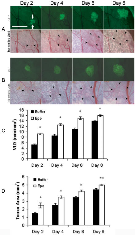
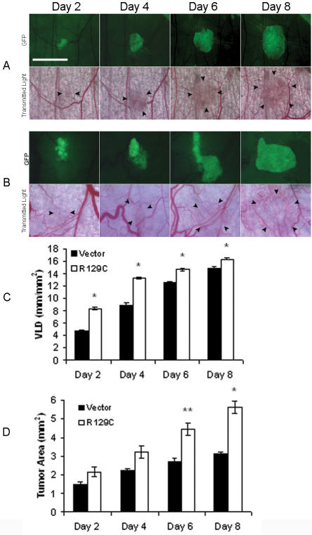
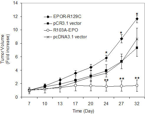
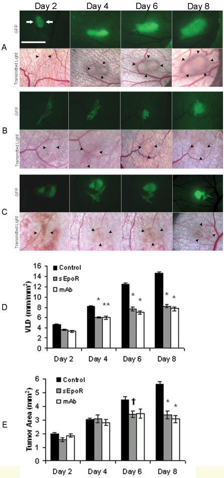
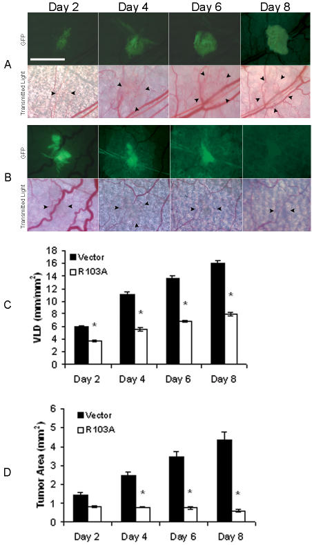
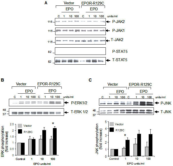
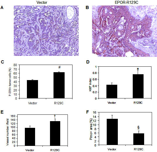
Methodology/principal findings: Fluorescently-labeled rodent mammary carcinoma cells were injected into dorsal skin-fold window chambers in mice, an angiogenesis model that allows direct, non-invasive, serial visualization and real-time assessment of tumor cells and neovascularization simultaneously using intravital microscopy and computerized image analysis during the initial stages of tumorigenesis. Erythropoietin or its antagonist proteins were co-injected with tumor cells into window chambers. In vivo growth of cells engineered to stably express a constitutively active erythropoietin receptor EPOR-R129C or the erythropoietin antagonist R103A-EPO were analyzed in window chambers and in the mammary fat pads of athymic nude mice. Co-injection of erythropoietin with tumor cells or expression of EPOR-R129C in tumor cells significantly stimulated tumor neovascularization and growth in window chambers. Co-injection of erythropoietin antagonist proteins (soluble EPOR or anti-EPO antibody) with tumor cells or stable expression of antagonist R103A-EPO protein secreted from tumor cells inhibited angiogenesis and impaired tumor growth. In orthotopic tumor xenograft studies, EPOR-R129C expression significantly promoted tumor growth associated with increased expression of Ki67 proliferation antigen, enhanced microvessel density, decreased tumor hypoxia, and increased phosphorylation of extracellular-regulated kinases ERK1/2. R103A-EPO antagonist expression in mammary carcinoma cells was associated with near-complete disruption of primary tumor formation in the mammary fat pad.

Conclusions/significance: These data indicate that erythropoietin is an important angiogenic factor that regulates the induction of tumor cell-induced neovascularization and growth during the initial stages of tumorigenesis. The suppression of tumor angiogenesis and progression by erythropoietin blockade suggests that erythropoietin may constitute a potential target for the therapeutic modulation of angiogenesis in cancer.


Conflict of interest statement

Competing Interests: The authors have declared that no competing interests exist.

Figures

Figure 1
Figure 1. Stimulation of tumor angiogenesis and growth in response to rEPO treatment.
Representative images of dorsal skin-fold window chambers implanted with R3230-GFP cells and local administration of (A) control buffer, or (B) recombinant EPO are shown (total n = 8 animals/group). Fluorescent (FITC) and transmitted light images were acquired serially on postoperative days 2, 4, 6, and 8. Scale bar = 2.5 mm. GFP-positive tumor area (green fluorescence) is indicated by white arrows and tumor-associated vasculature in transmitted light images is outlined by black arrowheads. (C) Quantification of tumor neovascularization as measured by vascular length density (VLD) in window chambers treated with EPO or control buffer revealed increased angiogenesis in EPO-treated chambers compared to controls, *P<0.001. (D) Quantification of tumor growth revealed significantly increased tumor area in EPO-treated chambers compared to controls, *P<0.001; ** P<0.01.
Figure 2
Figure 2. Stimulation of tumor angiogenesis and growth in response to tumor cell EPOR-R129C expression.
Representative images of dorsal skin-fold window chambers implanted with R3230-GFP cells transfected with (A) empty pCR3.1 vector control, or (B) EPOR-R129C expression vector are shown. Scale bar = 2.5 mm. Two independent single cell clones each of empty pCR3.1 vector and EPOR-R129C-transfected cells were tested for a total of 7 animals in each group. (C) Quantification of tumor neovascularization in window chambers implanted with EPOR-R129C or empty vector-transfected R3230-GFP cells revealed significantly increased VLD in EPOR-R129C expressing group compared to vector controls, *P<0.001. (D) Quantification of tumor growth in window chambers implanted with EPOR-R129C or vector transfected R3230-GFP cells revealed increased tumor area in EPOR-R129C expressing group compared to vector controls, *P<0.001; **P<0.01 (n = 7 animals/group).
Figure 3
Figure 3. In vivo tumor growth of R3230 mammary carcinoma cells in the mammary fat pad of nude mice.
Stably transfected R3230-GFP cells were injected into the mammary fat pad of mice, tumor volumes were measured and expressed as fold-increase of palpable tumor size at day 7. Three independent single cell clones of each cell line were analyzed and empty vector-transfected cells (pCR3.1 and pcDNA3.1) served as negative controls. Expression of EPOR-R129C was associated with a significant increase in tumor growth rate compared to pCR3.1 vector controls (*P<0.001, n = 19 tumors in EPOR-R129C and n = 16 in pCR3.1 group). No significant tumor growth was observed in animals injected with cells expressing R103A-EPO antagonist (n = 14) compared to pcDNA3.1 vector transfected cells (**P<0.001, n = 15 tumors in pcDNA3.1 group).
Figure 4
Figure 4. Erythropoietin blockade using soluble EPOR or anti-EPO mab suppress tumor angiogenesis and delay growth.
(A–C) Representative images of window chambers implanted with R3230-GFP cells co-injected with (A) control buffer or protein, (B) sEPOR, or (C) anti-EPO mab. Fluorescent and transmitted light images were acquired serially on postoperative days 2, 4, 6, and 8. Scale bar = 2.5 mm. (D) Quantification of tumor neovascularization revealed significantly decreased angiogenesis in sEPOR and mab-treated chambers compared to controls. Control buffer was PBS (n = 3) and control protein was mouse IgG1 (n = 4), *P<0.001; **P<0.01, (n = 7 animals/group). (E) Quantification of tumor growth revealed significantly decreased tumor area in sEPOR and mab-treated chambers compared to controls, *P<0.001; P<0.05, (n = 7 animals/group).
Figure 5
Figure 5. Expression of R103A-EPO antagonist inhibits induction of tumor angiogenesis and disrupts primary tumor growth.
Representative images of window chambers implanted with R3230-GFP cells transfected with (A) empty pcDNA3.1 vector, or (B) R103A-EPO antagonist expression vector. Note the constriction and near-complete disappearance of blood vessels in the areas surrounding the tumor which was observed in all window chambers injected with cells expressing R103A-EPO. Two independent single cell clones each of empty pcDNA3.1 vector or R103A-EPO-transfected cells were analyzed for a total of 7 animals in each group. (C) Quantification of tumor neovascularization revealed decreased angiogenesis in window chambers implanted with R103A-EPO secreting cells compared to vector controls, *P<0.001. (D) Quantification of tumor growth in window chambers implanted with R103A-EPO or vector transfected R3230-GFP cells. Compared to vector controls, decreased tumor area was observed in R103A-EPO expressing group with near-complete disappearance of tumor cells at day 8 in all chambers, *P<0.001 (n = 7).
Figure 6
Figure 6. Increased EPO-induced phosphorylation of ERK1/2 and JNK in mammary carcinoma cells expressing EPOR-R129C.
R3230-GFP cells transfected with empty pCR3.1 vector or EPOR-R129C were either left untreated as controls (C) or incubated with the indicated concentration of rEPO for 10 minutes. Whole cell lysates were analyzed by Western blotting using antibodies against phosphorylated or total proteins to detect (A) JAK2, JAK1 and STAT-5, (B) ERK1/2, and (C) JNK. Representative blots are shown. The phosphorylated (P) and total (T) proteins are indicated by the arrows. Comparable sample loading and protein integrity were confirmed by stripping the blots and hybridizing to respective antibodies to detect total protein amounts in the samples. The relative positions of the molecular weight markers are indicated in each blot. Quantitative representation of phosphorylated proteins using densitometry normalized to total protein levels are shown below the blots for ERK1/2 and JNK (n =  3 independent experiments using 3 different single cell clones, (*P<0.05).
Figure 7
Figure 7. Effect of tumor EPOR-R129C expression on in vivo ERK1/2 phosphorylation, Ki67 proliferation antigen expression, microvessel density and hypoxia.
Sections of pCR3.1 vector control and EPOR-R129C mammary fat pad tumors were analyzed by immunocytochemistry as described in Materials and Methods. (A–C) Phospho-ERK, (D) Ki67, (E) CD31, and (F) Pimonidazole hypoxia marker. #P<0.0001;*P = 0.0006; †P = 0.0008; § P = 0.026.

References

    1. Huang Q, Shan S, Braun RD, Lanzen J, Anyrhambatla G, et al. Noninvasive visualization of tumors in rodent dorsal skin window chambers. Nat Biotechnol. 1999;17:1033–1035. - PubMed
    1. Li CY, Shan S, Huang Q, Braun RD, Lanzen J, et al. Initial stages of tumor cell-induced angiogenesis: evaluation via skin window chambers in rodent models. J Natl Cancer Inst. 2000;92:143–147. - PubMed
    1. Folkman J. Incipient angiogenesis. J Natl Cancer Inst. 2000;92:94–95. - PubMed
    1. Cao Y, Li CY, Moeller BJ, Yu D, Zhao Y, et al. Observation of incipient tumor angiogenesis that is independent of hypoxia and hypoxia inducible factor-1 activation. Cancer Res. 2005;65:5498–5505. - PubMed
    1. Shan S, Robson ND, Cao Y, Qiao T, Li CY, et al. Responses of vascular endothelial cells to angiogenic signaling are important for tumor cell survival. Faseb J. 2004;18:326–328. - PubMed

Publication types

MeSH terms