. 2008 Jan;57(1):43-52.
doi: 10.1007/s00262-007-0348-6. Epub 2007 Jun 20.

A bispecific diabody directed against prostate-specific membrane antigen and CD3 induces T-cell mediated lysis of prostate cancer cells

Affiliations

A bispecific diabody directed against prostate-specific membrane antigen and CD3 induces T-cell mediated lysis of prostate cancer cells

P Bühler et al. Cancer Immunol Immunother. 2008 Jan.

Abstract

Background: Although cancer of the prostate is one of the most commonly diagnosed cancers in men, no curative treatment currently exists after its progression beyond resectable boundaries. Therefore, new agents for targeted treatment strategies are needed. Cross-linking of tumor antigens with T-cell associated antigens by bispecific monoclonal antibodies have been shown to increase antigen-specific cytotoxicity in T-cells. Since the prostate-specific membrane antigen (PSMA) represents an excellent tumor target, immunotherapy with bispecific diabodies could be a promising novel treatment option for prostate cancer.

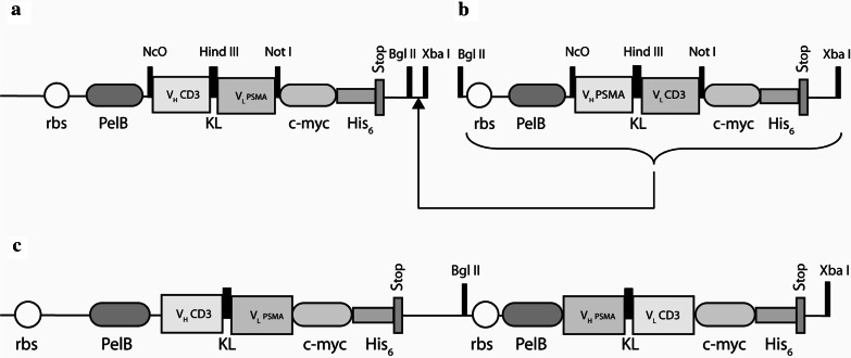
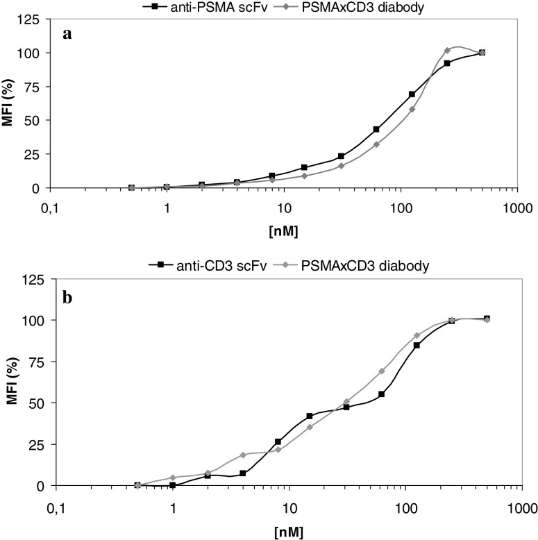
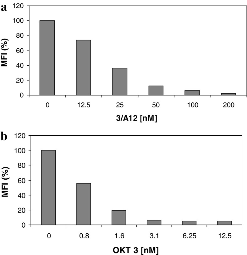
Methods: A heterodimeric diabody specific for human PSMA and the T-cell antigen CD3 was constructed from the DNA of anti-CD3 and anti-PSMA single chain Fv fragments (scFv). It was expressed in E. coli using a vector containing a bicistronic operon for co-secretion of the hybrid scFv V(H)CD3-V(L)PSMA and V(H)PSMA-V(L)CD3. The resulting PSMAxCD3 diabody was purified from the periplasmic extract by immobilized metal affinity chromatography (IMAC). The binding properties were tested on PSMA-expressing prostate cancer cells and PSMA-negative cell lines as well as on Jurkat cells by flow cytometry. For in vitro functional analysis, a cell viability test (WST) was used. For in vivo evaluation the diabody was applied together with human peripheral blood lymphocytes (PBL) in a C4-2 xenograft-SCID mouse model.

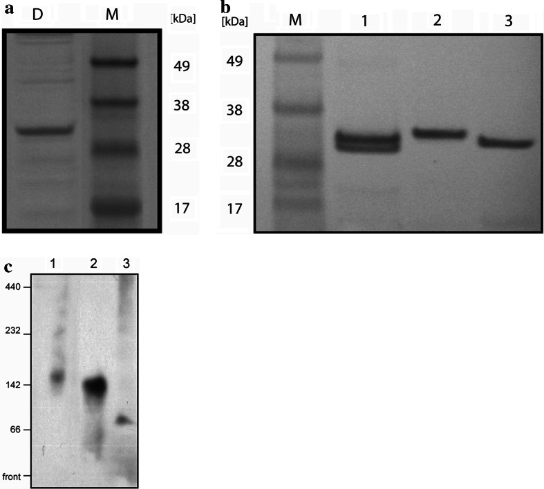
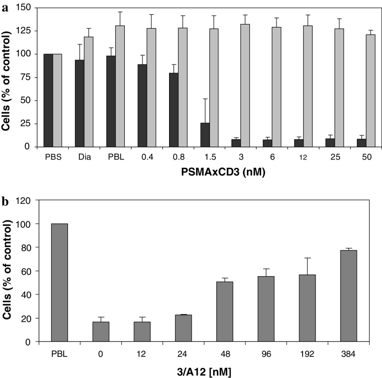
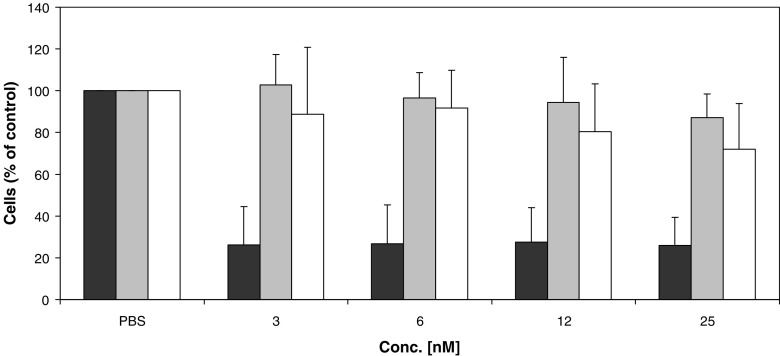
Results: By Blue Native gel electrophoresis, it could be shown that the PSMAxCD3 diabody is mainly a tetramer. Specific binding both to CD3-expressing Jurkat cells and PSMA-expressing C4-2 cells was shown by flow cytometry. In vitro, the diabody proved to be a potent agent for retargeting PBL to lyze C4-2 prostate cancer cells. Treatment of SCID mice inoculated with C4-2 tumor xenografts with the diabody and PBL efficiently inhibited tumor growth.

Conclusions: The PSMAxCD3 diabody bears the potential for facilitating immunotherapy of prostate cancer and for the elimination of minimal residual disease.


Figures

Fig. 1
Fig. 1
Construction scheme for the PSMAxCD3 diabody plasmid (c) from the hetero scFv VHCD3–VLPSMA (a) and VHPSMA-VLCD3 (b). The positions of the sequences of the variable regions of the light and heavy chains of the anti-PSMA-scFv and the anti-CD3-scFv, the ribosome binding sites (rbs), pelB leader sequences (pelB), c-myc epitopes (c-myc), hexa-histidine tags (His 6) and stop codons (stop) are indicated
Fig. 2
Fig. 2
a SDS-PAGE of the PSMAxCD3 diabody (lane D). The gel was stained with Coomassie brilliant blue. Lane M contains a molecular weight marker. b Gel electrophoresis under denaturing, reducing conditions of PSMAxCD3 diabody (lane 1), anti-PSMA scFv (lane 2) and anti-CD3 scFv (lane 3). Proteins were subjected to reducing SDS-PAGE and transferred to a Western blot membrane. The blot was developed with peroxidase-coupled anti-c-myc-mAb, and 3,3′-diamino-benzidine. Lane M contains a prestained molecular weight marker. c Gel electrophoresis under native conditions of PSMAxCD3 diabody (lane 1), anti-PSMA scFv (lane 2) and anti-CD3 scFv (lane 3). Proteins were separated by BN-PAGE and Western blotted with the peroxidase-coupled anti-c-myc-mAb and the ECL detection system. The molecular weights of the native protein standards are indicated on the left in kilo Daltons
Fig. 3
Fig. 3
Binding of anti-PSMA scFv (a), PSMAxCD3 diabody (b), hybrid scFv VHPSMA-VLCD3 (c) and hybrid scFv VHCD3–VLPSMA (d) to PSMA-positive C4-2 cells. Binding of anti-CD3 scFv (e), PSMAxCD3 diabody (f), hybrid scFv VHPSMA-VLCD3 (g) and hybrid scFv VHCD3–VLPSMA (h) to CD3-positive Jurkat cells. Cells were stained with diabody or scFv at 300 nM, mouse-anti-c-myc mAb and goat anti-mouse Ig-RPE. Histograms represent logarithms of PE-fluorescence on flow cytometer. Negative control (gray histograms) with mouse-anti-c-myc mAb, and goat anti-mouse Ig-RPE
Fig. 4
Fig. 4
Saturation curves of the PSMAxCD3 diabody and the parental anti-PSMA scFv to PSMA-positive C4-2 cells (a) and of PSMAxCD3 diabody and parental anti-CD3 scFv to CD3-positive Jurkat cells (b) at concentrations between 0.5 and 500 nM
Fig. 5
Fig. 5
Inhibition of the binding of PSMAxCD3 diabody to C4-2 cells by parental mAb 3/A12 (a) and to Jurkat cells by parental mAb OKT3 (b). Cells were preincubated with the mAb at different concentrations (5–200 nM) and then stained with PSMAxCD3 diabody at 200 nM, rat anti-c-myc mAb and goat anti-rat IgG(H + L)-RPE
Fig. 6
Fig. 6
a Effects of treatment of PSMA-positive C4-2 cells (black) and PSMA-negative DU 145 cells (gray) with PSMAxCD3 diabody and PBL effector cells (effector:target ratio 10:1). Cell viability was measured in WST assays after 48 h incubation as described. The results are expressed as the percentage of viable cells compared to the control (= cells incubated with PBS). Additionally, the data of treatment with diabody alone at 15 nM without PBL (Dia) and the data of treatment with PBL alone without diabody (PBL) are given. The results were corrected for the background of the effector cells. Data represent mean values of determinations with three different blood donors each in triplicate. Standard deviations (SD) are indicated by the error bars. b Blocking of the cytotoxic activity of the PSMAxCD3 diabody by an excess of parental anti-PSMA mAb. C4-2 cells were incubated with PBL and diabody at 12 nM together with parental mAb 3/A12 at different concentrations. Cell viability was measured in WST assays after 48 h incubation. Determination was with three different donors. SD are indicated by the error bars
Fig. 7
Fig. 7
Effects of treatment of PSMA-positive C4-2 cells with PSMAxCD3 diabody (black) or anti-PSMA scFv (gray) or anti-CD3 scFv (white) and PBL. (WST assay, E:T ratio 10:1, incubation time 48 h). The results are expressed as the percentage of viable cells compared to the control (= cells treated with PBS). Data represent mean values of three determinations with different donors. SD are indicated by the error bars
Fig. 8
Fig. 8
Treatment of C4-2 xenografted SCID mice with PSMAxCD3 diabody. Mice were inoculated with 2 × 106 C4-2 cells on day 0 and treated as follows: group a: 5 × 106 human PBL on day 6 after tumor inoculation and additionally 10 μg PSMAxCD3 diabody on day 6, 7, 10, 11 and 12; group b: 5 × 106 human PBL on day 6; group c: PBS vehicle. The mean tumor volume is shown for each group. SD are indicated by the error bars

References

    1. Bohlen H, Hopff T, Manzke O, Engert A, Kube D, Wickramanayake PD, Diehl V, Tesch H. Lysis of malignant B cells from patients with B-chronic lymphocytic leukemia by autologous T cells activated with CD3 × CD19 bispecific antibodies in combination with bivalent CD28 antibodies. Blood. 1993;82:1803–1812. - PubMed
    1. Bolhuis RL, Hoogenboom HR, Gratama JW. Targeting of peripheral blood T lymphocytes. Springer Semin Immunopathol. 1996;18:211–226. doi: 10.1007/BF00820667. - DOI - PubMed
    1. Canevari S, Stoter G, Arienti F, Bolis G, Colnaghi MI, Di Re EM, Eggermont AM, Goey SH, Gratama JW, Lamers CH, et al. Regression of advanced ovarian carcinoma by intraperitoneal treatment with autologous T lymphocytes retargeted by a bispecific monoclonal antibody. J Natl Cancer Inst. 1995;87:1463–1469. doi: 10.1093/jnci/87.19.1463. - DOI - PubMed
    1. Chambers CA, Allison JP. Costimulatory regulation of T-cell function. Curr Opin Cell Biol. 1999;11:203–210. doi: 10.1016/S0955-0674(99)80027-1. - DOI - PubMed
    1. Chang SS, Reuter VE, Heston WD, Bander NH, Grauer LS, Gaudin PB. Five different anti-prostate-specific membrane antigen (PSMA) antibodies confirm PSMA expression in tumor-associated neovasculature. Cancer Res. 1999;59:3192–3198. - PubMed

Publication types