

Preservation of glial cytoarchitecture from ex vivo human tumor and non-tumor cerebral cortical explants: A human model to study neurological diseases
- PMID: 17580092
- PMCID: PMC2744592
- DOI: 10.1016/j.jneumeth.2007.05.008
Preservation of glial cytoarchitecture from ex vivo human tumor and non-tumor cerebral cortical explants: A human model to study neurological diseases
Abstract
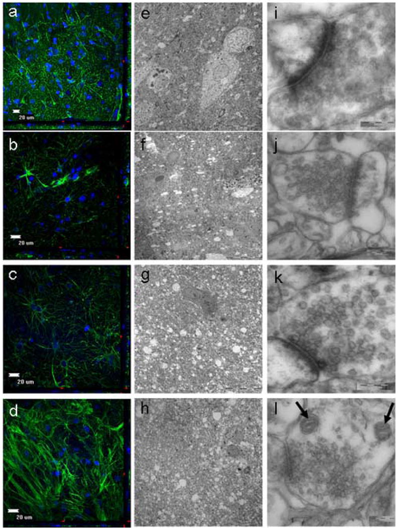
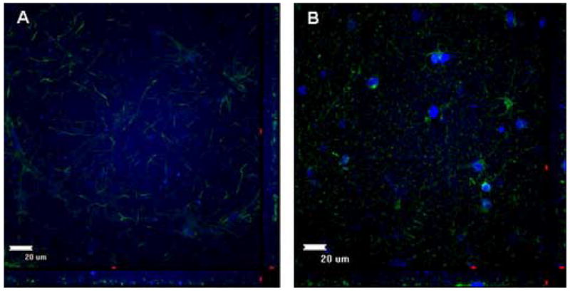
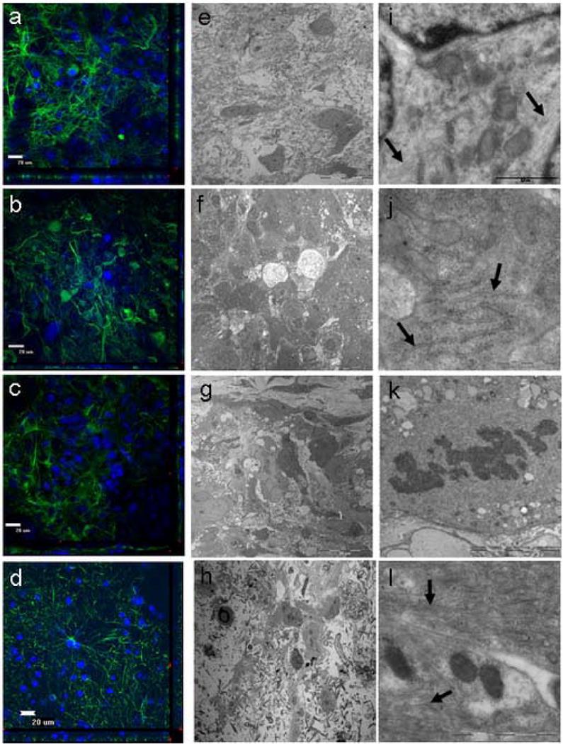
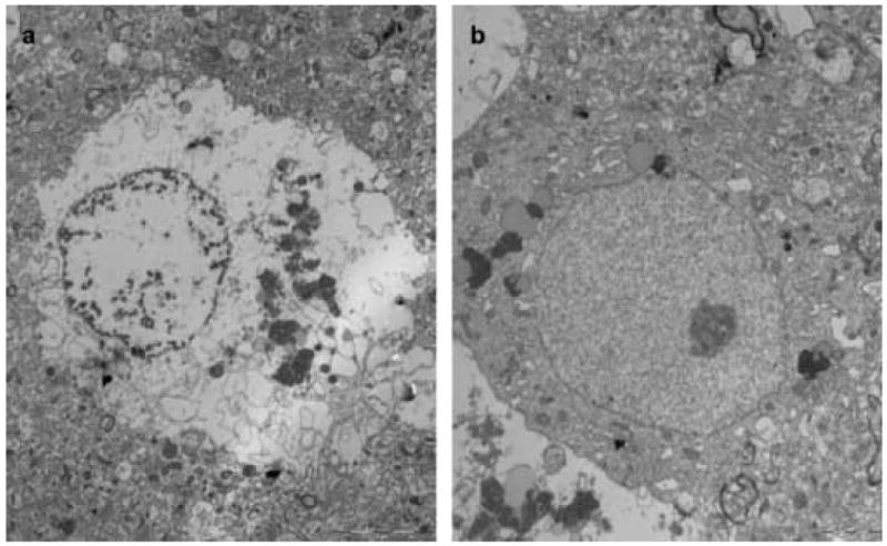
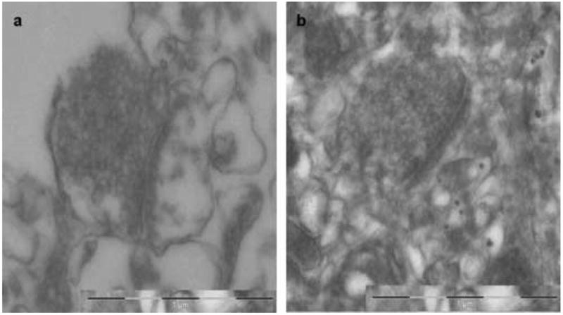
For the human brain, in vitro models that accurately represent what occurs in vivo are lacking. Organotypic models may be the closest parallel to human brain tissue outside of a live patient. However, this model has been limited primarily to rodent-derived tissue. We present an organotypic model to maintain intraoperatively collected human tumor and non-tumor explants ex vivo for a prolonged period of time ( approximately 11 days) without any significant changes to the tissue cytoarchitecture as evidenced through immunohistochemistry and electron microscopy analyses. The ability to establish and reliably predict the cytoarchitectural changes that occur with time in an organotypic model of tumor and non-tumor human brain tissue has several potential applications including the study of cell migration on actual tissue matrix, drug toxicity on neural tissue and pharmacological treatment for brain cancers, among others.
Figures
Similar articles
-
Time course modifications in organotypic culture of human neuroretina.Exp Eye Res. 2012 Nov;104:26-38. doi: 10.1016/j.exer.2012.08.012. Epub 2012 Sep 26. Exp Eye Res. 2012. PMID: 23022403
-
The astroblastoma and its possible cytogenic relationship to the tanycyte. An electron microscopic, immunohistochemical, tissue- and organ-culture study.Acta Neuropathol. 1989;78(5):472-83. doi: 10.1007/BF00687708. Acta Neuropathol. 1989. PMID: 2683559
-
Coculture system with an organotypic brain slice and 3D spheroid of carcinoma cells.J Vis Exp. 2013 Oct 9;(80):50881. doi: 10.3791/50881. J Vis Exp. 2013. PMID: 24145580 Free PMC article.
-
Organotypic 3D Ex Vivo Co-culture Model of the Macro-metastasis/Organ Parenchyma Interface.Methods Mol Biol. 2024;2764:165-176. doi: 10.1007/978-1-0716-3674-9_12. Methods Mol Biol. 2024. PMID: 38393595
-
Glial fibrillary acidic protein is a body fluid biomarker for glial pathology in human disease.Brain Res. 2015 Mar 10;1600:17-31. doi: 10.1016/j.brainres.2014.12.027. Epub 2014 Dec 25. Brain Res. 2015. PMID: 25543069 Review.
Cited by
-
Human organotypic brain slice culture: a novel framework for environmental research in neuro-oncology.Life Sci Alliance. 2019 Jun 27;2(4):e201900305. doi: 10.26508/lsa.201900305. Print 2019 Aug. Life Sci Alliance. 2019. PMID: 31249133 Free PMC article.
-
A dense poly(ethylene glycol) coating improves penetration of large polymeric nanoparticles within brain tissue.Sci Transl Med. 2012 Aug 29;4(149):149ra119. doi: 10.1126/scitranslmed.3003594. Sci Transl Med. 2012. PMID: 22932224 Free PMC article.
-
Divergent actions of physiological and pathological amyloid-β on synapses in live human brain slice cultures.Nat Commun. 2025 Apr 30;16(1):3753. doi: 10.1038/s41467-025-58879-z. Nat Commun. 2025. PMID: 40307257 Free PMC article.
-
Perivascular invasion of primary human glioblastoma cells in organotypic human brain slices: human cells migrating in human brain.J Neurooncol. 2023 Aug;164(1):43-54. doi: 10.1007/s11060-023-04349-9. Epub 2023 Jul 25. J Neurooncol. 2023. PMID: 37490233
-
Regulation of neural stem cell in the human SVZ by trophic and morphogenic factors.Curr Signal Transduct Ther. 2011 Sep 1;6(3):320-326. doi: 10.2174/157436211797483958. Curr Signal Transduct Ther. 2011. PMID: 22053150 Free PMC article.
References
-
- Bsibsi M, Persoon-Deen C, Verwer RW, Meeuwsen S, Ravid R, Van Noort JM. Toll-like receptor 3 on adult human astrocytes triggers production of neuroprotective mediators. Glia. 2006;53(7):688–695. - PubMed
-
- Cavaliere F, Dinkel K, Reymann K. The subventricular zone releases factors which can be protective in oxygen/glucose deprivation-induced cortical damage: An organotypic study. Exp Neurol 2006 - PubMed
-
- Cimarosti H, O'Shea RD, Jones NM, Horn AP, Simao F, Zamin LL, Nassif M, Frozza R, Netto CA, Beart PM, Salbego C. The effects of estradiol on estrogen receptor and glutamate transporter expression in organotypic hippocampal cultures exposed to oxygen--glucose deprivation. Neurochem Res. 2006;31(4):483–490. - PubMed
-
- Duveau V, Arthaud S, Serre H, Rougier A, Le Gal La Salle G. Transient hyperthermia protects against subsequent seizures and epilepsy-induced cell damage in the rat. Neurobiol Dis. 2005;19(12):142–149. - PubMed
MeSH terms
Substances
Grants and funding
LinkOut - more resources
Full Text Sources
Other Literature Sources
Medical
