. 2007 Aug 17;282(33):24109-19.
doi: 10.1074/jbc.M700821200. Epub 2007 Jun 19.

Acute lung injury edema fluid decreases net fluid transport across human alveolar epithelial type II cells

Affiliations

Acute lung injury edema fluid decreases net fluid transport across human alveolar epithelial type II cells

Jae W Lee et al. J Biol Chem. .

Abstract

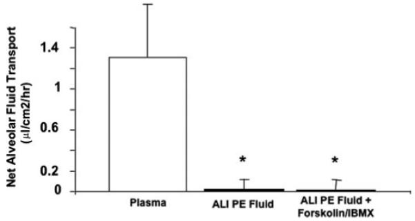
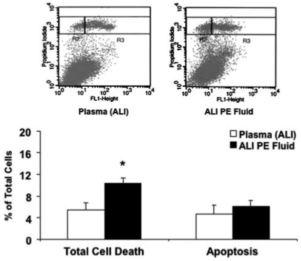
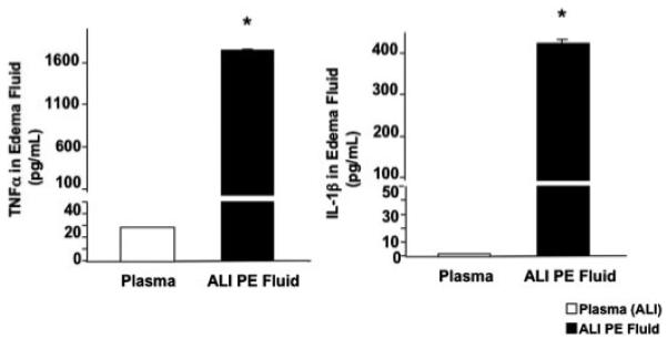
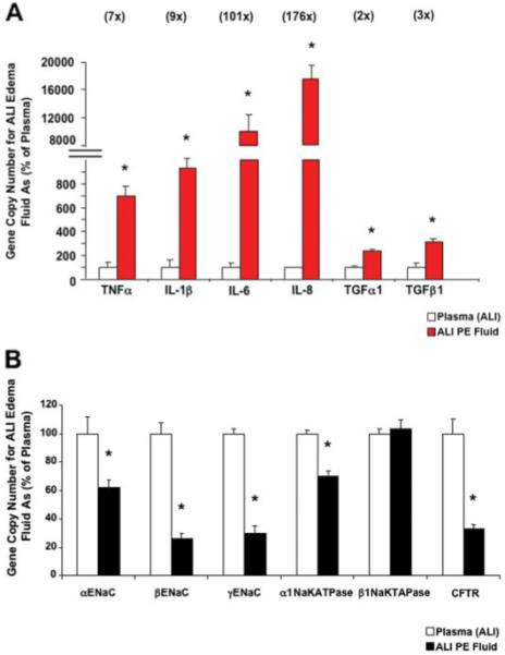
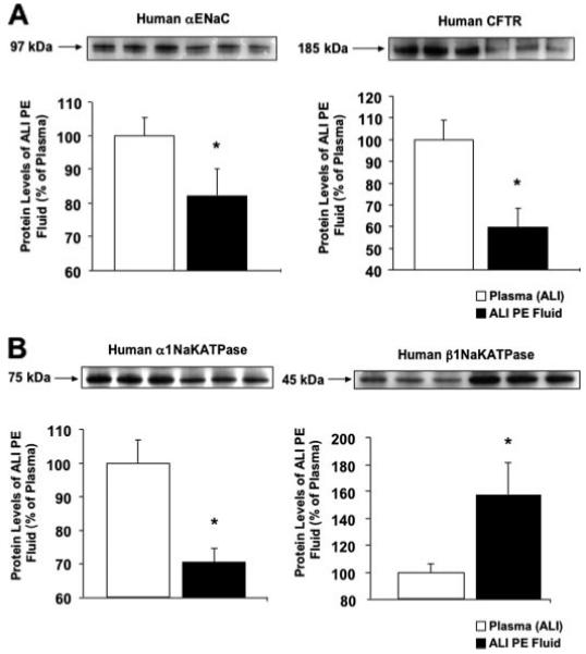
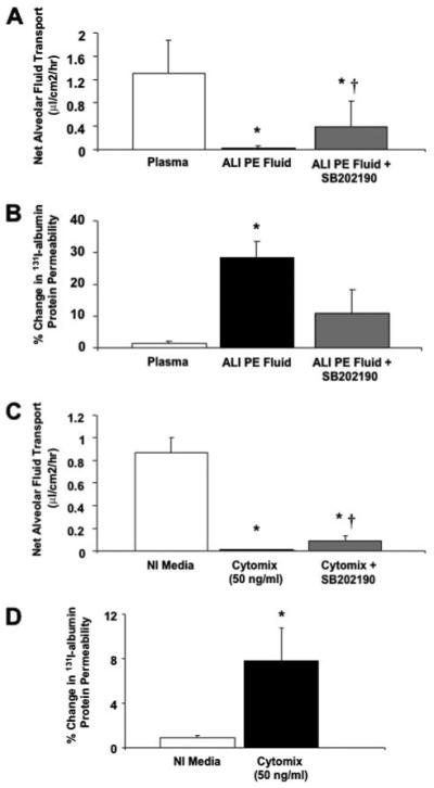
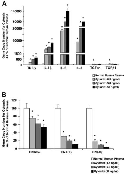
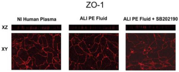
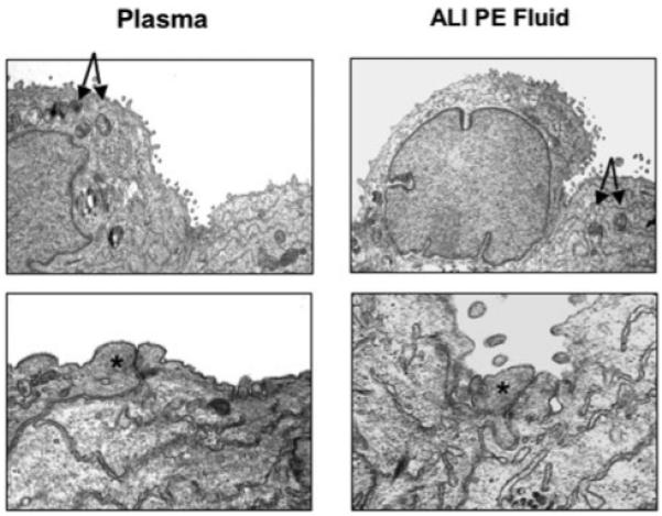
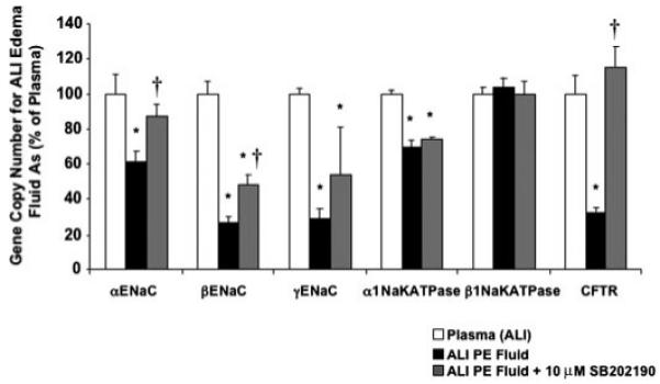
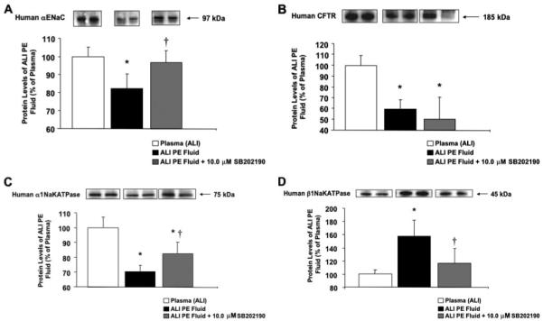
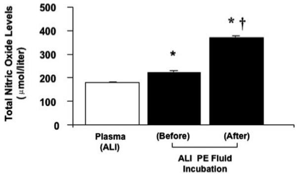
Most patients with acute lung injury (ALI) have reduced alveolar fluid clearance that has been associated with higher mortality. Several mechanisms may contribute to the decrease in alveolar fluid clearance. In this study, we tested the hypothesis that pulmonary edema fluid from patients with ALI might reduce the expression of ion transport genes responsible for vectorial fluid transport in primary cultures of human alveolar epithelial type II cells. Following exposure to ALI pulmonary edema fluid, the gene copy number for the major sodium and chloride transport genes decreased. By Western blot analyses, protein levels of alphaENaC, alpha1Na,K-ATPase, and cystic fibrosis transmembrane conductance regulator decreased as well. In contrast, the gene copy number for several inflammatory cytokines increased markedly. Functional studies demonstrated that net vectorial fluid transport was reduced for human alveolar type II cells exposed to ALI pulmonary edema fluid compared with plasma (0.02 +/- 0.05 versus 1.31 +/- 0.56 microl/cm2/h, p < 0.02). An inhibitor of p38 MAPK phosphorylation (SB202190) partially reversed the effects of the edema fluid on net fluid transport as well as gene and protein expression of the main ion transporters. In summary, alveolar edema fluid from patients with ALI induced a significant reduction in sodium and chloride transport genes and proteins in human alveolar epithelial type II cells, effects that were associated with a decrease in net vectorial fluid transport across human alveolar type II cell monolayers.


Figures

FIGURE 1
FIGURE 1. Net fluid transport across human alveolar epithelial type II cells exposed to plasma or ALI pulmonary edema fluid with or without forskolin + IBMX
Human alveolar epithelial type II cells cultured for 5 days were exposed to plasma or ALI pulmonary edema fluid with or without forskolin + IBMX, 48 h after the air-liquid interface was achieved, for 24 h. Net fluid transport was calculated, as done previously, by the change in weight and radioactivity of 131I-albumin in the upper compartment of the Transwell plate and expressed as mean (μl/cm2/h) ± S.D. of each sample run in triplicates. *, p < 0.02 compared with plasma. PE, pulmonary edema.
FIGURE 2
FIGURE 2. Effect of ALI pulmonary edema fluid on total cell death
Human alveolar epithelial type II cells cultured for 3 days were exposed to ALI pulmonary edema fluid or plasma for 24 h. The percentage of apoptotic cells was quantified by flow cytometry after double staining with propidium iodide and annexin V-fluorescein isothiocyanate of both the floating and detached cells in the supernatant and the plated cells. Total cell death was measured by propidium iodide staining alone. Data represent mean±S.D. of each sample measured in triplicates. *, p < 0.01 compared with plasma controls. In the panels above each graph, total cell death = R2 + R3 where R2 represents necrotic cells and R3 represents the apoptotic portion. PE, pulmonary edema.
FIGURE 3
FIGURE 3. Cytokine levels in ALI pulmonary edema fluid and plasma
The levels of TNFα and IL-1β were measured by enzyme-linked immunosorbent assay from the combined ALI pulmonary edema fluid and the corresponding plasma of 19 patients with ALI from sepsis. Data represent mean ± S.D. of each sample measured in triplicates. *, p < 0.005 compared with plasma controls. The level of TNFα in ALI pulmonary edema fluid was 62× and the level of IL-1β was 220× the level found in the corresponding plasma. PE, pulmonary edema.
FIGURE 4
FIGURE 4. A and B, effect of ALI pulmonary edema fluid on the gene expression of the major inflammatory cytokines and chemokines (A) and sodium and chloride transport proteins (B) by human alveolar epithelial type II cells exposed to ALI pulmonary edema fluid or plasma
Human alveolar epithelial type II cells cultured for 3 days were exposed to ALI pulmonary edema fluid or plasma for 24 h. The gene expression was measured by two-step multiplex quantitative RT-PCR and expressed as the percent GCN of plasma ± S.D. of each sample measured in triplicates. *, p < 0.01 compared with plasma controls. The number above each cytokine/chemokine represents the multiple times the GCN of ALI pulmonary edema exposed cells exceeded the GCN of plasma exposed cells. PE, pulmonary edema.
FIGURE 5
FIGURE 5. A and B, effect of ALI pulmonary edema fluid on the protein expression of the major sodium and chloride transport proteins by human alveolar epithelial type II cells exposed to ALI pulmonary edema fluid or plasma
Human alveolar epithelial type II cells cultured for 3 days were exposed to ALI pulmonary edema fluid or plasma for 24 h. Protein levels were measured by Western blot analyses and expressed as the percent protein levels of plasma±S.D. of each sample measured in triplicates. *, p < 0.05 compared with plasma controls. A representative Western blot is depicted above each graph for the major sodium and chloride transport proteins. PE, pulmonary edema.
FIGURE 6
FIGURE 6. A-D, effect of ALI pulmonary edema fluid on the net fluid transport (A and C) and paracellular protein permeability (B and D) by human alveolar epithelial type II cells exposed to ALI pulmonary edema fluid or cytomix with and without pretreatment with a p38 MAPK inhibitor or plasma
Human alveolar epithelial type II cells cultured for 5 days were exposed to ALI pulmonary edema fluid, plasma, or cytomix, 48 h after the air-liquid interface was achieved, for 24 h. Another set of human type II cells were pretreated with 10.0 μm SB202190 for 30 min prior to exposure with ALI pulmonary edema fluid or cytomix. Net fluid transport was calculated, as done previously, by the change in weight and radioactivity of 131I-albumin in the upper compartment of the Transwell plate and expressed as mean (μl/cm2/h) ± S.D. of each sample run in triplicates. Paracellular protein permeability was calculated, as done previously, by the passage of 131I-albumin from the upper compartment to the lower compartment of the Transwell plate and expressed as percent change in 131I-albumin permeability±S.D. of each sample run in triplicates. *, p < 0.02 compared with plasma controls; †, p < 0.01 compared with ALI pulmonary edema or cytomix-exposed cells. PE, pulmonary edema; NI, normal.
FIGURE 7
FIGURE 7. A and B, the dose response of human alveolar epithelial type II cells exposed to cytomix or normal human plasma on the expression of the major inflammatory cytokines and chemokines (A) and sodium transport proteins (B)
Human alveolar epithelial type II cells cultured for 3 days were exposed to different concentrations of cytomix (0.5-50 ng/ml) or plasma for 24 h. Gene expression was measured by two-step multiplex quantitative RT-PCR and expressed as the percent GCN of plasma ± S.D. of each sample measured in triplicates. *, p < 0.05 versus normal human plasma.
FIGURE 8
FIGURE 8. Effect of ALI pulmonary edema fluid on ZO-1 distribution in primary cultures of human alveolar epithelial type II cells
Human alveolar type II cells were grown on Transwell plates for 5 days and exposed to plasma, ALI pulmonary edema fluid, or ALI pulmonary edema fluid pretreated with 10 μm SB202190, a p38 MAPK inhibitor, for 24 h. The cells were fixed and stained with anti-ZO-1 antibodies and then analyzed by confocal microscopy. En face sections (x-y) were generated from selected planes in the vertical (x-z) sections. PE, pulmonary edema; NI, normal.
FIGURE 9
FIGURE 9. Effect of ALI pulmonary edema fluid on the ultrastructure of cultured human alveolar epithelial type II cells
Transmission electron microscopy demonstrated no significant changes in the characteristic features of alveolar epithelial type II cells on exposure to ALI pulmonary edema fluid compared with plasma: the cuboidal shape or the presence of lamellar bodies and microvillus. More significantly, no signs of apoptosis were detected, and the tight junctions appeared intact. The arrows point toward lamellar bodies, and the asterisks depict the location of tight junctions. PE, pulmonary edema.
FIGURE 10
FIGURE 10. Changes in the gene expression of the major sodium and chloride transport proteins by human alveolar epithelial type II cells by pretreatment with a p38 MAPK inhibitor prior to exposure to ALI pulmonary edema fluid
Human alveolar epithelial type II cells cultured for 3 days were exposed to ALI pulmonary edema fluid or plasma for 24 h. Another set of human type II cells were pretreated with 10.0 μm SB202190 for 30 min prior to exposure with ALI pulmonary edema fluid. The gene expression was measured by two-step multiplex quantitative RT-PCR and expressed as the percent GCN of plasma ± S.D. of each sample measured in triplicates. *, p < 0.05 compared with plasma controls; †, p is significant by analysis of variance with Bonferroni correction. PE, pulmonary edema.
FIGURE 11
FIGURE 11. A-D, changes in the protein levels of the major sodium and chloride transport proteins by human alveolar epithelial type II cells by pretreatment with a p38 MAPK inhibitor prior to exposure to ALI pulmonary edema fluid
Human alveolar epithelial type II cells cultured for 3 days were exposed to ALI pulmonary edema fluid or plasma for 24 h. Another set of human type II cells were pretreated with 10.0 μm SB202190 for 30 min prior to exposure with ALI pulmonary edema fluid. Protein levels were measured by Western blot analyses and expressed as the percent protein levels of plasma±S.D. of each sample measured in triplicates. *, p < 0.05 compared with plasma controls; †, p < 0.02 compared with ALI pulmonary edema-exposed cells. A representative Western blot is depicted above each column for the major sodium and chloride transport proteins. PE, pulmonary edema.
FIGURE 12
FIGURE 12. Nitric oxide levels of the ALI pulmonary edema fluid before and after incubation with human alveolar epithelial type II cells for 24 h
The total nitric oxide level was measured by the Griess reaction and expressed in μmol/liter±S.D. of each sample measured in triplicates. *, p < 0.001 compared with plasma controls; †,p<0.0005 compared with ALI pulmonary edema prior to exposure to alveolar epithelial type II cells. PE, pulmonary edema.

Similar articles

Cited by

References

    1. Matthay MA, Wiener-Kronish JP. Am. Rev. Respir. Dis. 1990;142:1250–1257. - PubMed
    1. Ware LB, Matthay MA. Am. J. Respir. Crit. Care Med. 2001;163:1376–1383. - PubMed
    1. Matthay MA, Folkesson HG, Clerici C. Physiol. Rev. 2002;82:569–600. - PubMed
    1. Folkesson HG, Matthay MA. Am. J. Respir. Cell Mol. Biol. 2006;35:10–19. - PMC - PubMed
    1. Matalon S, O’Brodovich H. Annu. Rev. Physiol. 1999;61:627–661. - PubMed

Publication types