. 2007 Aug;14(16):1189-98.
doi: 10.1038/sj.gt.3302974. Epub 2007 Jun 21.

Control of human mesothelin-expressing tumors by DNA vaccines

Affiliations

Control of human mesothelin-expressing tumors by DNA vaccines

C-L Chang et al. Gene Ther. 2007 Aug.

Abstract

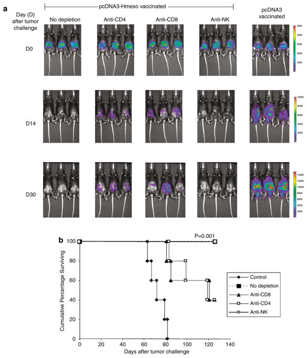
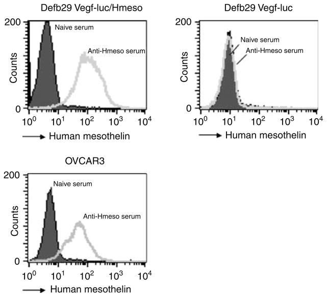
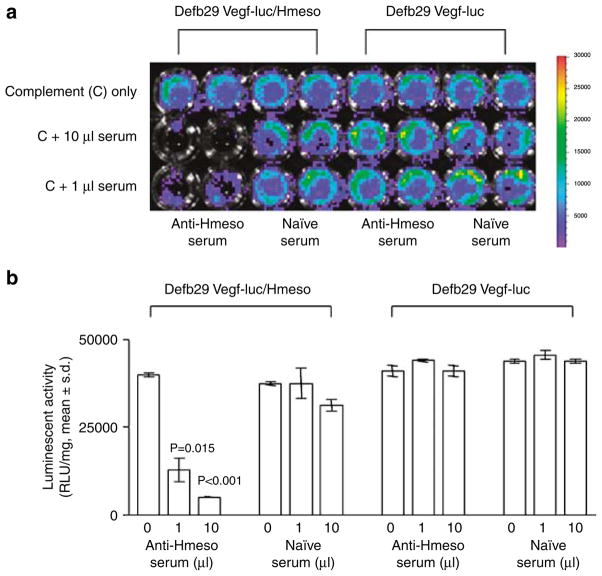
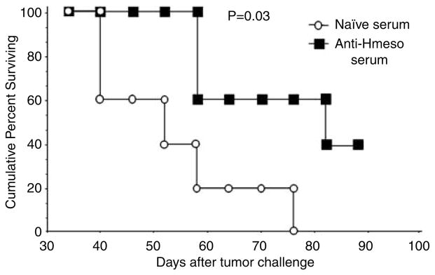
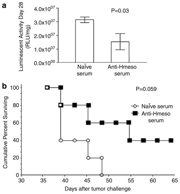
Mesothelin has been implicated as a potential ideal target antigen for the development of antigen-specific cancer immunotherapy for the control of mesothelin-expressing cancers such as ovarian cancer, mesothelioma and pancreatic adenocarcinoma. In the current study, we utilized a DNA vaccine encoding human mesothelin (pcDNA3-Hmeso) to treat C57BL/6 mice challenged with luciferase-expressing, Hmeso-expressing ovarian cancer cell line, Defb29 Vegf-luc/Hmeso. The therapeutic effect of the tumor-challenged mice was followed by noninvasive bioluminescence imaging systems. The mechanism of the antitumor effect was characterized by depletion of subsets of lymphocytes as well as adopted transfer of serum from pcDNA3-Hmeso-vaccinated mice. We found that vaccination with pcDNA3-Hmeso DNA vaccine generates a significant antitumor effect and promotes survival in mice challenged with Defb29 Vegf-luc/Hmeso. Furthermore, we found CD4+ and CD8+ T-cell immune responses as well as the humoral immune responses are important for the observed antitumor effects in vaccinated mice. Our data indicated that vaccination with DNA vaccine targeting Hmeso could generate potent antitumor effects against mesothelin-expressing tumors through both T cell-mediated immunity as well as antibody-mediated immunity.


Figures

Figure 1
Figure 1
Flow cytometry analysis to characterize the expression of human mesothelin (Hmeso) in Defb29 Vegf-luc/Hmeso cell line. Characterization of Hmeso expression was performed in Defb29 Vegf-luc/Hmeso and Defb29 Vegf-luc cells using flow cytometry analysis. The cell lines were stained with the Hmeso-specific mouse monoclonal antibody CAK-1, followed by flow cytometry analysis. Mouse IgG1 isotype was used as a control.
Figure 2
Figure 2
Characterization of antitumor effects generated by treatment with human mesothelin (Hmeso)-expressing DNA vaccine. C57BL/6 mice (five per group) were challenged with 5 × 10⁵/mouse of Defb29 Vegf-luc/Hmeso cells (day 0). Three days after tumor challenge, mice with established Defb29 Vegf-luc/Hmeso tumors were treated with DNA vaccine encoding Hmeso (pcDNA3-Hmeso) via gene gun. An empty vector vaccine (pcDNA3) was used as a control. Mice were imaged using the IVIS Imaging System Series 200. Bioluminescence signals were acquired for 1 min. (a) Luminescence images of representative Defb29 Vegf-luc/Hmeso-challenged mice treated with pcDNA3-Hmeso or pcDNA3 DNA vaccines from day 3 and 60 after tumor challenge. (b) Bar graph depicting the luminescence activity (tumor load) of tumor-bearing mice treated with pcDNA3-Hmeso DNA or pcDNA3 DNA from day 3 and 60 after tumor challenge. (c) Kaplan–Meier survival analysis of the tumor-challenged mice treated with pcDNA3-Hmeso or pcDNA3 DNA vaccines. The mice were tumor challenged on day 0.
Figure 3
Figure 3
In vivo antibody depletion experiment. C57BL/6 mice (five per group) were intraperitoneally (i.p.) immunized with pcDNA3-Hmeso twice at a 1-week interval via gene gun. One week after the last vaccination, the pcDNA3-Hmeso-vaccinated mice were depleted of either CD8, CD4 or NK cells using relevant antibodies every other day for 1 week and then once every week, as described in the Materials and methods section. A group of nondepleted pcDNA3-Hmeso-vaccinated mice was used as a control. Two weeks after vaccination, depleted and nondepleted mice were challenged with 1 × 10⁶/mouse of Defb29 Vegf-luc/Hmeso tumor cells (day 0). Mice were imaged using the IVIS Imaging System Series 200. Bioluminescence signals were acquired for 1 min. pcDNA3-vaccinated mice challenged with Defb29 Vegf-luc/Hmeso cells were used as a control. (a) Luminescence images of representative mice challenged with Defb29 Vegf-luc/Hmeso cells without depletion or with CD4 depletion, CD8 depletion or NK depletion from days 0, 14 and 30 after tumor challenge. (b) Kaplan–Meier survival analysis of the pcDNA3-Hmeso-vaccinated mice challenged with Defb29 Vegf-luc/Hmeso tumor cells without depletion or with CD4 depletion, CD8 depletion or NK depletion. The mice were tumor challenged on day 0. NK, natural killer.
Figure 4
Figure 4
Flow cytometry analysis to characterize the expression of human mesothelin (Hmeso) in murine and human ovarian cancer cell lines. Hmeso-specific antibody containing serum was generated by immunization of C57BL/6 mice with pcDNA3-Hmeso DNA vaccine 3 times at 1-week intervals via gene gun. One week after vaccination, blood sera were collected from immunized mice and used to stain murine and human cancer cell lines. The characterization of Hmeso expression in Defb29 Vegf-luc/Hmeso, Defb29 Vegf-luc and OVCAR3 was performed with flow cytometry analysis using sera collected from pcDNA3-Hmeso-immunized mice. Sera from naive mice were used as a negative control.
Figure 5
Figure 5
Complement-dependent cytotoxicity assay using human mesothelin (Hmeso)-specific antibodies from pcDNA3-Hmeso-immunized mice. 1 × 10⁴ Defb29 Vegf-luc/Hmeso cells were seeded in 96-well plate. Defb29 Vegf-luc cells were used as a negative control. Cell viability was determined after adding serum and complement using the IVIS Imaging System Series 200. Sera obtained from either pcDNA3-Hmeso immunized mice or naive mice were added in amounts of 0, 1 and 10 μl/well to both cell lines. Rabbit serum (complement) was added to all wells at a 1:5 dilution. Bioluminescence signals were acquired for 1 min. (a) Representative figures of luminescence images of 96-well plates showing complement-mediated lysis effect on Defb29 Vegf-luc/Hmeso or Defb29 Vegf-luc cells. Note: significant lysis was demonstrated by decrease of luminescence activity. (b) Bar graph depicting the quantification of luminescence in Defb29 Vegf-luc/Hmeso or Defb29 Vegf-luc tumor cells mixed with sera from pcDNA3-Hmeso-immunized mice or sera from naive mice.
Figure 6
Figure 6
Adoptive serum transfer experiments in tumor-bearing C57BL/6 mice. C57BL/6 mice (five per group) were challenged with 5 × 10⁴/mouse of Defb29 Vegf-luc/Hmeso cells. Five days later, the tumor-bearing mice were treated with sera from pcDNA3-Hmeso-immunized mice or sera from naive mice intraperitoneally (i.p.) every 3 days for 4 times. Kaplan–Meier survival analysis of the tumor-bearing mice was performed. The days indicated follow from day 0 of tumor challenge. Hmeso, human mesothelin.
Figure 7
Figure 7
Serum transfer experiments in tumor-bearing immunocompromised mice. Athymic nude mice (five per group) were challenged with 5 × 10⁴/mouse of Defb29 Vegf-luc/Hmeso cells (day 0). Three days later, the tumor-bearing mice were treated with sera from pcDNA3-Hmeso-immunized mice or sera from naive mice intraperitoneally (i.p.) every 3 days for 4 times. Tumor load in treated mice was monitored using the IVIS Imaging System Series 200. Bioluminescence signals were acquired for 1 min. (a) Representative luminescence images of tumor-bearing athymic nude mice that received sera from naive mice or sera from pcDNA3-Hmeso immunized mice. (b) Bar graph depicting the luminescence activity (tumor load) on day 28 after tumor challenge of tumor-bearing athymic nude mice treated with sera from naive mice or sera from pcDNA3-Hmeso-immunized mice. (c) Kaplan–Meier survival analysis of tumor-bearing athymic nude mice that received sera from naive mice or sera from pcDNA3-Hmeso-immunized mice. Hmeso, human mesothelin.
Figure 8
Figure 8
Serum transfer experiments in human ovarian cancer bearing immunocompromised mice. Athymic nude mice (five per group) were challenged with 5 × 10⁴/mouse of OVCAR3-luc/Hmeso cells (day 0). Three days later, the tumor-bearing mice were treated with sera from pcDNA3-Hmeso-immunized mice or sera from naive mice intraperitoneally (i.p.) every 3 days for 4 times. Tumor load in treated mice was monitored using the IVIS Imaging System Series 200. Bioluminescence signals were acquired for 1 min. (a) Bar graph depicting the luminescence activity (tumor load) on day 28 after tumor challenge of tumor-bearing athymic nude mice treated with sera from naive mice or sera from pcDNA3-Hmeso-immunized mice. (b) Kaplan–Meier survival analysis of tumor-bearing athymic nude mice that received sera from naive mice or sera from pcDNA3-Hmeso-immunized mice. The days indicated follow from day 0 of tumor challenge. Hmeso, human mesothelin.

References

    1. Greenlee RT, Murray T, Bolden S, Wingo PA. Cancer statistics, 2000. CA Cancer J Clin. 2000;50:7–33. - PubMed
    1. Zellos LS, Sugarbaker DJ. Multimodality treatment of diffuse malignant pleural mesothelioma. Semin Oncol. 2002;29:41–50. - PubMed
    1. Nowak AK, Lake RA, Kindler HL, Robinson BW. New approaches for mesothelioma: biologics, vaccines, gene therapy, and other novel agents. Semin Oncol. 2002;29:82–96. - PubMed
    1. Jhala N, Jhala D, Vickers SM, Eltoum I, Batra SK, Manne U, et al. Biomarkers in diagnosis of pancreatic carcinoma in fine-needle aspirates. Am J Clin Pathol. 2006;126:572–579. - PubMed
    1. Shedlock DJ, Weiner DB. DNA vaccination: antigen presentation and the induction of immunity. J Leukoc Biol. 2000;68:793–806. - PubMed

Publication types

MeSH terms