. 2007 Jul 25;26(14):3283-95.
doi: 10.1038/sj.emboj.7601768. Epub 2007 Jun 21.

Latent TGF-beta-binding protein 2 binds to DANCE/fibulin-5 and regulates elastic fiber assembly

Affiliations

Latent TGF-beta-binding protein 2 binds to DANCE/fibulin-5 and regulates elastic fiber assembly

Maretoshi Hirai et al. EMBO J. .

Abstract

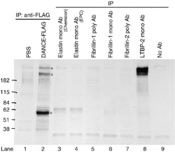
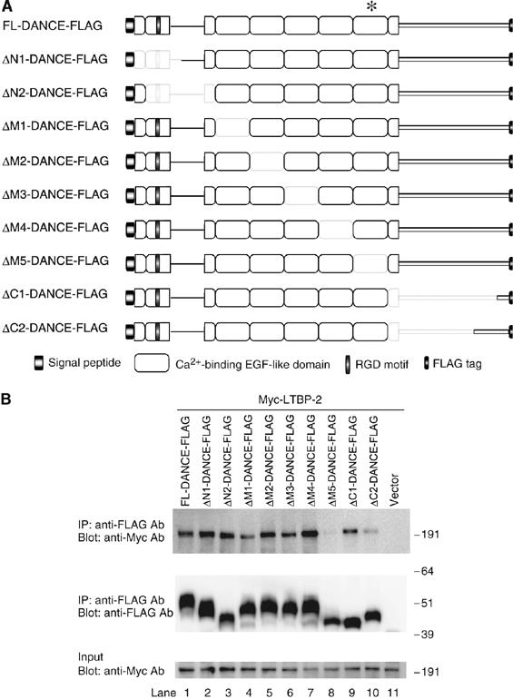
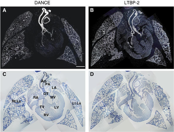
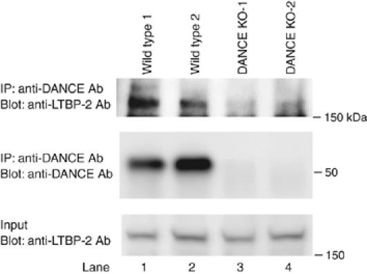
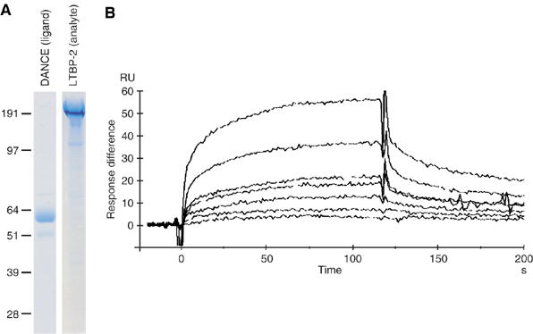
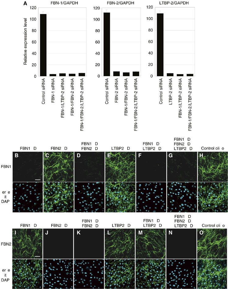
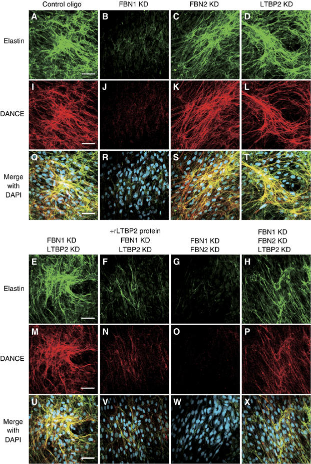
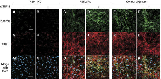
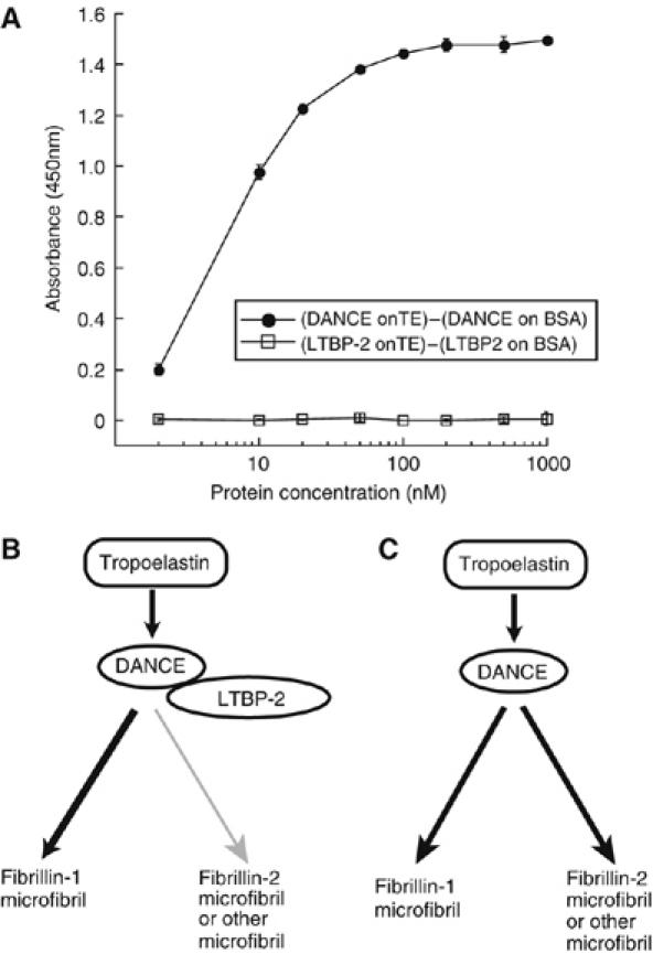
Elastic fibers play the principal roles in providing elasticity and integrity to various types of human organs, such as the arteries, lung, and skin. However, the molecular mechanism of elastic fiber assembly that leads to deposition and crosslinking of elastin along microfibrils remains largely unknown. We have previously shown that developing arteries and neural crest EGF-like protein (DANCE) (also designated fibulin-5) is essential for elastogenesis by studying DANCE-deficient mice. Here, we report the identification of latent transforming growth factor-beta-binding protein 2 (LTBP-2), an elastic fiber-associating protein whose function in elastogenesis is not clear, as a DANCE-binding protein. Elastogenesis assays using human skin fibroblasts reveal that fibrillar deposition of DANCE and elastin is largely dependent on fibrillin-1 microfibrils. However, downregulation of LTBP-2 induces fibrillin-1-independent fibrillar deposition of DANCE and elastin. Moreover, recombinant LTBP-2 promotes deposition of DANCE onto fibrillin-1 microfibrils. These results suggest a novel regulatory mechanism of elastic fiber assembly in which LTBP-2 regulates targeting of DANCE on suitable microfibrils to form elastic fibers.


Figures

Figure 1
Figure 1
Latent TGF-β-binding protein-2 (LTBP-2) as a candidate DANCE-binding protein. Bovine smooth muscle cells were metabolically labeled with 35S-cysteine/methionine, and conditioned media were subjected to immunoprecipitation. Several DANCE-binding proteins are detected (lane 2, asterisks). The sizes of the bands larger than 182 kDa are similar to those of proteins precipitated with anti-LTBP-2 antibody (compare lanes 2 and 8). Another 60-kDa band is inferred to be DANCE, which is abundantly expressed in aortic smooth muscle cells (lane 2).
Figure 2
Figure 2
Schematic representation of the DANCE deletion mutants, and mapping of the binding domain of DANCE with LTBP-2. (A) Domain structure of the full-length DANCE and the DANCE deletion mutants used for the in vitro binding assay. These mutants were expressed as C-terminally FLAG-tagged proteins. We prepared expression vectors encoding FLAG-tagged DANCE (FL-DANCE-FLAG) or DANCE deletion mutants, including N-terminal domain deletion mutants (ΔN1- and ΔN2-DANCE-FLAG), central calcium-binding EGF (cbEGF)-like repeat domain deletion mutants (ΔM1-, ΔM2-, ΔM3-, ΔM4-, and ΔM5-DANCE-FLAG), and C-terminal domain deletion mutants (ΔC1- and ΔC2-DANCE-FLAG). The asterisk indicates the sixth cbEGF-like domain of DANCE, where LTBP-2 binds. (B) The sixth cbEGF-like domain of DANCE interacts with LTBP-2. 293T cells were transiently transfected with the vectors shown in A or mock vector. Expression vectors for Myc-tagged full-length LTBP-2 (LTBP-2-Myc) were also independently transfected into 293T cells. Transfected cells were cultured in serum-free medium for 48 h, and then the cell lysates and the conditioned media were harvested and mixed. After incubation of LTBP-2-Myc with a set of FLAG-tagged DANCE deletion mutants, each mixture was subjected to immunoprecipitation with anti-FLAG antibody. These immunoprecipitates were then separated by SDS–PAGE, and analyzed by Western blotting with a monoclonal anti-Myc antibody.
Figure 3
Figure 3
Schematic representation of the LTBP-2 truncation mutants, and mapping of the binding domain of LTBP-2 with DANCE. (A) Domain structure of the full-length LTBP-2 and the LTBP-2 truncation mutants used for the in vitro binding assay. These mutants were expressed as N-terminally FLAG-tagged proteins flanked by the preprotrypsin signal sequence, except for the full-length LTBP-2. The LTBP-2-H fragment is prone to degradation, so we constructed C-terminal fusion proteins with the constant region of human IgG to prevent the degradation (LTBP-2-G-Ig, LTBP-2-H-Ig, and LTBP-2-I-Ig). The characteristics of each domain are described below the figure. The asterisk indicates the second four-cystein domain of LTBP-2, where DANCE binds. (B) Left panel, DANCE interacts with the N-terminal domain of LTBP-2 (LTBP-2-A). Right panel, DANCE interacts with the second four-cysteine domain of LTBP-2 (LTBP-2-I). The expression vector of each LTBP-2 truncation mutant was transfected into 293T cells. Mixtures of the media and cell lysates of FLAG-tagged LTBP-2 truncation mutants were incubated with DANCE-Myc, and then these reactants were subjected to immunoprecipitation with anti-FLAG antibody. The immunoprecipitates were separated by SDS–PAGE, and analyzed by Western blotting with anti-Myc antibody. (C) Calcium dependency of the LTBP-2–DANCE interaction. The expression vector for Myc-tagged full-length LTBP-2 was transfected into 293T cells. Mixtures of the conditioned media and cell lysates of Myc-LTBP-2 were incubated with the conditioned media of DANCE-FLAG in the presence of EDTA (0, 1, 2, 5, or 10 mM). These cocktails were subjected to immunoprecipitation with anti-FLAG antibody. The immunoprecipitates were separated by SDS–PAGE, and analyzed by Western blotting with anti-Myc antibody.
Figure 4
Figure 4
Expression patterns of DANCE (A) and LTBP-2 (B) transcripts in neonatal mice as shown by in situ hybridization with dark field (A, B) and bright field (C, D) views. Ao, aorta; PA, pulmonary artery; OT, cardiac outflow tract; MV, mitral valve; TV, tricupsid valve, LV, left ventricle; RV, right ventricle; LA, left atrium; RA, right atrium; Rt.Lu, right lung; Lt.Lu, left lung. Scale bar; 500 μm.
Figure 5
Figure 5
DANCE interacts with LTBP-2 in vivo. Whole lungs were dissected from two lines each of wild-type and DANCE-deficient mice. Proteins were extracted using a homogenizer on ice. Immunoreactive DANCE protein was precipitated from lung extracts with anti-DANCE antibody, followed by Western blotting with a polyclonal anti-LTBP-2 antibody.
Figure 6
Figure 6
Quantification of the affinity of DANCE binding to LTBP-2 by surface plasmon resonance. (A) Recombinant full-length DANCE and full-length LTBP-2 proteins were purified by chelating chromatography, separated by SDS–PAGE, and stained with Coomassie Blue. (B) LTBP-2 was injected over a DANCE-immobilized sensor chip surface. Each sensorgram shows seven different analyte concentrations of 15, 30, 60, 90, 180, 240, and 360 μg/ml. Response difference; the difference between experimental and control flow cells in response units (RU). Time is shown in seconds (s).
Figure 7
Figure 7
Quantitative PCR analysis of gene knockdown in skin fibroblasts (A), and fibrillin-1 and -2 knockdown specifically abolishes the meshwork of fibrillin-1 and -2 microfibrils, respectively (BO). (A) Total RNA from siRNA-transfected skin fibroblasts was extracted 9 days after transfection. Complementary DNA was synthesized and was subjected to quantitative real-time PCR for the expression of fibrillin-1, fibrillin-2, LTBP-2, and glyceraldehyde-3-phosphate dehydrogenase (GAPDH) transcripts. A graphic presentation of the results obtained by qPCR is shown. Levels of GAPDH transcript are used to normalize the cDNA levels. The relative amount of the PCR product amplified from control siRNA-treated fibroblasts is set at 100. Data are presented as the means±s.e. of three independent experiments, each performed in duplicate. (B–O) HSFs were transfected with each RNAi oligo as indicated and cultured in 10% serum containing media. Cells were stained with anti-fibrillin-1 (BH) or anti-fibrillin-2 antibody (I–O). Lower panels are superimpositions of upper panels with DAPI (4,6-diamidino-2-phenylindole) nuclear staining. KD, knockdown; FBN-1, fibrillin-1; FBN-2, fibrillin-2. Scale bars; 60 μm.
Figure 8
Figure 8
DANCE and elastin deposition is dependent on fibrillin-1 microfibril, but LTBP-2 knockdown induces fibrillin-1-independent deposition of DANCE and elastin. HSFs were transfected with each RNAi oligo as indicated, cultured in 10% serum containing media for 14 days and fixed. Cells were stained with anti-elastin (A–H) and anti-DANCE (I–P) antibodies. In (F) and (N), recombinant LTBP-2 (rLTBP2) was added to the culture medium to cancel the knockdown effect of LTBP-2. (Q–X) Superimpositions of (A–H) and (I–P) with DAPI nuclear staining, showing that DANCE colocalizes with elastic fibers. Scale bars; 60 μm.
Figure 9
Figure 9
Recombinant LTBP-2 markedly increases deposition of DANCE onto fibrillin-1 microfibrils in fibrillin-2-knockdown cells. HSFs were transfected with each RNAi oligo as indicated, cultured in 5% serum containing media for 14 days and fixed. Two days after transfection, recombinant LTBP-2 protein (16 μg/ml) or mock was added to the culture medium. Cells were stained with anti-DANCE (A–F) and anti-fibrillin-1 (G–L) antibodies. (MR) Superimpositions of (A–F) and (G–L) with DAPI nuclear staining. Scale bars; 60 μm.
Figure 10
Figure 10
DANCE strongly interacts with tropoelastin, whereas LTBP-2 does not directly interact with tropoelastin (A), and working hypothesis on the role of LTBP-2 in elastic fiber assembly (B, C). (A) Solid phase binding assay on recombinant tropoelastin or bovine serum albumin (BSA) was performed using recombinant FLAG-tagged LTBP-2 or DANCE as a soluble ligand. LTBP-2 does not interact with tropoelastin at all, whereas DANCE definitely interacts with tropoelastin. Signals detected for binding on BSA are subtracted as nonspecific background. All measurements were performed in triplicate, and values shown are means±s.d. (B, C) Schematic illustration of our hypothesis. LTBP-2 promotes deposition of DANCE onto fibrillin-1 microfibrils, whereas inhibiting deposition of DANCE on fibrillin-2 or other potential microfibrils. LTBP-2 thus actively regulates the differential usage of microfibrils in elastic fiber assembly.

Similar articles

Cited by

References

    1. Arteaga-Solis E, Gayraud B, Lee SY, Shum L, Sakai L, Ramirez F (2001) Regulation of limb patterning by extracellular microfibrils. J Cell Biol 154: 275–281 - PMC - PubMed
    1. Bailey AJ (2001) Molecular mechanisms of ageing in connective tissues. Mech Ageing Dev 122: 735–755 - PubMed
    1. Carta L, Pereira L, Arteaga-Solis E, Lee-Arteaga SY, Lenart B, Starcher B, Merkel CA, Sukoyan M, Kerkis A, Hazeki N, Keene DR, Sakai LY, Ramirez F (2006) Fibrillins 1 and 2 perform partially overlapping functions during aortic development. J Biol Chem 281: 8016–8023 - PMC - PubMed
    1. Charbonneau NL, Dzamba BJ, Ono RN, Keene DR, Corson GM, Reinhardt DP, Sakai LY (2003) Fibrillins can co-assemble in fibrils, but fibrillin fibril composition displays cell-specific differences. J Biol Chem 278: 2740–2749 - PubMed
    1. Chaudhry SS, Gazzard J, Baldock C, Dixon J, Rock MJ, Skinner GC, Steel KP, Kielty CM, Dixon MJ (2001) Mutation of the gene encoding fibrillin-2 results in syndactyly in mice. Hum Mol Genet 10: 835–843 - PubMed

Publication types

MeSH terms