. 2007 Jul 11;26(13):3238-49.
doi: 10.1038/sj.emboj.7601754. Epub 2007 Jun 21.

The FANCJ/MutLalpha interaction is required for correction of the cross-link response in FA-J cells

Affiliations

The FANCJ/MutLalpha interaction is required for correction of the cross-link response in FA-J cells

Min Peng et al. EMBO J. .

Abstract

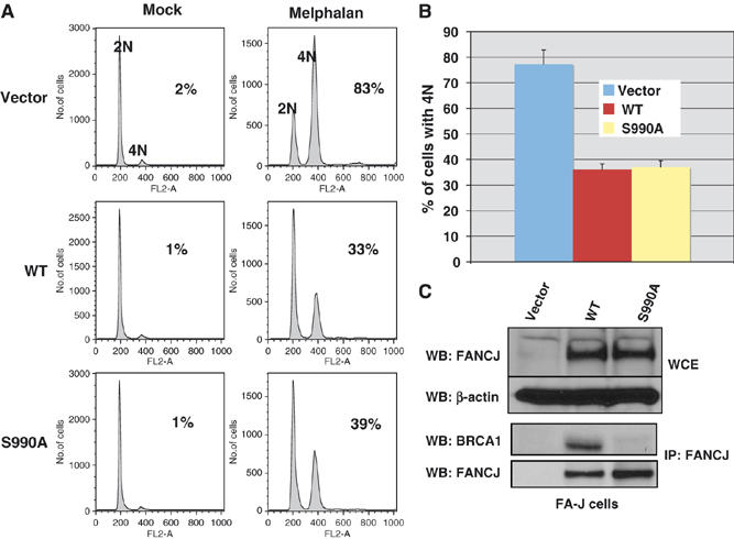
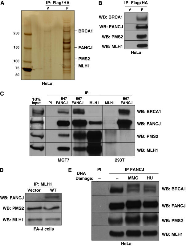
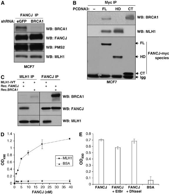
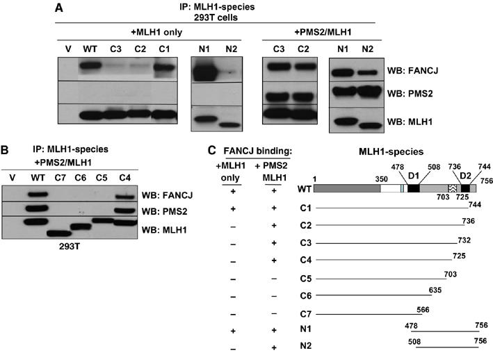
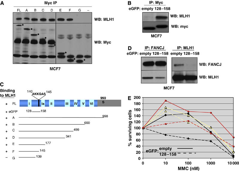
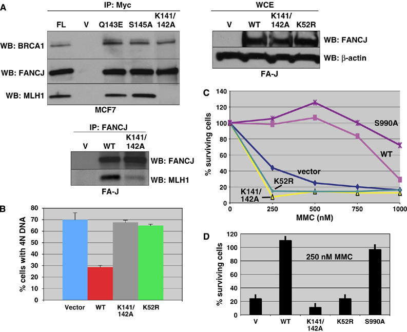
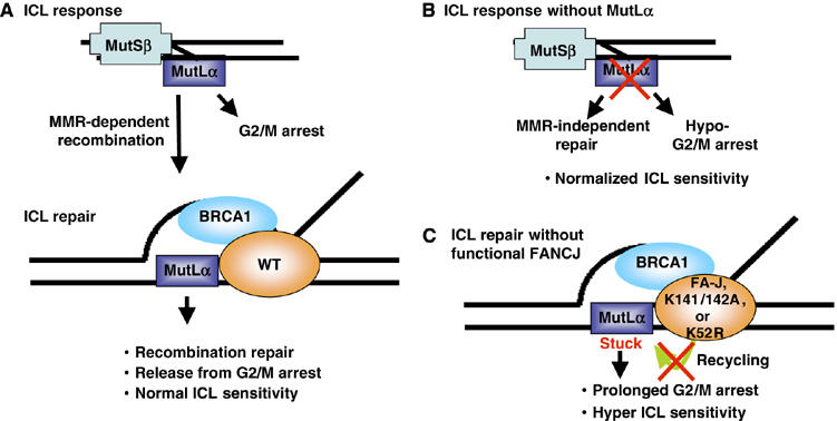
FANCJ also called BACH1/BRIP1 was first linked to hereditary breast cancer through its direct interaction with BRCA1. FANCJ was also recently identified as a Fanconi anemia (FA) gene product, establishing FANCJ as an essential tumor suppressor. Similar to other FA cells, FANCJ-null (FA-J) cells accumulate 4N DNA content in response to DNA interstrand crosslinks (ICLs). This accumulation is corrected by reintroduction of wild-type FANCJ. Here, we show that FANCJ interacts with the mismatch repair complex MutLalpha, composed of PMS2 and MLH1. Specifically, FANCJ directly interacts with MLH1 independent of BRCA1, through its helicase domain. Genetic studies reveal that FANCJ helicase activity and MLH1 binding, but not BRCA1 binding, are essential to correct the FA-J cells' ICL-induced 4N DNA accumulation and sensitivity to ICLs. These results suggest that the FANCJ/MutLalpha interaction, but not FANCJ/BRCA1 interaction, is essential for establishment of a normal ICL-induced response. The functional role of the FANCJ/MutLalpha complex demonstrates a novel link between FA and MMR, and predicts a broader role for FANCJ in DNA damage signaling independent of BRCA1.


Figures

Figure 1
Figure 1
The FANCJ/BRCA1 interaction is dispensable for correction of the 4N DNA accumulation defect in FA-J cells. (A) FA-J cells were reconstituted with vector, WT or S990A, and FANCJ expression was analyzed in whole-cell extracts (WCE) by Western blot. β-Actin served as a loading control for the WCE samples. (B) Immunoprecipitations with FANCJ (E67) were analyzed by Western blot with the indicated Abs. (C) FA-J cells reconstituted with vector, WT, or S990A FANCJ were either left untreated or treated with melphalan, and the percentage of cells with 4N DNA content was analyzed by FACS. The percentage of cells with 4N DNA content after ICL-treatment was averaged for each cell line from four independent experiments, with standard deviation (s.d.) indicated by error bars.
Figure 2
Figure 2
FANCJ interacts with the MMR proteins MLH1 and PMS2. (A) Silver-stained gel of the WT FANCJ (F) compared to vector (V)-purified complexes from HeLa S3 cells by consecutive Flag and HA purification steps (Flag/HA). Identified unique bands are indicated and FANCJ is observed as two species, the 140 kDa band is labeled. (B) Western blot detection of Flag/HA-purified FANCJ complexes. (C) Immunoprecipitations with either FANCJ (E67 or E47) or MLH1 Abs from MCF7 or 293T cells were analyzed by Western blot with the indicated Abs. (D) Western blot shows the presence of the indicated proteins from MLH1 IPs from FA-J cells reconstituted with vector or WT FANCJ. (E) HeLa cells were either left untreated or treated with 1 mM HU for 24 h or 2.4 μg/ml MMC for 1 h. HeLa cell lysates were IPed with PI or FANCJ Abs followed by Western blot analysis with the indicated Abs.
Figure 3
Figure 3
FANCJ helicase domain associates with the MutLα complex independent of BRCA1 and through a direct interaction with MLH1. (A) MCF7 cells were stably infected with a lentivirus encoding shRNA for either eGFP or BRCA1. FANCJ IP was performed followed by Western blot for the indicated proteins. (B) MCF7 cells were transiently transfected with pCDNA3 vectors containing no insert (−), full-length FANCJ (FL), helicase domain including amino-acid residues 1–882 (HD) or C-terminus including residues 882–1249 (CT) of FANCJ, and then IPed with the Myc Ab (9E10). Arrows indicate the respective FANCJ myc-tagged species. Immunoglobulin (IgG) is shown. (C) Western blot of the indicated IP experiments in which in vitro translated MLH1 was incubated with recombinant FANCJ or BRCA1 proteins. (D) Purified recombinant MLH1 or BSA was coated onto ELISA plates. Following blocking with 3% BSA, the wells were incubated with increasing concentrations of purified recombinant FANCJ (0–40 nM) for 1 h at 30°C, and bound FANCJ was detected by ELISA using a rabbit polyclonal Ab against FANCJ, followed by incubation with secondary horseradish peroxidase (HRP)-labeled Abs and OPD substrate. Data points are the mean of three independent experiments performed in duplicate, with s.d. indicated by error bars. (E) ELISA was performed as described in panel D using 4.9 nM FANCJ alone or in the presence of EtBr (50 μg/ml) or DNaseI (2 μg/ml). BSA (3%) was used as a control instead of MLH1 during the coating step.
Figure 4
Figure 4
PMS2 facilitates the FANCJ interaction with the MLH1 C-terminus. (A) MLH1 or Myc (9E10) IP experiments were performed from 293T cells that were transfected with vector alone (V), full-length MLH1 (WT) or MLH1 species, alone or in combination with PMS2 (C1–C3, N1, N2). IP products were analyzed by Western blot with FANCJ, PMS2, and MLH1 Abs. (B) MLH1 IP experiments were performed from 293T cells that were transfected with vector alone (V), full-length MLH1 (WT) or MLH1 species, in combination with PMS2 (C4–C7). IP products were analyzed by Western blot with FANCJ, PMS2, and MLH1 Abs. (C) Schematic representation of the MLH1/FANCJ dimer domains (D1, D2), and the region between 703–725 is highlighted as an essential element for maintaining the MLH1/PMS2/FANCJ complex.
Figure 5
Figure 5
Expression of FANCJ residues 128–158 disrupts the FANCJ/MLH1 interaction to generate ICL sensitivity. (A) Myc (9E10) IP experiments were performed from MCF7 cells that were transfected with vector alone (−), full-length FANCJ (FL) and the different FANCJ constructs (A–G) shown in panel C, followed by Western blot with MLH1, and Myc Abs. The asterisk denotes the migration of the different myc-tagged FANCJ species. (B, D) Myc IP experiments were performed from MCF7 cells that were transfected with either eGFP empty vector or the 128–158 FANCJ-eGFP constructs, followed by Western blot with the indicated Abs. (C) The different FANCJ constructs are indicated with a positive (+) or negative (−) to indicate binding to MLH1. (E) MCF7 cells transfected with vector alone or the 128–158 FANCJ-eGFP construct, treated with increasing concentrations of MMC and incubated for 4–5 days. Cell growth was measured by ATP content. Three independent representative experiments are shown and depicted by lines with squares, triangles, and diamonds. Solid lines represent cells transfected with empty-eGFP vector and hatched lines represent cells transfected with 128–158-eGFP vector (for color figure see online version).
Figure 6
Figure 6
MLH1 binding to FANCJ is essential to correct FA-J cells. (A) Myc IP experiments were performed from MCF7 cells that were transfected with vector alone (−), FL, V, Q143E, S145A, and K141/142A FANCJ constructs, followed by Western blot with the indicated Abs. FA-J cells were reconstituted with empty vector, WT, K141/142A, or K52R FANCJ vectors, and FANCJ expression was analyzed by whole-cell extracts; β-actin served as a loading control for the WCE samples. Western blot shows the presence of the indicated proteins from FANCJ IPs from FA-J cells reconstituted with vector, WT, or K141/142A FANCJ. (B) FA-J cell lines reconstituted with empty vector, WT, K141/142A, or K52R FANCJ were either left untreated or treated with melphalan. The percentage of cells with 4N DNA content after ICL treatment was averaged for each cell line from four independent experiments, with standard deviation (s.d.) indicated by error bars. (C) FA-J cells reconstituted with vector, WT, K141/142A, K52R, or S990A FANCJ were seeded on 24-well plates and incubated overnight under normal growth conditions. The cells were then treated with the indicated doses of MMC and incubated for 8 days. On the final day, the cells were counted and the percentage of live cells was calculated. Experiments were performed in triplicate and a representative plot is shown. (D) The IC50 dose for the FA-J vector (250 nM) was compared for all mutants, and error bars represent the standard deviation.
Figure 7
Figure 7
Model depicting how FANCJ and MutLα proteins function to mediate the ICL response. (A) The normal ICL response is proposed to include MMR proteins. This is supported by the findings that the MMR machinery has been shown to specifically bind crosslinked DNA adducts (Duckett et al, 1996; Yamada et al, 1997; Zhang et al, 2002), mediate ICL recombination repair (Zheng et al, 2006) and induce a G2/M arrest (Cejka et al, 2003). Furthermore, MMR proteins including the MutLα complex, similar to BRCA1 and FANCJ are essential for recombination processes (de Wind et al, 1995; Jasin, 2002; Mohindra et al, 2002; Litman et al, 2005). (B) The ICL response without MutLα is predicted to lead to a MMR-independent non-recombination-based mechanism with a minimal G2/M arrest, so that ICL sensitivity is normalized. (C) ICL repair without functional FANCJ is predicted to be directed to recombination as in panel A, but generates a prolonged G2/M arrest, due to absent or dysfunctional FANCJ protein. In the absence of FANCJ protein, helicase activity or MLH1 binding, FANCJ is unable to displace MutLα from recombination intermediates, and consequently, the MutLα complex remains stuck or tethered to DNA for a longer time period, delaying the exit from the G2/M arrest and enhancing ICL sensitivity.

References

    1. Akkari YM, Bateman RL, Reifsteck CA, D'Andrea AD, Olson SB, Grompe M (2001) The 4N cell cycle delay in Fanconi anemia reflects growth arrest in late S phase. Mol Genet Metab 74: 403–412 - PubMed
    1. Andreassen PR, D'Andrea AD, Taniguchi T (2004) ATR couples FANCD2 monoubiquitination to the DNA-damage response. Genes Dev 18: 1958–1963 - PMC - PubMed
    1. Aquilina G, Ceccotti S, Martinelli S, Hampson R, Bignami M (1998) N-(2-chloroethyl)-N′-cyclohexyl-N-nitrosourea sensitivity in mismatch repair-defective human cells. Cancer Res 58: 135–141 - PubMed
    1. Bridge WL, Vandenberg CJ, Franklin RJ, Hiom K (2005) The BRIP1 helicase functions independently of BRCA1 in the Fanconi anemia pathway for DNA crosslink repair. Nat Genet 37: 953–957 - PubMed
    1. Brown KD, Rathi A, Kamath R, Beardsley DI, Zhan Q, Mannino JL, Baskaran R (2003) The mismatch repair system is required for S-phase checkpoint activation. Nat Genet 33: 80–84 - PubMed

MeSH terms