Review
. 2007 Aug 29;362(1484):1421-36.
doi: 10.1098/rstb.2007.2125.

Heart valve macro- and microstructure

Affiliations
Review

Heart valve macro- and microstructure

Martin Misfeld et al. Philos Trans R Soc Lond B Biol Sci. .

Abstract

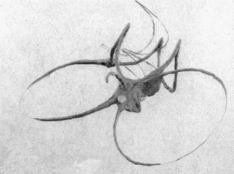
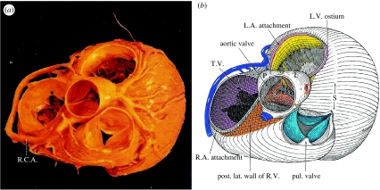
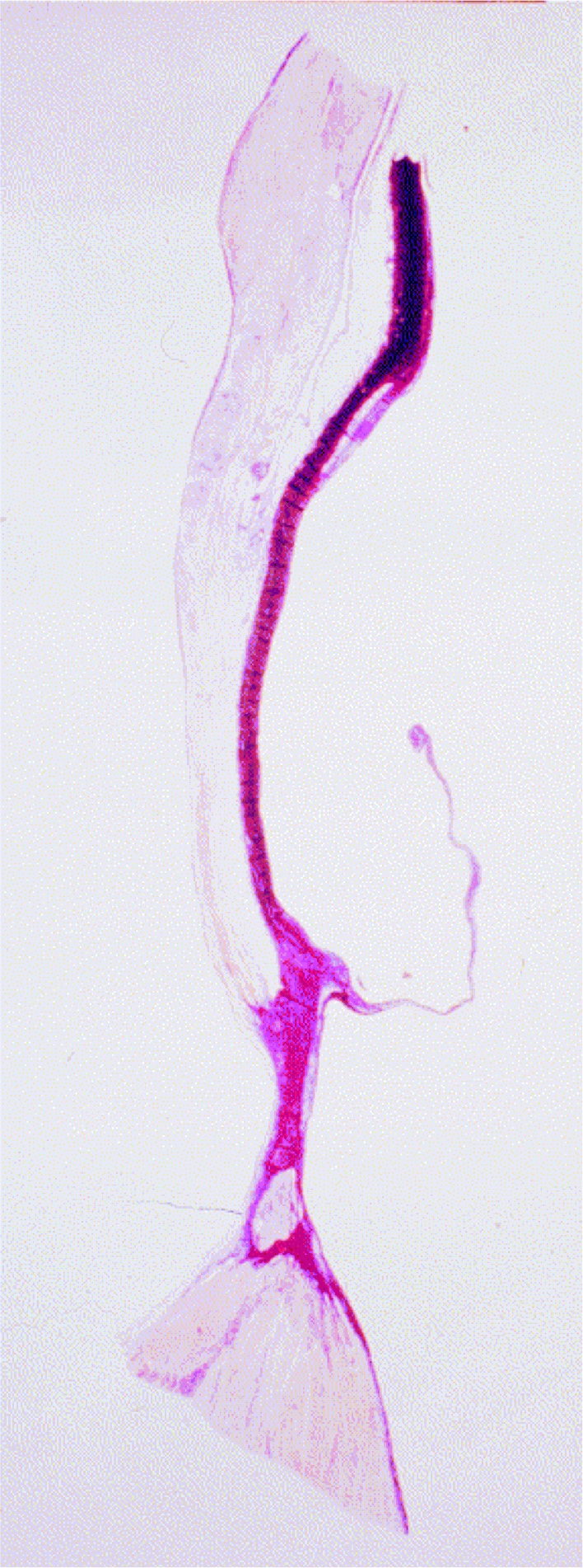
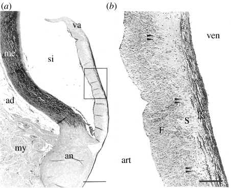
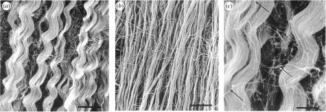
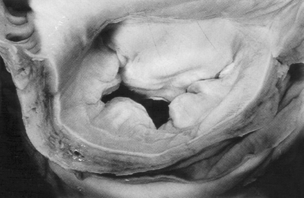
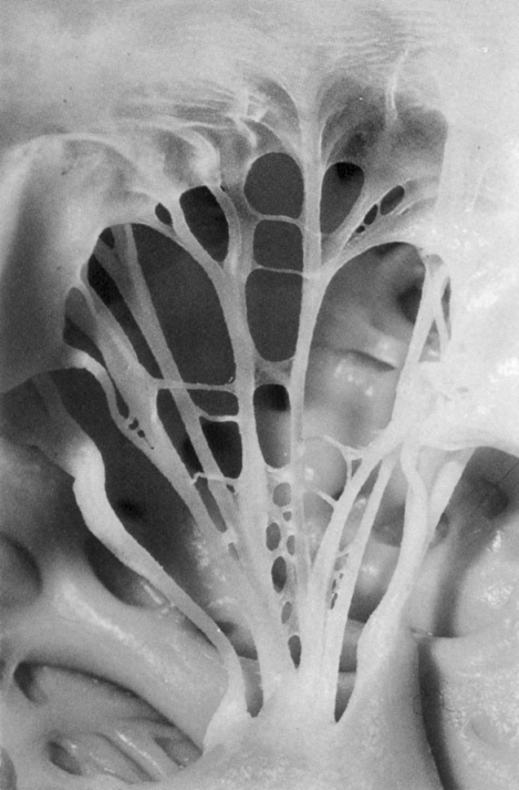
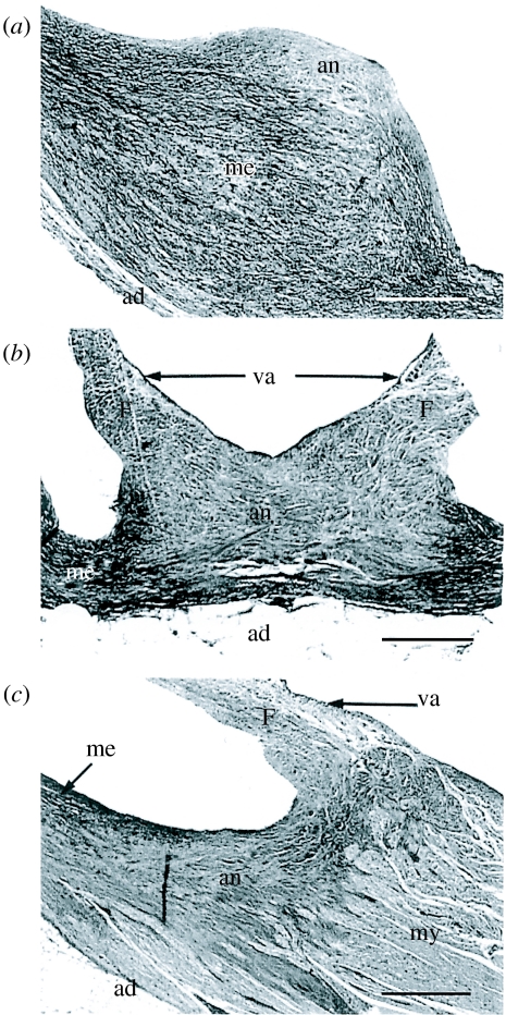
Each heart valve is composed of different structures of which each one has its own histological profile. Although the aortic and the pulmonary valves as well as the mitral and the tricuspid valves show similarities in their architecture, they are individually designed to ensure optimal function with regard to their role in the cardiac cycle. In this article, we systematically describe the structural elements of the four heart valves by different anatomical, light- and electron-microscopic techniques that have been presented. Without the demand of completeness, we describe main structural features that are in our opinion of importance in understanding heart valve performance. These features will also have important implications in the treatment of heart valve disease. They will increase the knowledge in the design of valve substitutes or partial substitutes and may participate to improve reconstructive techniques. In addition, understanding heart valve macro- and microstructure may also be of benefit in heart valve engineering techniques.


Figures

Figure 1
Figure 1
View on the four heart valves from superior (adapted from Anderson & Becker 1982, p. 18).
Figure 2
Figure 2
Schematic drawing of the fibrous skeleton of the heart (adapted from Zimmerman & Bailey 1962).
Figure 3
Figure 3
(a) View of the four heart valves from superior and (b) schematic drawing of the relationship to the right and left ventricle (S, summit of the left ventricle (L.V.), R.C.A., right coronary artery; T.V., tricuspid valve; L.A., left atrium; R.A., right atrium; R.V., right ventricle (adapted from McAlpine 1975, p. 6).
Figure 4
Figure 4
Schematic drawing of aortic root structures after longitudinal opening of the root.
Figure 5
Figure 5
Section through the aortic root (adapted from Yacoub et al.1999).
Figure 6
Figure 6
(a,b) View of the mitral valve (M.V.) and aortic root after removal of the atria. Note the large intervalvular trigone (I.V.T). R.P.O., right posterior oblique; T.V., tricuspid valve; R.A., right atrium; L.A., left atrium; J, junction between the atrioventricular valves. (adapted from McAlpine 1975, p. 13).
Figure 7
Figure 7
View of the right coronary leaflet. The lannula is approximately one-third of the leaflet height. The nodulus of Arantii forms a triangle (adapted from McAlpine 1975, p. 22).
Figure 8
Figure 8
Electron micrograph of annulus tissue. Inside a network of collagenous fibrils and fibroblasts, a non-myelinated nerve can be identified. 1a, fibroblast; 2, collagenous fibrils; 5, non-myelinated nerve (magnification × 6900).
Figure 9
Figure 9
Electron micrograph of commissure tissue in the area of the endothelial cell layer. The endothelial cells are separated by the basal layer from the elastic fibres and collagenous fibrils. The endothelial cells show microvilli at their surface, which increase the overall surface area for an increased exchange of substances. 2, collagenous fibrils; 3, endothelial cell; 3a, basal layer; *, microvilli (magnification × 6900).
Figure 10
Figure 10
(a) Section through the aortic root showing the attachment of the leaflet to the annulus. (b) Magnification of the leaflet demonstrating the three layers. art, arterial side; ven, ventricular side; si, sinus; va, valvula; me, tunica media aortae; ad, adventitia; my, myocardium; an, annulus; F, lamina fibrosa; S, lamina spongiosa; R, lamina radialis; arrows indicate the transmission zone between the lamina spongiosa and fibrosa. Scale bars, 144 μm (adapted from Fastenrath 1995, p. 35).
Figure 11
Figure 11
Electron micrograph of the arrangement of collagenous fibrils of the lamina radialis of an aortic valve leaflet at different magnifications. Arrows indicate non-directional fibrils surrounding helical arranged collagenous fibrils. (a,b) Scale bars, 8 μm and (c) 3 μm (adapted from Fastenrath 1995, p. 43).
Figure 12
Figure 12
Photomicrographs showing immuno-histochemical staining of an aortic valve leaflet with antibodies against smooth muscle cell alpha-actin. Arrows indicate muscle fibres (a, magnification × 25; b, magnification × 40).
Figure 13
Figure 13
Section through the left ventricle demonstrating the mitral valve apparatus and its relation to the aortic valve (adapted from Anderson & Becker 1982, p. 72).
Figure 14
Figure 14
Non-planar model of the mitral valve annulus showing saddle-shaped three-dimensional geometry (adapted from Salgo et al. 2002).
Figure 15
Figure 15
Section through the posterior mitral annulus showing the insertion of the posterior mitral leaflet (adapted from Anderson & Becker 1982, p. 70).
Figure 16
Figure 16
View of the mitral valve leaflets from the atrial side. The three parts of the posterior leaflet and its relation to the anterior leaflet are shown (adapted from Anderson & Becker 1982, p. 68).
Figure 17
Figure 17
View of the mitral valve apparatus. The lateral papillary muscle and the superior commissure (S.C.) are shown where the anterior (A.M.V.) and posterior leaflet (P.M.V.) confluent (adapted from McAlpine 1975, p. 40).
Figure 18
Figure 18
View of the commissural chordae tendinae inserting at the free margin of two adjoined leaflets (adapted from Anderson & Becker 1982, p. 47).
Figure 19
Figure 19
View of the pulmonary valve. L.A.L., left anterior leaflet; R.A.L., right anterior leaflet; Posterior, posterior leaflet; arrows indicate the three commissures (adapted from McAlpine 1975, p. 74).
Figure 20
Figure 20
Section through the pulmonary root. The area of attachment of the pulmonary leaflet can be seen (adapted from Anderson & Becker 1982, p. 49).
Figure 21
Figure 21
Sections through the annulus of the pulmonary root. (a) Upper part of the commissure, (b) middle part with the attachment of two leaflets, (c) basal part of the annulus. an, annulus; me, tunica media; ad, adventitia; my, myocardium; va, valvula; F, lamina fibrosa. Scale bar, 517 μm (adapted from Fastenrath 1995, p. 65).
Figure 22
Figure 22
(a) Section through the pulmonary root, va, valvula; me, tunica media; si, sinus; ad, adventitia; my, myocardium; arrows, zone of the annulus; scale bar, 1.1 mm. (b) Enlargement of the leaflet showing the different layers of the leaflet, ven, ventricular side; art, arterial side; V, lamina ventricularis; R, lamina radialis; S, lamina spongiosa; F, lamina fibrosa; arrows, area between lamina spongiosa and fibrosa. Scale bar, 83 μm (adapted from Fastenrath 1995, p. 35).
Figure 23
Figure 23
(a,b) Electronmicroscopic section of collagenous fibrils of the lamina fibrosa of the pulmonary leaflet. The bundles are not strictly arranged in a parallel fashion (arrows on the right), but interweave with each other in an acute-angle (adapted from Fastenrath 1995, p. 53). Scale bars: (a) 83 μm, (b) 17 μm.
Figure 24
Figure 24
View of the tricuspid valve (adapted from Anderson & Becker 1982, p. 60).
Figure 25
Figure 25
View of an isolated tricuspid valve showing part of the subvalvular apparatus. The septal leaflet (X) is supported by chordae tendinae originating from the inferior papillary muscle IPM and the anterior papillary muscle APM (adapted from McAlpine 1975, p. 74).
Figure 26
Figure 26
Section through the tricuspid valve (adapted from Anderson & Becker 1982, p. 60).

References

    1. Adamczyk M.M, Lee T.C, Vesely I. Biaxial strain properties of elastase-digested porcine aortic valves. J. Heart Valve Dis. 2000;9:445–453. - PubMed
    1. Aktas E.O, Govsa F, Kocak A, Boydak B, Yavuc I.C. Variations in the papillary muscle of normal tricuspid valves and their clinical relevance in medicolegal autopsies. Saudi Med. J. 2004;25:1176–1185. - PubMed
    1. Anderson R.H. Clinical anatomy of the aortic root. Heart. 2000;84:670–673. doi:10.1136/heart.84.6.670 - DOI - PMC - PubMed
    1. Anderson R.H, Becker A.E. Thieme; Stuttgart, NY: 1982. Anatomy of the heart.
    1. Anderson R.H, Devine W.A, Ho S.Y, Smith A, McKay R. The myth of the aortic annulus: the anatomy of the subaortic outflow tract. Ann. Thorac. Surg. 1991;52:640–646. - PubMed

LinkOut - more resources