. 2007 Jul 3;104(27):11298-303.
doi: 10.1073/pnas.0703723104. Epub 2007 Jun 20.

Hyaluronic acid hydrogel for controlled self-renewal and differentiation of human embryonic stem cells

Affiliations

Hyaluronic acid hydrogel for controlled self-renewal and differentiation of human embryonic stem cells

Sharon Gerecht et al. Proc Natl Acad Sci U S A. .

Abstract

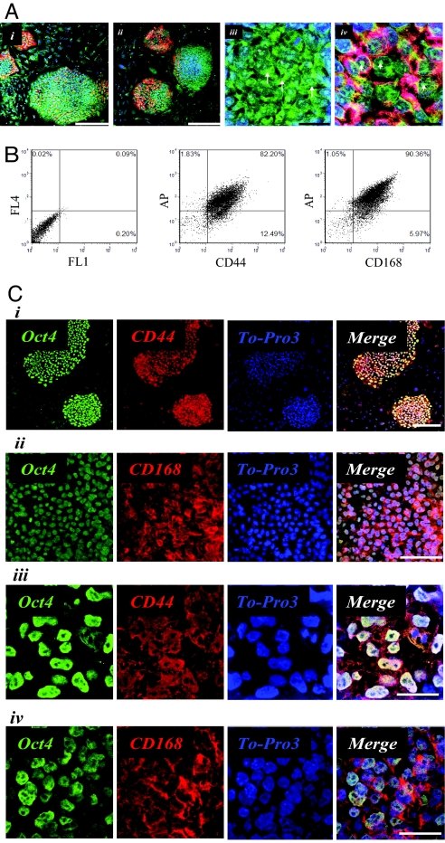
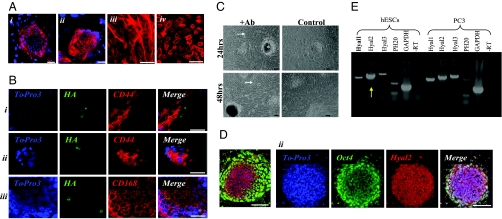
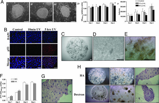
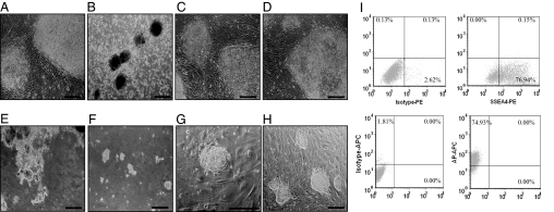
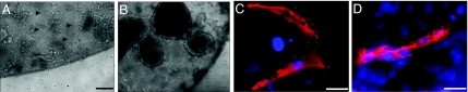
Control of self-renewal and differentiation of human ES cells (hESCs) remains a challenge. This is largely due to the use of culture systems that involve poorly defined animal products and do not mimic the normal developmental milieu. Routine protocols involve the propagation of hESCs on mouse fibroblast or human feeder layers, enzymatic cell removal, and spontaneous differentiation in cultures of embryoid bodies, and each of these steps involves significant variability of culture conditions. We report that a completely synthetic hydrogel matrix can support (i) long-term self-renewal of hESCs in the presence of conditioned medium from mouse embryonic fibroblast feeder layers, and (ii) direct cell differentiation. Hyaluronic acid (HA) hydrogels were selected because of the role of HA in early development and feeder layer cultures of hESCs and the controllability of hydrogel architecture, mechanics, and degradation. When encapsulated in 3D HA hydrogels (but not within other hydrogels or in monolayer cultures on HA), hESCs maintained their undifferentiated state, preserved their normal karyotype, and maintained their full differentiation capacity as indicated by embryoid body formation. Differentiation could be induced within the same hydrogel by simply altering soluble factors. We therefore propose that HA hydrogels, with their developmentally relevant composition and tunable physical properties, provide a unique microenvironment for the self-renewal and differentiation of hESCs.


Conflict of interest statement

The authors declare no conflict of interest.

Figures

Fig. 1.
Fig. 1.
HA plays a role during hESC culture on MEFs. (A) Staining of hESCs (H1 line) grown on MEFs for HA binding site (green), undifferentiated membrane marker TRA-1–81 (red), and nuclei (blue): Intracellular localization of HA (Ai and Aii), including perinuclear areas (arrows) (Aiii) and nuclei (∗), and nucleoli (arrowheads) (Aiv). (B) FACS analysis revealed that compared with isotype control (Left), the majority of undifferentiated hESCs were found to express HA receptors CD44 (82%) (Center) and CD168 (90%) (Right). (Ci and Cii) By using immunofluorescence staining, undifferentiated hESC colonies were easily detected with undifferentiated cell markers Oct4 (green) and CD44 or CD168 (red), respectively (nuclei: blue). (Ciii and Civ) Higher magnification suggests intracellular expression of CD44 and either membrane or intracellular expression of CD168. (Scale bars: Ai, Aii, Ci, and Cii, 100 μm; Aiii, Ciii, and Civ, 25 μm; Aiv, 10 μm.)
Fig. 2.
Fig. 2.
HA interaction with hESCs. (A) Localization of HA receptors in response to addition of human FL-HA to the growth medium of hESCs (H9 line) cultured on MEFs. (Ai and Aii) Confocal analysis suggests relocalization of HA receptors in cell membranes of both CD44 (Ai) and CD168 (shown in red, nuclei shown in blue) (Aii). (Aiii and Aiv) Higher magnification of CD168 localization is shown with (Aiii) and without (Aiv) the addition of human HA. (B) HA uptake by hESC (H9 line) colonies. Undifferentiated hESCs (H1 line) grown on MEFs were incubated overnight with fluorescein-HA and further stained for CD44 and CD168. (Bi) Edge of colony suggests internalizing FL-HA via CD44. (Bii and Biii) Intracellular localization of FL-HA. (C) Seeding of hESCs (H9 line) in the presence of anti-CD44 and CD168 resulted in less and differentiated colonies (both at the edge and center of the colonies, as indicated by arrows and asterisks, respectively), whereas control cultures contain expanding undifferentiated colonies. (D) Human ESC (H13 line) colonies grown on MEFs positive for Oct4 (green) express Hyal 1 (Di) or Hyal 2 (Dii) (red; nuclei are shown in blue) mainly in densely packed areas of the colonies. (E) RT-PCR analysis revealed high expression levels of a hyaluronidase isomer, Hyal 2, in undifferentiated hESCs (H9 line). PC3 line served as positive control. (Scale bars: 100 μm.)
Fig. 3.
Fig. 3.
HA hydrogels support the maintenance of viable hESCs in their undifferentiated state. (Ai–Aiv) Undifferentiated hESCs (H9 line) were passaged and recultured on feeder layers for 4 days in culture medium containing: no macromer (Ai), 10 μl/ml macromer (corresponding to the hydrogel containing 80% nonpolymerized monomer) (Aii), or 50 μl/ml macromer (corresponding to completely nonpolymerized monomer) (Aiii). Toxic effects were detected only at the macromer concentration of 50 μl/ml. (Aiv and Av) XTT assay (Aiv) and cell count (Av) revealed no negative effect of macromer on cell viability at a concentration of 10 μl/ml and a slight decrease in hESC viability at a macromer concentration of 50 μl/ml. Results are presented as means ± SD (∗, P < 0.05). (B) Low basal levels of p53 are expressed by hESCs (H13 line) 12 h postincubation after 10 minutes of UV exposure, whereas accumulation of p53 in hESCs was observed 12 h postincubation after 5 h UV exposure. (C and D) Light microscopy revealed uniform distribution of hESC colonies in HA gels (C) with a range of colony sizes (D). (E) Incubation with XTT revealed orange dye in metabolically active hESCs (H13 line) encapsulated in HA hydrogels. (F) XTT assay shows comparable growth rates of hESCs encapsulated in HA hydrogels and on Matrigel. (G) A representative histological section of hESC-HA constructs (H9 line) cultured for 20 days demonstrates uniform morphology (H&E stain) of undifferentiated colony within 3D networks (folding of the hydrogel is indicated by an asterisk). (H) Encapsulation of hESCs (H13 line) in HA hydrogels was compared with dextran hydrogels after 15 days of culture. Light microscope images of both cultures at low and high magnifications and histological sections (H&E stain) demonstrate EB formation in dextran hydrogels vs. colony arrangements of undifferentiated hESCs in HA hydrogels. (Scale bars: A, C–F, and H, 100 μm; B and G, 25 μm.)
Fig. 4.
Fig. 4.
Cell release from hydrogels and cell karyotyping. (A–D) hESCs (H13 line) grown on MEFs were incubated for 24 h in growth medium (A), 1% collagenase solution in growth medium (B), 1,000 units/ml hyaluronidase solution in growth medium (C), and 2,000 units/ml hyaluronidase solution in growth medium (D). To release hESCs from HA hydrogel, constructs were incubated with 2,000 units/ml hyaluronidase in growth medium. (E) After 18 h, small particles of hydrogels remained that trapped hESCs. (F) After 24 h, hESCs colonies were completely released from the hydrogel. (G and H) hESCs (H9 line) released from the hydrogel after 30 days of encapsulation and cultured on MEFs formed small colonies of undifferentiated cells after 24 h (G) and were propagated on MEFs for three passages (H). (I) FACS analyses of released cells after 20 days of HA culture revealed high levels of SSEA4 and alkaline phosphatase. (Scale bars: 100 μm.)
Fig. 5.
Fig. 5.
Differentiation. H9 line cells were cultured in conditioned medium for 1 week followed by the replacement of medium containing VEGF. (A and B) Cell sprouting was observed after 48 h in gels transferred to medium containing VEGF (arrows) (A) compared with gels continuously cultured in conditioned medium (B). (C and D) After 1 week of differentiation, sprouting elongating cells were mainly positive for vascular α-smooth muscle actin (C), whereas some were positive for early stage endothelial marker (D). CD34 (in situ 3D staining of gels). (Scale bars: A and B, 100 μm; C and D, 25 μm.)

References

    1. Thomson JA, Itskovitz-Eldor J, Shapiro SS, Waknitz MA, Swiergiel JJ, Marshall VS, Jones JM. Science. 1998;282:1145–1147. - PubMed
    1. Xu C, Inokuma MS, Denham J, Golds K, Kundu P, Gold JD, Carpenter MK. Nat Biotechnol. 2001;19:971–974. - PubMed
    1. Richards M, Fong CY, Chan WK, Wong PC, Bongso A. Nat Biotechnol. 2002;20:933–936. - PubMed
    1. Stoikovic P, Lako M, Przyborski S, Stewart R, Armstong L, Evans J, Zhang X, Stojkpvic M. Stem Cells. 2005;23:895–902. - PubMed
    1. Itskovitz-Eldor J, Schuldiner M, Karsenti D, Eden A, Yanuka O, Amit M, Soreq H, Benvenisty N. Mol Med. 2000;6:88–95. - PMC - PubMed

Publication types