Comparative Study
. 2007 Jun 20;27(25):6751-9.
doi: 10.1523/JNEUROSCI.1337-07.2007.

NKCC1 phosphorylation stimulates neurite growth of injured adult sensory neurons

Affiliations
Comparative Study

NKCC1 phosphorylation stimulates neurite growth of injured adult sensory neurons

Simon Pieraut et al. J Neurosci. .

Abstract

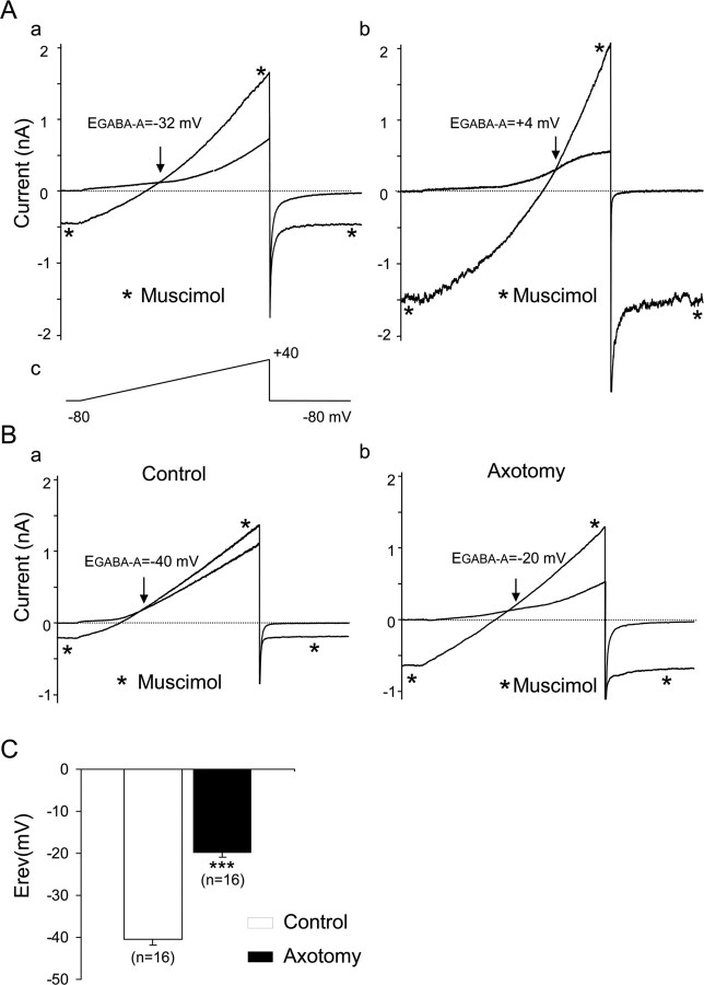
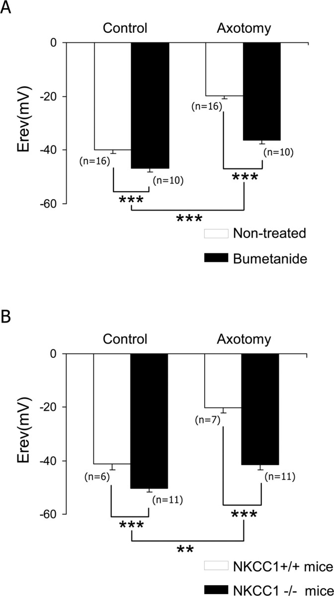
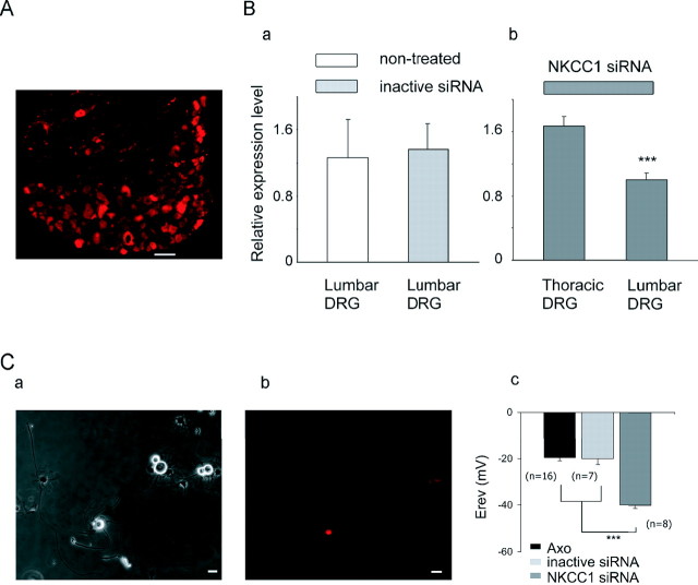
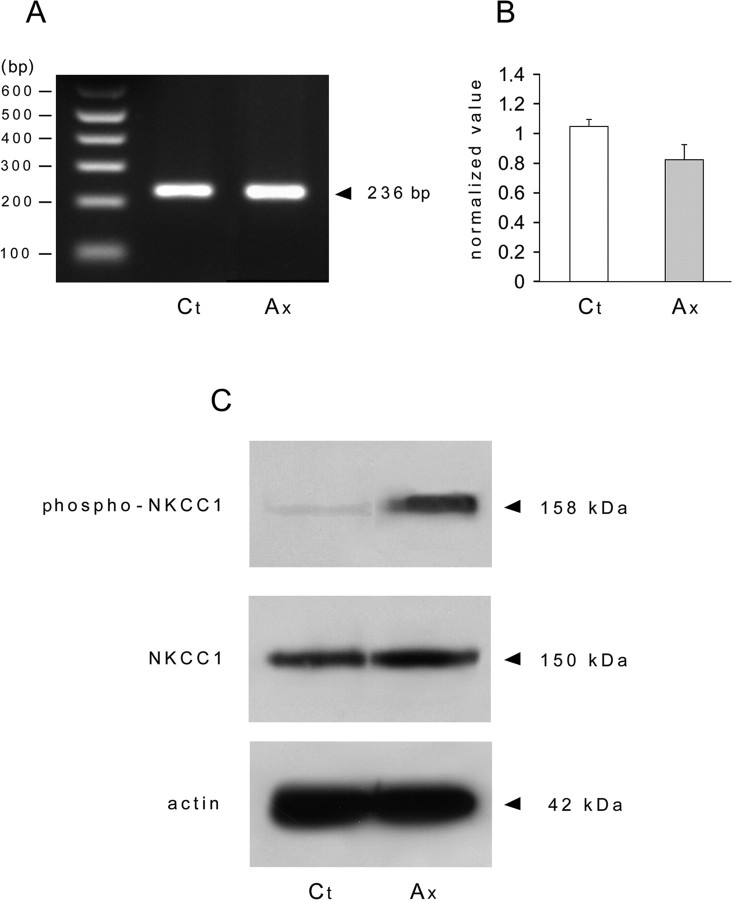
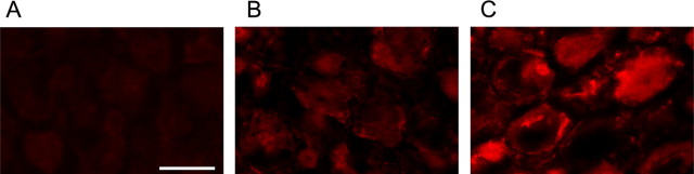
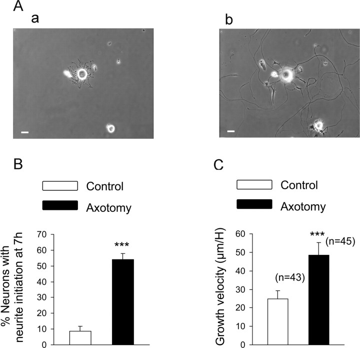
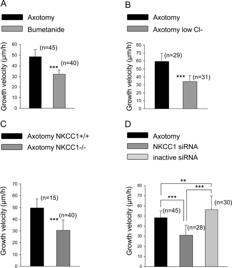
Peripheral nerve section promotes regenerative, elongated neuritic growth of adult sensory neurons. Although the role of chloride homeostasis, through the regulation of ionotropic GABA receptors, in the growth status of immature neurons in the CNS begins to emerge, nothing is known of its role in the regenerative growth of injured adult neurons. To analyze the intracellular Cl- variation after a sciatic nerve section in vivo, gramicidin perforated-patch recordings were used to study muscimol-induced currents in mice dorsal root ganglion neurons isolated from control and axotomized neurons. We show that the reversal potential of muscimol-induced current, E(GABA-A), was shifted toward depolarized potentials in axotomized neurons. This was attributable to Cl- influx because removal of extracellular Cl- prevented this shift. Application of bumetanide, an inhibitor of NKCC1 cotransporter and E(GABA-A) recordings in sensory neurons from NKCC1-/- mice, identified NKCC1 as being responsible for the increase in intracellular Cl- in axotomized neurons. In addition, we demonstrate with a phospho-NKCC1 antibody that nerve injury induces an increase in the phosphorylated form of NKCC1 in dorsal root ganglia that could account for intracellular Cl- accumulation. Time-lapse recordings of the neuritic growth of axotomized neurons show a faster growth velocity compared with control. Bumetanide, the intrathecal injection of NKCC1 small interfering RNA, and the use of NKCC1-/- mice demonstrated that NKCC1 is involved in determining the velocity of elongated growth of axotomized neurons. Our results clearly show that NKCC1-induced increase in intracellular chloride concentration is a major event accompanying peripheral nerve regeneration.


Figures

Figure 1.
Figure 1.
Reversal potential of GABAA receptor-mediated chloride current in control and axotomized mouse sensory neurons. A, Conventional whole-cell patch-clamp recordings with a patch electrode containing 30 mm Cl (a) and 140 mm Cl (b). Ramp protocols from −80 to +40 mV were applied every 5 s in a Na-free, K-free, 147 mm TEA-Cl extracellular solution (c). To reduce Ca2+ currents amplitude, 100 μm NiCl2 and CdCl2 were added. Under these experimental conditions, the voltage ramp elicited a small-amplitude outwardly rectifying conductance. Application of 100 μm muscimol to activate the GABAA current induced an increase in slope conductance. Reversal potential of GABAA current, EGABA-A, was determined at the intersection point between control and muscimol conductance. Consistent with a muscimol-induced chloride current, EGABA-A was −32 mV in 30 mm (a) and +4 mV in 140 mm intracellular Cl (b) in these neurons. B, Gramicidin-perforated patch recordings were used to determine [Cl]i. EGABA-A was − 40 mV in a control neuron (a) and −20 mV in an axotomized neuron (b). C, The mean EGABA-A in control was significantly less depolarized than in axotomized neurons (***p < 0.001, Student's t test). The Nernst equation was used to calculate [Cl]i, averaging 30.7 ± 1.6 mm, n = 16 and 68.4 ± 3.2 mm, n = 16 in control and axotomized neurons, respectively.
Figure 2.
Figure 2.
NKCC1 is responsible for [Cl]i increase in axotomized neurons. A, Neuronal cultures pretreated for 1 h with 10 μm bumetanide showed a shift of EGABA-A toward hyperpolarized values in both control and axotomized neurons, and the main effect of bumetanide was associated with axotomy. EGABA-A remains significantly more depolarized in bumetanide-treated axotomized neurons than in bumetanide-treated control neurons (p < 0.001; data not shown). B, Analysis of the NKCC1−/− mouse demonstrated a shift in EGABA-A toward hyperpolarized values in control and axotomized neurons, and the main effect of NKCC1 deletion was associated with axotomy. EGABA-A remains significantly more depolarized in NKCC1−/− axotomized neurons than in NKCC1−/− control neurons (p < 0.001; data not shown). **p < 0.01, ***p < 0.001, two-way ANOVA.
Figure 3.
Figure 3.
NKCC1 is responsible for [Cl]i increase in axotomized neurons. Gene inhibition of NKCC1 was achieved after NKCC1 siRNA/dextran–rhodamine intrathecal injection. Transfection was estimated by visualization of fluorescent neurons in DRG or in culture. A, Neuron counting in a slice of L5 DRG shows a near 40% of bright fluorescent neurons. Scale bar, 100 μm. B, qRT-PCR analysis of NKCC1 transcripts in lumbar and thoracic DRG. a, Control siRNA-injected mice shows similar level of NKCC1 transcripts than noninjected mice (n = 4). b, Intrathecal injection of NKCC1 siRNA demonstrates the decrease in NKCC1 transcripts in lumbar compared with thoracic DRG (n = 4; ***p < 0.001, Mann–Whitney U test). C, Phase-contrast photograph of axotomized neurons at 1 d in vitro from L4–L5 DRG after intrathecal siRNA/dextran injection (a). Half of these neurons express rhodamine fluorescence (red) (b). Scale bar, 20 μm. Recordings of [Cl]i with the gramicidin-perforated patch-clamp method demonstrating that most of the fluorescent axotomized neurons showed EGABA-A to be significantly more hyperpolarized in NKCC1 siRNA than in control siRNA-treated animals (c). ***p < 0.001, one-way ANOVA.
Figure 4.
Figure 4.
Nerve injury induces NKCC1 phosphorylation. A, RT-PCR was performed on control and axotomized DRG. The NKCC1 primer amplified a product of the expected size (236 bp) in control (Ct) and axotomized (Ax) DRG whose molecular identity was confirmed by sequencing. B, qRT-PCR analysis of NKCC1 transcript levels in lumbar L4–L5 DRG from control and axotomized mice shows that peripheral nerve injury does not quantitatively modify NKCC1 mRNA (n = 3). C, Western blot of protein expression with T4 (NKCC1) and R5 (phospho-NKCC1) antibodies shows an increase in protein phosphorylation in axotomized DRG. SDS-PAGE was conducted using 20 μg of protein extract from uninjured DRG (control) and 5 d after sciatic nerve section (axotomized DRG). The apparent molecular size markers are indicated on the right. The same extracts were probed for β-actin.
Figure 5.
Figure 5.
Phospho-NKCC1 immunohistochemistry in adult DRG. Phospho-NKCC1 protein expression was studied by immunohistochemistry using the rabbit polyclonal antibody R5 directed against a diphosphopeptide containing Thr212 and Thr217. A, R5 staining in DRG from NKCC1−/− mice is used as negative control for specific staining. B, Control DRG neurons show a weak staining of phospho-NKCC1 (red) in their cytoplasm and at the cell membrane. C, At 5 d after peripheral nerve injury, intense R5 staining is expressed in sensory neurons. The R5 signal (red) is located in the cytoplasm and at the cell membrane. Scale bar, 50 μm.
Figure 6.
Figure 6.
Analysis of the regenerative neurite growth. Time-lapse video microscopy was used to analyze the neuritic growth velocity of control and axotomized sensory neurons. A, Phase-contrast photograph showing neurons from a sciatic nerve injury conditioned culture 3 h (a) and 24 h (b) after plating. Scale bar 20 μm. B, Neurite initiation was evaluated 7 h after plating and estimated as a neurite length longer than one cell diameter. The total number of neurons counted per well ranged in between 20 and 30; eight wells were analyzed per conditions. The percentage of neurons having initiated neurite growth was significantly greater after an axotomy. C, Growth velocity, estimated as the length of a neurite measured each hour for 24 h, is significantly faster in axotomized neurons compared with control neurons. ***p < 0.001, Student's t test.
Figure 7.
Figure 7.
NKCC1 is involved in the regenerative neurite growth. A, Bumetanide at 10 μm was added 2 h after plating, and the culture was processed for time-lapse microscopy. Bumetanide treatment significantly decreased growth velocity of regenerating neurons. B, Axotomized neuron growth velocity was significantly decreased when recorded in a low external Cl solution (in mm: 140 Na-methanesulfonate, 2 CaCl2, 1.5 MgCl2, 5 KCl, 10 glucose, and 10 HEPES). C, Regenerative neuritic growth velocity from NKCC1−/− is significantly faster compared with NKCC1+/+ mice. D, Silencing NKCC1 expression with an intrathecal injection of NKCC1 siRNA once a day for 5 d significantly decreased growth velocity of rhodamine-labeled regenerating neurons. No modification of growth velocity was seen with control siRNA compared with nontreated axotomized neurons. **p < 0.01, ***p < 0.001, one way ANOVA.

References

    1. Alessandri-Haber N, Yeh JJ, Boyd AE, Parada CA, Chen X, Reichling DB, Levine JD. Hypotonicity induces TRPV4-mediated nociception in rat. Neuron. 2003;39:497–511. - PubMed
    1. Alvarez-Leefmans FJ, Leon-Olea M, Mendoza-Sotelo J, Alvarez FJ, Anton B, Garduno R. Immunolocalization of the Na+-K+-2Cl− cotransporter in peripheral nervous tissue of vertebrates. Neuroscience. 2001;104:569–582. - PubMed
    1. Andre S, Boukhaddaoui H, Campo B, Al-Jumaily M, Mayeux V, Greuet D, Valmier J, Scamps F. Axotomy-induced expression of calcium-activated chloride current in subpopulations of mouse dorsal root ganglion neurons. J Neurophysiol. 2003;90:3764–3773. - PubMed
    1. Araki T, Nagarajan R, Milbrandt J. Identification of genes induced in peripheral nerve after injury. Expression profiling and novel gene discovery. J Biol Chem. 2001;276:34131–34141. - PubMed
    1. Ben-Ari Y. Excitatory actions of gaba during development: the nature of the nurture. Nat Rev Neurosci. 2002;3:728–739. - PubMed

Publication types

MeSH terms

Substances

LinkOut - more resources