Comparative Study
. 2007 Jun 20;27(25):6815-22.
doi: 10.1523/JNEUROSCI.0598-07.2007.

Focal stimulation of the posterior parietal cortex increases the excitability of the ipsilateral motor cortex

Affiliations
Comparative Study

Focal stimulation of the posterior parietal cortex increases the excitability of the ipsilateral motor cortex

Giacomo Koch et al. J Neurosci. .

Abstract

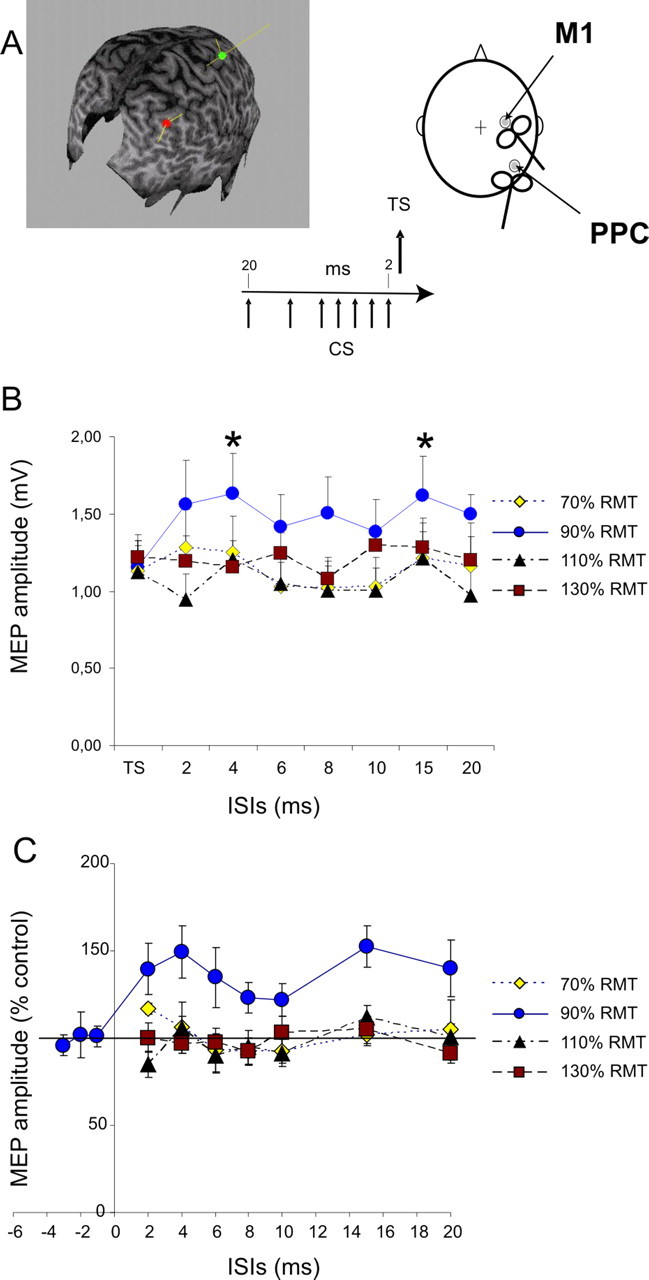
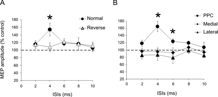
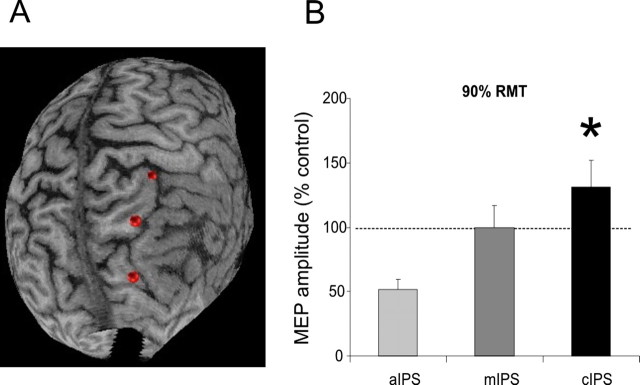
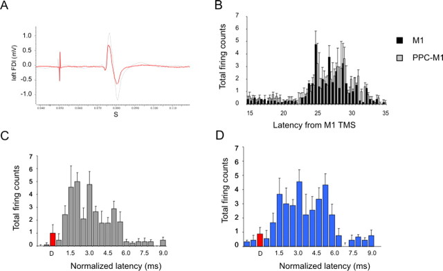
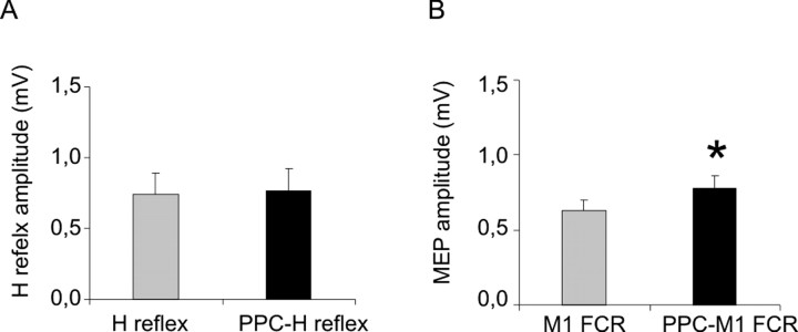
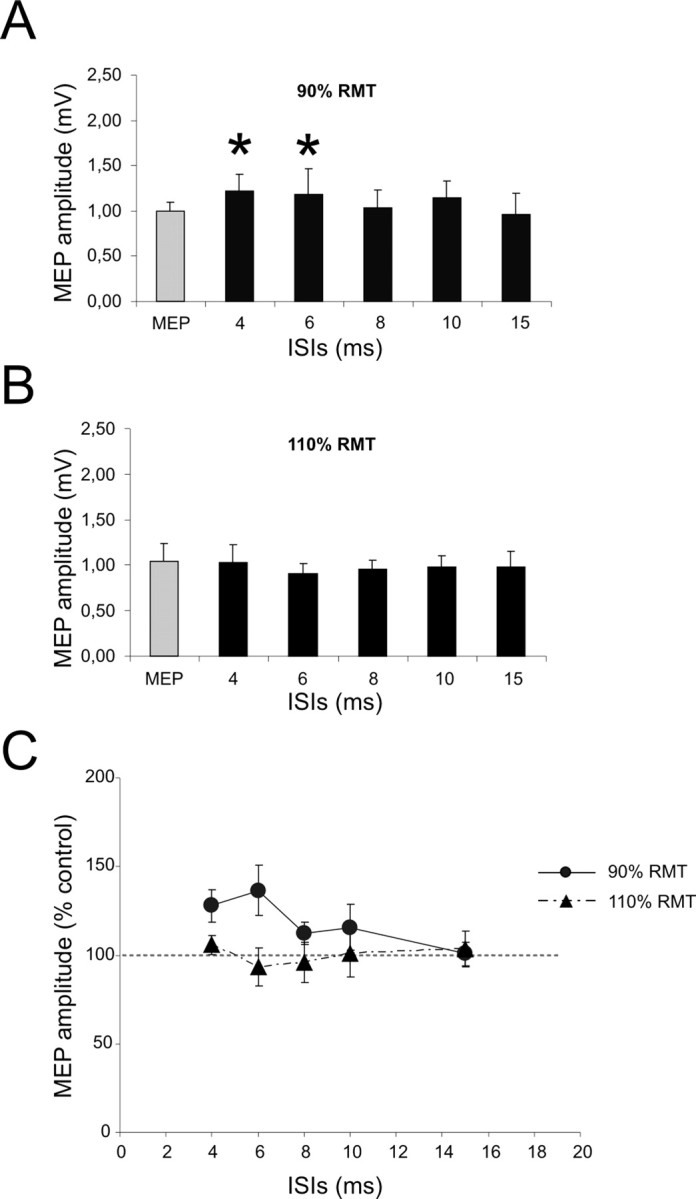
Paired-pulse transcranial magnetic stimulation (TMS) has been applied as a probe to test functional connectivity within distinct cortical areas of the human motor system. Here, we tested the interaction between the posterior parietal cortex (PPC) and ipsilateral motor cortex (M1). A conditioning TMS pulse over the right PPC potentiates motor evoked-potentials evoked by a test TMS pulse over the ipsilateral motor cortex, with a time course characterized by two phases: an early peak at 4 ms interstimulus interval (ISI) and a late peak at 15 ms ISI. Activation of this facilitatory pathway depends on the intensity of stimulation, because the effects are induced with a conditioning stimulus of 90% resting motor threshold but not at lower or higher intensities. Similar results were obtained testing the ipsilateral interaction in the left hemisphere with a slightly different time course. In control experiments, we found that activation of this facilitatory pathway depends on the direction of induced current in the brain and is specific for stimulation of the caudal part of the inferior parietal sulcus (cIPS) site, because it is not observed for stimulation of adjacent scalp sites. Finally, we found that by using poststimulus time histogram analysis of single motor unit firing, the PPC conditioning increases the excitability of ipsilateral M1, enhancing the relative amount of late I wave input recruited by the test stimulus over M1, suggesting that such interaction is mediated by specific interneurons in the motor cortex. The described facilitatory connections between cIPS and M1 may be important in a variety of motor tasks and neuropsychiatric disorders.


Figures

Figure 1.
Figure 1.
Effects of CS applied over the right PPC at different intensities on MEPs obtained by right M1 stimulation with subjects at rest. CS preceded TS applied over M1 by different ISIs ranging from 20 to 2 ms. The relative orientations of the coils are shown. In each case, the monophasic current pulse flows into the handle of the coil, inducing current in the opposite direction in the underlying cortex. In the MRI reconstruction, the most anterior point (green dot) is at the junction with the postcentral sulcus (hand area of the motor cortex), and the posterior point (red dot) lies over the angular gyrus. The yellow lines represent the ideal trajectories of the magnetic fields; these lines terminate at the presumed site of stimulation. A, Right PPC conditioning exerted potentiation over ipsilateral motor cortex. A single CS applied over the right PPC changed the amplitude of MEP obtained for ipsilateral M1 stimulation selectively when intensity of CS was set at 90% RMT with significant peaks obtained for ISIs of 4 and 15 ms. B, No significant change was observed for lower (70% RMT) or higher intensities of CS (110%, 130% RMT). Absolute values of MEPs in different conditions (mV) are shown in B, whereas data in C are normalized and expressed as percentages of control test conditions. Data obtained in experiment 3, when TS preceded by 1, 2, and 3 ms the CS set at 90% RMT intensity, are included in C. Errors bars indicate 1 SEM. Asterisks indicate a p value <0.05 at post hoc analysis.
Figure 2.
Figure 2.
The effect of PPC conditioning is sensitive to the direction of current circulating in the coil. A, In fact, potentiation of MEPs was obtained at 90% RMT only when the direction of the coil was the same as in experiment 1 but not when it was delivered with the opposite direction. B, There was facilitation selectively when CS was applied over P4 but not 2 cm medially or laterally. Errors bars indicate 1 SEM. Asterisks indicate a p value <0.05 at post hoc analysis.
Figure 3.
Figure 3.
A, A three-dimensional structural MRI of one representative subject. The red dots represent the three cortical sites for the conditioning stimulus: top dot, aIPS; middle dot, mIPS; bottom dot, cIPS. B, Facilitation of ipsilateral motor cortex was found selectively for stimulation of the cIPS. Errors bars indicate 1 SEM. Asterisks indicate a p value <0.05 at post hoc analysis.
Figure 4.
Figure 4.
PPC conditioning exerts facilitation of ipsilateral M1 trough corticocortical projections that interact with the late components of MEPs. A, Average surface MEPs from a representative subject for TS (red line) and CS (90% RMT with 4 ms ISI, gray line). The same slope was observed in the early phase, with a late increase in amplitude after 3 ms. The effect of PPC conditioning on MEP evoked by right M1 TMS were then studied with PSTHs recorded trough single unit EMG activity. B, The different firing probability at various latencies from M1 stimulation averaged across subjects when TS was delivered alone (black bars) or was preceded by PPC CS (gray bars). PPC conditioning significantly modified the firing probability of PSTHs components. C, D, Compared with TS alone (C), there was more late period-related firing probability for PPC conditioning (D). Error bars indicate SEM.
Figure 5.
Figure 5.
Effects of PPC conditioning on H-reflexes and MEPs recorded from the FCR muscle. Average data from five subjects. A, No significant modulation of H-reflex was found during PPC conditioning. B, FCR MEP amplitude was potentiated during PPC conditioning (90% intensity; ISI, 4 ms). Error bars indicate 1 SEM. Asterisks indicate a p value <0.05 at post hoc analysis.
Figure 6.
Figure 6.
Effects of CS applied over left PPC at different intensities on MEPs obtained by left M1 stimulation. CS preceded TS applied over M1 by different ISIs ranging from 15 to 4 ms. A, A single CS applied over the left PPC potentiated the amplitude of MEP obtained for ipsilateral M1 stimulation selectively when intensity of CS was set at 90% RMT with significant peaks obtained for ISIs of 4 and 6 ms. B, No significant change was observed for higher intensities of CS (110% RMT). Data in C are normalized and expressed as a percentage of control test condition. Errors bars indicate 1 SEM. Asterisks indicate a p value <0.05 at post hoc analysis.

References

    1. Battaglia-Mayer A, Caminiti R, Lacquaniti F, Zago M. Multiple levels of representation of reaching in the parieto-frontal network. Cereb Cortex. 2003;13:1009–1022. - PubMed
    1. Baumer T, Bock F, Koch G, Lange R, Rothwell JC, Siebner HR, Munchau A. Magnetic stimulation of human premotor or motor cortex produces interhemispheric facilitation through distinct pathways. J Physiol (Lond) 2006;572:857–868. - PMC - PubMed
    1. Bestmann S, Baudewig J, Siebner HR, Rothwell JC, Frahm J. Functional MRI of the immediate impact of transcranial magnetic stimulation on cortical and subcortical motor circuits. Eur J Neurosci. 2004;19:1950–1962. - PubMed
    1. Bestmann S, Baudewig J, Siebner HR, Rothwell JC, Frahm J. BOLD MRI responses to repetitive TMS over human dorsal premotor cortex. NeuroImage. 2005;28:22–29. - PubMed
    1. Bonato C, Miniussi C, Rossini PM. Transcranial magnetic stimulation and cortical evoked potentials: a TMS/EEG co-registration study. Clin Neurophysiol. 2006;117:1699–1707. - PubMed

Publication types

LinkOut - more resources